Hepatoprotective Effect of Solanum melongena/Eggplant against Acute
Hepatitis
Nabhan Komara1, Herri S. Sastramihardja2, Afiati3
1Faculty of Medicine, Universitas Padjadjaran, 2Department of Pharmacology and Therapy,
Faculty of Medicine, Universitas Padjadjaran, 3Department of Pathology Anatomy, Faculty of
Medicine, Universitas Padjadjaran/Dr. Hasan Sadikin General Hospital, Bandung
Abstract
Background: Hepatitis is a liver inflammation that can be acute or chronic and may cause damage to
hepatocytes such as necrosis. Hepatocyte necrosis can be inhibited by antioxidants such as flavonoids
found in Solanum melongena fruit. This study aimed to determine the hepatoprotective effect of Solanum
melongena fruit infusion to inhibit hepatocytes damage in CCl4-induced rats.
Methods: Twenty five male Wistar rats were randomly divided into five groups, and adapted for 7 days
before the experimental study. Negative and positive groups were given aquadest, Group III−V were given
Solanum melongena fruit infusion containing 1.125 grams, 2.25 grams and 4.5 grams orally for 9 days. At
the 9th day, all rats were induced by 8 mL/kgBW of 10% CCl4 in paraffin, except for the negative group. Rats were sacrificed on the 11th day, and liver biopsy preparations were made. Hepatocyte necrosis was counted and was analyzed by Kruskal-Wallis test and Mann-Whitney test.
Results: The study showed that the percentage of necrotic hepatocytes in group III, IV a nd V were lower
than in the positive group. Using Mann-Whitney test, there were significant differences in negative group, group III, and group V (p<0.05). Meanwhile, unsignificant difference was seen between the positive group and group IV (p>0.05). Kruskal-Wallis test showed that there weresignificantly differences among groups (p<0.05).
Conclusions: Solanum melongena fruit infusion has hepatoprotective effects against acute hepatitis in rat model histopathologically.
Key words: CCl4, flavonoids, hepatocyte, necrosis, Solanum melongena
Correspondence: Nabhan Komara, Faculty of Medicine, Universitas Padjadjaran, Jalan Raya Bandung-Sumedang Km.21,
Jatinangor, Sumedang, Indonesia, Phone: +6285220095758 Email: [email protected]
Introduction
Hepatitis is an inflammatory condition of the liver. Two billion people have been infected by hepatitis B virus around the world and about 350 million people are at
risk to have cirrhosis, liver failure and liver cancer. Moreover, hepatitis causes 1 million death each year. 1 Hepatitis can be acute or chronic. It is called acute hepatitis if less than
6 months, and chronic hepatitis if it is more
than 6 months. Hepatitis is also classified by
the causes, such as hepatitis virus infection, autoimmune hepatitis, and hepatitis due
to chemical or hepatotoxicity, which one of agent toxicity is carbon tetrachloride.2,3
Carbon tetrachloride (CCl4) is a synthetic chemical compound that can be converted to free radical compounds that are not stable and
can cause damage to the liver. The damage
of the liver progresses rapidly can be either
degeneration or necrosis of hepatocyte.3 Rubinstein and Suja et al. in Orhan et al.4 reported that the use of CCl4 inducing
hepatotoxicity model is frequently conducted
to investigate hepatoprotective drugs effect
and plant extract, because the present of liver
damage is similar to acute viral hepatitis.
Solanum melongena L (Solanaceae)/ Eggplant is a common subtropic and tropic vegetable, and consumed throughout the world. It is known to contain phenolic and flavonoids substances, which have antioxidant
activity.5,6 Cao et al. in Akanitapichat et al.5
reported that the whole eggplant fruits
possess antioxidant activities and are ranked among the top 10 vegetables in terms of antioxidant capacity. Nasunin, an anthocyanin
isolated from the peel of purple eggplant fruit,
superoxide scavenging activity.5
The study aimed to determine the hepatoprotective effect of Solanum melongena
to inhibit hepatocytes damage in CCl4-induced
rats
Methods
An experimental study was conducted at Animal Laboratory of Department of
Pharmacology and Therapy in Dr. Hasan Sadikin General Hospital Bandung during the
period of September to October 2012. Solanum melongena fruits were procured from Lembang, Bandung, and were botanically identified at the Herbarium Jatinangor, Universitas Padjadjaran. Twenty five healthy male Wistar rats (2−3 months old) weighing 150−250 gram were used as subjects. All rats were procured from Pusat Antar Universitas, Institut Teknologi
Bandung; and were adapted in homogenous
temperature and dark-light cycle for 7 days
with an access to food and drink. The procedure
was conducted in accordance with Russel and Burch’s principle of experimental ethics : reduction, refinement, and replacement.7
Solanum melongena fruit infusion was
prepared from sliced Solanum melongena
fruit weighing according to dosage and 100
mL of water for every treatment group. Doses were obtained from Paget and Barnes’s table
conversion, and were made to ½, 1 and 2 times dosage.8 Mixed raw material was heated
in an infusion pan for 15 minutes after the
temperature reached 90˚C and stirred every 5 minutes. Furthermore, the mixture was filtered using flanel fabric into measuring cup to produce infusion. If the infusion did not reach 100 mL, extra boiling water was added
to the infusion, stirred for one minute and then
filtered again into the measuring cup until it reached the required volume.
Negative and positive groups were given aquades, group III−V were given Solanum
melongena fruit infusion with a row that contained 1.125 grams, 2.25 grams and 4.5 grams orally for 9 days every morning. At afternoon of 9th day, all rats were induced
8 ml/kg of 10% CCl4 in paraffin, except negative group. Rats were sacrificed on
11th day using ketamine hydrochloride 0.4
mL. After laparotomy, blood in rat liver was drained by physiological saline through the
left ventricle. The liver was taken and put into formalin solution then cut into size of
1 cm x 0.5 cm x 0.5 cm and the liver chunks were made into paraffin block. The liver was sliced with thickness of 5 µm and stained by
haematoxylin-eosin. Hepatocyte necrosis in
each group was counted and calculated into
percentage value by comparing the amount of
hepatocyte necrosis with amount of normal hepatocyte in the negative group. Data were
statistically analyzed using Kruskal-Wallis and Mann-Whitney non-parametric tests.9
Results
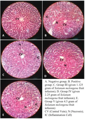
Histopathological appearance of liver biopsy can be seen in Figure 1. The negative group
showed hepatocytes arranged radially surrounded the central vein. Hepatocytes
looked good with round nuclei and basophils, homogeneous cytoplasm, clear cell borders,
indicating that liver tissue was still in normal circumstances, although found a few hepatocytes that underwent necrosis. The positive group showed a defect in hepatic tissue. Hepatocyte appeared unstructured radially,
with abundant infiltration of inflammatory
cells such as lymphocytes in the portal area
(zone 1) and extends toward the central
vein. There were found many degeneration
cells such as ballooning degeneration and acidophilic degeneration. Cells undergoing
necrosis were found in almost all zones, with the worst damage in zone 1. However, normal
hepatocytes could be still found in zones 2 and 3. The group III, IV and V showed a defect in hepatic tissue but not as bad as in the positive
group. Hepatocyte appeared unstructured
radially but not as bad as in the positive group, with a few of infiltrations of inflammatory cells such as lymphocytes in the portal area (zone 1). There were rarely found degeneration cells either ballooning degeneration or acidophilic degeneration. Cells undergoing necrosis were
found in zone 1, and normal hepatocytes were more common than in the positive group.
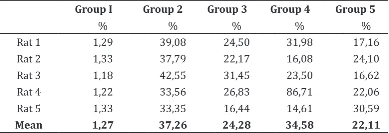
The number and percentage of necrotic hepatocyte in each group can be seen in Table
1. The negative group had the fewest necrotic
hepatocyte (1.27%). Meanwhile, the most
necrotic hepatocyte were seen in positive
group (37.26%). All groups given Solanum melongena fruit infusion in 9 days before induction by CCl4 had necrotic hepatocytes fewer than positive group.
This result showed that Solanum
melongena fruit infusion provided protection for hepatocyte against further damage, or in other word it had hepatoprotective effect in
CCl4-induced rats.
To determine hepatoprotective effect
A. Negative group; B. Positive group; C. Group III (given 1.125 gram of Solanum melongena fruit infusion); D. Group IV (given 2.25 gram of Solanum melongena fruit infusion); E. Group V (given 4.5 gram of Solanum melongena fruit infusion)
CV (Central Vein); N (Necrosis); IC (Inflammation Cell)
CV
A
CV N IC
B
CV
N
IC
E
N CV
D CV
IC N
C
Figure 1 Histopathology of liver biopsy with 400x magnification
positive group. The result of this test were
significant different in the negative group,
the group given 1.125 gram, and 4.5 gram of Solanum melongena fruit infusion with
p=0.005 (p<0.05). Meanwhile, insignificant difference was seen between the positive
group and the group who given 2.25 gram of Solanum melongena fruit infusion with
p=0.058 (p>0.05).
To determine the significance effect, Kruskal-Wallis test was exerted, and the result was significantly with p=0.002 (p<0.05).
Discussion
In this study, based on comparison with normal
cells in the negative group, it was found that
in the positive group which is only induced by CCl4 showed that the amount of hepatocyte
necrosis is 37.26%. CCl4 is a chemical activated
by cytochrome p450 to form free radicals. Initially, CCl4 converted to trichloromethyl
radical (CCl3-) will form trichloromethylperoxy radical (CCl3O2-), which can cause lipid
peroxidation if it reacts with oxygen. Although lipid peroxidation caused by CCl3O2- is more
dominant, CCl3- can also directly cause
lipid peroxidation by binding with fat. Lipid peroxidation is the main cause of tissue damage, including inflammation. In this study, liver
tissue damage was found in accordance with a
previous study by Brattin and Shi which stated that CCl4 can cause damage to hepatocytes.10,11
Table 1 The percentage of necrotic hepatocyte in each group
Group I Group 2 Group 3 Group 4 Group 5
% % % % %
Rat 1 1,29 39,08 24,50 31,98 17,16
Rat 2 1,33 37,79 22,17 16,08 24,10
Rat 3 1,18 42,55 31,45 23,50 16,62
Rat 4 1,22 33,56 26,83 86,71 22,06
Rat 5 1,33 33,35 16,44 14,61 30,59
Mean 1,27 37,26 24,28 34,58 22,11
I = Negative Group; II = Positive Group; III = Dosage ½ x; IV = Dosage 1 x; V = Dosage 2 x.
previous study by Shi and Badria, liver tissue damage induced by CCl4 naturally suggests that
hepatocyte necrosis is more abundant in zone 3 (centrilobular necrosis), because in zone 3
there are many cytochrome p450 that activate
CCl4 into CCl3- which are free radicals. In this
study, hepatocytes that undergo necrosis
are more common in zone 1 (periportal necrosis).11,12 In addition to hepatocyte damage caused by CCl3- which is attached directly to fat
causing lipid peroxidation, necrosis can also be caused by the CCl3O2- which is a product of
a reaction between CCl3- and oxygen. CCl3O2-
has a more reactive form and predominantly
causes lipid peroxidation.3 Zone 1 is the richest zone with oxygen, therefore reaction of CCl3- with oxygen to form CCl3O2- is more common in this zone. Thus, it is possible for
more necrosis in zone 1 as found in this study.
It is also found in another study by Domitrovic (2009) that the results showed the presence of
liver tissue damage in zone 1.13
Cells that suffered from damage caused by unstable molecules such as free radicals can be protected by a compound called antioxidants. The antioxidants can slow or prevent the formation of free radical oxidation. One of the antioxidants contained in Solanum melongena fruit is flavonoids. Flavonoids will give electrons to CCl3-, thus prevent the
formation of CCl3O2- and prevent covalent bonds with fat. Therefore, the process of lipid peroxidation can be inhibited, so there is no tissue damage to the liver.14 In this study, the
groups that were given Solanum melongena fruit infusion, which are group III (24.28%), group IV (34.58%), and group V (22.11%)
have fewer hepatocyte necrosis compared to the positive group which has more necrosis
(37.26%). This is consistent with the results of the study by Akanitapichat which showed that
the Solanum melongena fruit has antioxidant
and hepatoprotective activities. Another study
by Arhoghro also showed that flavonoids have
hepatoprotective activities although not from the Solanum melongena fruit.15
Based on these results, it can be concluded
that administration of Solanum melongena fruit infusion in all rat groups III and V can provide hepatoprotective effect against CCl4 -induced acute hepatitis. In group IV, there is a rat with 86.71% necrosis which has more
necrosis than the mean in the positive group
(37.26%), although the mean necrosis in group IV (34.58%) is still below the mean of positive group. This could happen because
according to the theory, each rat has different level of immunity as well as different immunity of each individual human. Therefore, although this rat has given Solanum melongena fruit infusion which has hepatoprotective effect on other groups, hepatoprotective effect in this rat does not appear.
This study analyzed only one variety of eggplant, meanwhile there are many
varieties that can be planted in Indonesia and frequently consumed by Indonesian people. Those varieties should be analyzed its
hepatoprotective effect as well.
It concluded that Solanum melongena fruit infusion has hepatoprotective effects against acute hepatitis rat model.
References
1. Ministry of Health Republic of Indonesia.
Saatnya lawan hepatitis. 2012 [Cited 2012 April 8]; Available from: http://www. depkes.go.id/index.php/berita/press-release/1557-saatnya-lawan-hepatitis.
html.
2. Fauci AS, Braunwald E, Kasper DL, Hauser
3. Hodgson E, editor. A Textbook of Modern
Toxicology. 3rd ed. New Jersey: John Wiley & Sons, Inc.; 2004.
4. Hariana A. 812 Resep untuk mengobati 235 penyakit. 2nd ed. Depok: Penebar
Swadaya; 2005.
5. Akanitapichat P, Phraibung K, Nuchklang
K, Prompitakkul S. Antioxidant and hepatoprotective activities of five eggplant varieties. Food Chem Toxicol. 2010;48(10):3017−21.
6. Tiwari A, Jadon RS, Tiwari P, Nayak S. Phytochemical investigation of crown of
solanum melongena fruit. International Journal of Phytomedicine. 2009;1:9−11. 7. Hanafiah MJ. Etika kedokteran dan hukum
kesehatan. 4th ed. Jakarta: EGC; 2009.
8. Hashem MM, Atta AH, Arbid MS, Nada
SA, Asaad GF. Immunological studies on Amaranth, Sunset Yellow and Curcumin as food colouring agent in albino rats. Food Chem Toxicol. 2010;48(6):1581−86.
9. Dahlan MS. Statistik untuk kedokteran dan kesehatan. 4th ed. Jakarta: Salemba
Medika; 2009.
10. Brattin WJ, Glende Jr EA, Recknagel RO.
Pathological mechanisms in carbon
tetrachloride hepatotoxicity. J Free Radic Biol Med. 1985;1(1):27−38.
11. Shi J, Aisaki K, Ikawa Y, Wake K. Evidence of hepatocyte apoptosis in rat liver after
the administration of carbon tetrachloride. Am J Pathol. 1998;153(25):515−25.
12. Badria AF, El-Belbasi HI, Sobh MM, Badria
FA. Parallelism study between biochemical,
immunological and histochemical
parameter of liver injury induced by carbon tetrachloride on rats. Journal of American Science. 2011;7(5):581−91.
13. Domitrovic R, Jakovac H, Tomac J, Sain
I. Liver fibrosis in mice induced by carbon tetrachloride and its reversion by luteolin. Toxicol App Pharmacol. 2009;241(3):311−21.
14. Hamid AA, Aiyelaagbe OO, Usman LA,
Ameen OM, Lawal A. Antioxidants:
its medicinal and pharmacological
applications. Afr J Pure App Chem. 2010;4(8):142−51.
15. Arhoghro EM, Ekpo Ke, Ibeh GO. Effect