© 2013 Savvy Science Publisher
The Effect of Two Bulk Fill Resin Composites on Microleakage in
Endodontically Treated Teeth
Safa Tuncer
1, Mustafa Demirci
1,*, Neslihan Tekçe
2, Aysun Kara Tuncer
3and
Harika Gözükara Ba
41
Department of Conservative Dentistry, Faculty of Dentistry, Istanbul University, Istanbul, Turkey
2
Ataehir Oral Health Hospital, Istanbul Turkey 3
Department of Endodontics, Faculty of Dentistry, Bezmialem Vakıf University, Istanbul, Turkey 4
Department of Biostatistics, Faculty of Medicine, Inonu University, Malatya, Turkey
Abstract: Purpose: To evaluate the effect of two bulk fill resin composites on microleakage in endodontically treated
Class II restorations.
Materials and Methods: Twenty-four non-carious molars were selected and randomly divided into three groups (n = 8).
The Class II cavity preparations were made with the cervical margin 1 mm below the cementum-enamel junction, and endodontic treatment was performed using a resin-based sealer and gutta-percha points. In Group 1 (Control), resin composite (G-aenial Posterior; GC Corp., Tokyo, Japan) was applied to the interproximal wall, and then restorative material was applied incrementally. In Group 2, resin composite was applied to the interproximal wall, followed by a 4-mm-layer of a bulk fill flowable composite (x-tra base; Voco, GmbH, Cuxhaven, Germany) and the remaining occlusal part of the cavity was filled with resin composite. In Group 3, resin composite was applied to the interproximal wall, followed by a 4mm layer of a bulk fill fiber-reinforced composite (everX Posterior; GC Corp., Tokyo, Japan), and the remaining occlusal part of the cavity was filled with a resin composite. The restorations were then subjected to 500 thermal cycles, each with a dwell time of 20 seconds at 5 and 55 oC. The adaptation at the cervical margin was evaluated by dye penetration, and one tooth was used to evaluate the restorative material interface using SEM. The data were statistically analyzed using the Kruskal Wallis test (p < 0.05).
Results: No significant difference in dye penetration was found between the control and the experimental groups.
Microleakage was significantly higher on enamel margins compared with the dentin margins for all of the groups.
Conclusion: The use of bulk fill restorative materials under resin composites does not affect the sealing ability of
restorations.
Keywords: Short fiber composite, Leakage, Resin based material.
INTRODUCTION
Endodontic treatment is generally performed on teeth significantly affected by caries, multiple repeat restorations and/or fractures [1]. Kirkevang et al. evaluated the relationship between the quality of endodontic and coronal restorations and periapical status, demonstrating the significance of coronal restoration quality in the incidence of apical periodontitis as well as the significance of root canal treatment quality [2]. Endodontically treated teeth can be restored with either indirect or direct restorations. With direct resin composite restorations, polymerization shrinkage and shrinkage stress can cause gap formation and microleakage, which can result in secondary caries, post-operative sensitivity, and clinical failure of the restoration. New resin composites and adhesive materials have been developed to improve marginal adaptation, reduce polymerization shrinkage
*Address correspondence to this author at the Department of Conservative Dentistry, Faculty of Dentistry, Istanbul University, Istanbul, Turkey; Tel: +90 212 414 20 20; Fax: +90 212 525 00 75; E-mail: md.demirci@gmail.com
and shrinkage stress and work effectively on enamel, dentin and cement in Class II direct composite restorations [3,4].
conventional resin-based composite materials, with a reported depth of cure in excess of 4 mm [8-11].
Recently, a short fiber-reinforced composite material (everX Posterior, GC Corp., Tokyo, Japan) was introduced for use with the bulk filling technique [12]. This restorative material is intended to be used as a base-filling material in high stress-bearing areas, especially in large cavities of vital and non-vital posterior teeth [12]. The restoration base should be a maximum of 4 mm thick (“cavity lining”) for Class I and II composite restorations and must be covered with a wear- resistant, polishable restorative composite, such as GC G–ænial Posterior or GC Kalore. The composite layer should be 1-2 mm on the occlusal surface [13]. EverX Posterior consists of a combination of a resin matrix, randomly orientated E-glass fibers and inorganic particulate fillers [5]. The resin matrix contains bis-GMA, TEGDMA and PMMA, forming a matrix referred to as a semi-interpenetrating polymer network (semi-IPN), which provides good bonding properties and improves the toughness of the polymer matrix [14,15].
The low-viscosity, flowable composite x-tra base (Voco, GmbH, Cuxhaven, Germany) (filler content: 75% by weight) is a flowable composite with reduced shrinkage on a traditional methacrylate base. It is indicated for use in the bulk filling technique to introduce a maximum 4-mm-thick restoration base (“cavity lining”) in Class I and II composite restorations. After curing, x-tra base must be covered in the region of the occlusal anatomy by another layer of a methacrylate-based hybrid composite at least 2 mm in thickness and suitable for posterior teeth, such as the nanoparticle-modified hybrid composite GrandioSO (Voco, GmbH, Cuxhaven, Germany). X-tra base can also be applied in a first thin layer as a cavity liner in Class I and II cavities [10,16].
An elastic liner between the tooth structure and composite resin may compensate for contraction stresses and prevent gap formation [17]. The performance of dentin adhesives or a low-viscosity, low-modulus intermediate resin as an elastic barrier between the dentin adhesive and resin-based restorative material has been investigated previously [18,19]. However, the use of flowable resin did not produce gap-free resin margins in Class II cavities or in bulk filled restorations [20].
The restoration of endodontically treated teeth must be designed to maximize the strength of these teeth and increase their longevity [21]. Bulk fill flowable
resin-based composite bases being marketed for use beneath conventional resin-based composite materials have also been recommended for use with endodontically treated teeth [10,13]. Therefore, the aim of this study was to evaluate the effect of two bulk fill resin composites on microleakage in endodontically treated Class II (MO) restorations of premolar teeth.
MATERIALS & METHODS
Twenty-four sound human upper premolars with fully developed apexes extracted for orthodontic reasons were selected for this study. After extraction, they were hand-scaled to remove tissue remnants and stored at 4 oC in a 0.5% aqueous chloramine T solution until further use.
Before preparing the teeth, an outline of the cavity was drawn with a lead pencil, and parallel-sided standardized MO cavities were prepared using a diamond bur (835-012-4, Diatech, Switzerland) in a high-speed hand piece under an air–water spray. The buccolingual width of the cavity was 4 ± 0.1 mm on the occlusal and gingival sides. The gingival wall was 3.5 ± 0.2 mm deep to the axial wall. The bur was replaced after every fourth cavity preparation to ensure high cutting efficacy. The facial and lingual walls of the occlusal segment were prepared in parallel. The cavosurface margins were prepared at 90°, and all of the internal angles were rounded.
After the preparations, a conservative endodontic access was performed on the pulp chamber wall. Next, all of the canals were prepared using a Pro-Taper Ni-Ti Rotary System (Dentsply Malleiffer) and obturated with an AH 26 sealer (Dentsply; DeTrey, Konstanz, Germany) and gutta-percha using a lateral compaction technique. Following the endodontic treatments, the coronal root canal openings were sealed with a thin layer of conventional glass ionomer cement (Ketac Molar Easy Mix, 3M Espe, St. Paul, MN, USA), and then the teeth were randomly divided into three groups (n = 7) according to the restorative materials that were used.
Group 1 (Controls)
and light cured for 10 s with a quartz-tungsten-halogen (QTH) curing light (Optilux 501, SDS/Kerr, Danbury, CT, USA) according to the manufacturer’s instructions. After adhesive polymerization, a metal matrix band (Adapt SuperCap Matrices, Kerr, Bioggio, Switzerland) was placed around the tooth. A thin layer of a resin composite (G-aenial Posterior GC Corp., Tokyo, Japan) was applied toward the metallic matrix contacting the cavosurface of the proximal box up to one third of the occlusal-cervical extension. The other two layers were applied over the previous increment contacting the cavosurface margin of the proximal box and forming the marginal ridge. Using the centripetal technique, the mesial wall (approximately 1 mm thick) of the restoration was completed, and the cavity was turned into a class I thereafter. Then, three increments of resin composite were applied horizontally. Each increment was light-cured for 20 s.
Group 2
G-aenial Bond was applied to the enamel and dentin surfaces according to the manufacturer’s instructions. After the matrix application, the mesial wall of the cavity was created with G-aenial Posterior as previously described. The bulk fill flowable composite x-tra base (Voco GmbH, Cuxhaven Germany) was applied at a 4-mm thickness horizontal to the resulting class I cavity and cured for 20 s. After the flowable composite application, the remaining cavity was restored with resin composite.
Group 3
G-aenial Bond was applied to the enamel and dentin surfaces according to the manufacturer’s instructions. After the matrix application, the mesial wall of the cavity was created with G-aenial Posterior as previously described for Group 1. The bulk fill fiber-reinforced composite everX Posterior (GC Corp., Tokyo, Japan) was applied at a 4-mm thickness horizontal to the resulting class I cavity and cured for 20 s. After the fiber-reinforced composite application, the remaining cavity was restored with resin composite.
The restorations were polished using a one-step finishing/polishing system (One Gloss, Shofu Inc., Kyoto, Japan) The restorations were stored in distilled water at 37°C for 24 h and then thermocycled 500 times between 5 and 55 °C with a dwell time of 30 s. After thermocycling, the apices of all of the teeth were sealed with amalgam (YDA Amalgam Alloy Capsules, Hangzhou Yinya New Materials Co. Ltd., Hangzhou, China), and 2 coats of nail polish were applied to within
approximately 1 mm of the tooth/composite interface. Next, the specimens were immersed in a 0.5% aqueous solution of methylene blue at room temperature for 24 h. After removal from the solution, any surface-adhered dye was carefully rinsed away with tap water.
On each restoration, two mesio-distal cuts were prepared longitudinally with a diamond saw mounted in a cutting machine (Isomet, Buehler; Lake Bluff, Illinois, USA). These preparations yielded four evaluation surfaces (28 evaluation surfaces per group) for each restoration. The sections were observed under a stereomicroscope (Olympus SZ61, Olympus Corporation, Tokyo, Japan) at 40 magnification, and microleakage at the occlusal and gingival walls in each section was evaluated by two independent operators according to the following scoring system:
Scores for cervical margins were:
0 = no penetration;
1 = leakage extending within first 1/2 of the cavity wall;
2 = leakage extending beyond 1/2, but not as far as the cervical cavity floor;
3 = leakage extending beyond the cervical cavity Wall and reaching the cavity floor.
For enamel margins, the scores were:
0= no penetration;
1 = leakage extending within first of the enamel wall;
2 = leakage extending beyond 1/2, but not as far as the dentin-enamel junction;
3 = leakage extending beyond the enamel-dentin junction. [22]
dried for 10 seconds. Afterwards, specimens were treated with 10% sodium hypochlorite for 30 seconds, rinsed thoroughly with water and fixed in glutaraldehyde solution (pH 7.4) for two hours. Then they were dehydrated through ascending series of ethanol (25% to 100%) and dried at room temperature for 24 hours. Following the drying procedure, the samples were sputter-coated with gold (Emitech K-550X sputter coater; Emitech, Ashford,UK), operating at 20kV under SEM (JEOL JCM-5000 NeoScope™ , JEOL, Tokyo, Japan) with various magnifications.
Statistical analysis was carried out using the Kruskal-Wallis test to determine statistically significant differences in leakage at the occlusal and gingival margins separately among groups for each dentin adhesive, and between the three dentin adhesives for the same groups. An intergroup comparison of occlusal versus gingival margin locations was completed using the Wilcoxon signed-rank test. All the statistical tests were performed at a p<0.05 level of significance.
RESULTS
The distribution data, mean values and standard deviations for the enamel and dentin microleakage for each group as well as pairwise comparisons are shown in Table 1.
For the enamel and dentin margins, there were no significant differences among the tested groups. In contrast, a significant difference between the enamel and dentin microleakage was observed in all three groups (Figure 1).
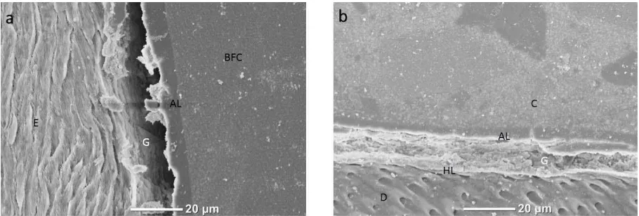
The SEM images of the interfaces treated with G-Aenial Bond for the three groups are shown in Figures
2-4. In Group 1, the adhesive layer (AL) was uniform;
however, there were gap formations between the AL and the enamel (Figure 2a). Figure 2b shows that the hybrid layer (HL) was very thin, although the AL was uniform and thick. In addition, a uniform contact was observed between the AL and the composite, although
voids in the AL and resin tags were clearly visible. In Group 2, there was a gap between the AL and enamel (Figure 3a). In the dentin margins, the AL, HL and resin tag formation were visible, although there was a breakdown in the AL. In Group 3, gap formation occurred in the enamel margin (Figure 4a), as was observed for the other groups. In Figure 4b, a non-uniform HL and AL were observed with gap formations noted between these 2 layers.
Figure 1: Typical dye leakage; enamel score 3, cervical score 1.
DISCUSSION
Different restorative materials and techniques are used for the coronal restoration of endodontically treated teeth. A coronal restoration after endodontic treatment can prevent the movement of bacteria and their products and also reinforce the residual tooth structure. Therefore, the long-term prognosis of endodontically treated teeth depends on the quality of the final restoration [23-26]. In endodontically treated teeth, the volume of the restoration is larger and more resin increments are required to fill the cavity preparation. Therefore, endodontic treatment causes Table 1: Data of distribution, the mean values and standard deviations of enamel and dentin microleakage for each
group and pairwise comparisons. Capital letters are the comparison of the groups within the enamel and dentin margins (p>0.05). Within a row, values having different lower case exhibited statistically significant difference (p<0.05); comparison of the same groups between enamel and dentin
Enamel leakage scores Dentin leakage scores Adhesive
system
Groups
n 0 1 2 3 Mean SD * 0 1 2 3 Mean SD *
G1 28 0 5 8 15 2.36 .78 Aa 6 9 6 7 1.5 1.11 Ab
G2 28 1 3 13 11 2.21 .77 Aa 8 5 6 9 1.57 1.23 Ab
G-aenial Bond
Figure 2: (a) SEM image enamel-G-Aenial Bond -resin composite interface in Group 1. (C-Composite, AL-Adhesive Layer, E-Enamel); (b) SEM image of dentin- G-Aenial Bond interface in Group 1. (black arrow-Voids, C-Composite, D-Dentin, AL-Adhesive Layer, HL-Hybrid Layer).
Figure 3: (a) SEM image enamel-G-Aenial Bond -resin composite interface in Group 2. (FRC-Fiber reinforced composite, AL-Adhesive Layer, E-Enamel, G-Gap formation); (b) SEM image of dentin- G-Aenial Bond interface in Group 2. (black arrow-Voids, FRC-Fiber reinforced composite, C-Composite, D-Dentin, AL-Adhesive Layer, HL-Hybrid Layer, G-Gap formation).
Figure 4: (a) SEM image enamel-G-Aenial Bond -resin composite interface in Group 3. (BFC-Bulk fill composite, AL-Adhesive Layer, E-Enamel, G-Gap formation); (b) SEM image of dentin- G-Aenial Bond interface in Group3. (C-Composite, D-Dentin, AL-Adhesive Layer, HL-Hybrid Layer).
the loss of the roof of the pulp chamber and may flex due to shrinkage stresses. All these factors may affect the marginal quality of bonded restorations in endodontically treated teeth [22]. In the present study, there were no statistically significant differences observed among the three groups with respect to
capacity of restorations in class II cavities. Consistent with these findings, it has been reported that a bulk fill resin-based composite used as a 4 mm bulk fill dentine replacement performs well with respect to marginal quality [27]. Also in support of the present study findings, Moorthy et al. demonstrated that class II cavities filled with a resin-based composite (GrandioSO) with an oblique increment revealed no significant difference compared with a bulk fill flowable resin-based composite (e-xtra base) with a 2mm single increment [8]. It has been reported that the placement of a flowable compomer as a liner beneath its packable counterpart results in the least amount of overall leakage compared with other material combinations when a flowable composite is used as a liner [28]. Furthermore, the C-factor may be reduced by the use of flowable materials as a liner underneath resin composites where the C-factor is the ratio of bonded to unbonded surfaces linked by the increment of composite being cured. Increments linking fewer surfaces are considered to have a reduced C-factor, leading to a reduction in polymerization stress and associated problems. Reducing the C-factor may lower the internal stresses within a restoration [29-31]. These observations may explain the findings of the present study that the levels of microleakage associated with bulk fill resin-based materials were similar to that of the control group.
In the present study, there were statistically significant differences observed between the microleakage of the occlusal and gingival margins for all three of the treatment groups; the microleakage scores for the gingival margins were significantly lower than the microleakage scores for the occlusal margins. In agreement this finding, Demirci et al. demonstrated that G-aenial Bond resulted in higher microleakage for enamel margins compared with dentinal margins [32]. One-bottle one-step self-etching adhesives were less effective for bonding to enamel than etch-and-rinse adhesives, showing inferior marginal quality scores [33]. In addition, based on the results of this study, it may be concluded that bulk fill resin-based materials reduce microleakage in the gingival margins of class II cavities. Consistent with these results, it has been showed that the use of a flowable resin composite or compomer may reduce microleakage at the gingival margin of a deep Class II composite restoration that extends apical to the cemento-enamel junction [29]. In the present study, bulk fill fiber-reinforced composites (Groups 2 and 3) were applied at a 4-mm thickness horizontal to the resulting class I cavity and cured for 20 s. After the fiber-reinforced composite application,
the remaining cavity was restored with resin composite. Different cavity models have been investigated using finite element analysis, leading to the conclusion that due to the lack of wall deformation in class I and small class II MO cavities, the maximum stresses are generated along the tooth restoration interface [34]. In the control group with conventionally layered resin composite restorations, the resin composites were applied using a simplified horizontal incremental technique [27]. Horizontal layering in the proximal box as described here is easier to perform than more sophisticated layering techniques [35]. However, horizontal layering has been reported to result in unfavorable configuration factors of the individual increment, being added up to the top of the proximal box resin composite modellation [27]. The fact that the bulk fill resin-based materials were also applied in a 4 mm horizontal increment in this study may have contributed to the study outcomes. In addition, a new category of flowable resin-based composites was introduced as bulk fill material and as a liner in class I and II restorations. This new material category is may allow restorative material to be applied in 4 mm thick bulks instead of using the current incremental placement technique without negatively affecting polymerization shrinkage, cavity adaptation or the degree of conversion [37]. Moreover, manufacturers state that the polymerization shrinkage of these materials is lower than the commonly used flowable and conventional resin-based composites [37], and a 4-mm-placement of these bulk fill materials decreased the volume of resin composite which covered the entire restoration. Decreased composite volume may help reduce polymerization shrinkage. Together, this information may explain the findings of the present study that bulk fill resin-based composite reduced microleakage in the gingival margins.
CONCLUSION
REFERENCES
[1] Berman LH, Hartwell GR. Diagnosis. In: Pathways of the Pulp, Ninth Edition. Cohen S, Hargreaves KM, eds. St. Louis: Mosby, Inc., 2006 pp. 2-39.
[2] Kirkevang LL, Ørstavik D, Hörsted-Bindslev P, Wenzel A . Periapical status and quality of root fillings and coronal restorations in a Danish population. Int Endod J 2000; 33(6): 509-15.
http://dx.doi.org/10.1046/j.1365-2591.2000.00381.x
[3] Atlas AM, Raman P, Dworak M, Mante F, Blatz MB. Effect of delayed light polymerization of a dual-cured composite base on microleakage of Class 2 posterior composite open-sandwich restorations. Quintessence Int 2009; 40(6): 471-7. [4] Nayif MM, Nakajima M, Aksornmuang J, Ikeda M, Tagami J.
Effect of adhesion to cavity walls on the mechanical properties of resin composites. Dent Mater 2008; 24(1): 83-9. http://dx.doi.org/10.1016/j.dental.2007.02.008
[5] Garoushi S, Säilynoja E, Vallittu PK, Lassila L. Physical properties and depth of cure of a new short fiber reinforced composite. Dent Mater 2013; 29(8): 835-41.
http://dx.doi.org/10.1016/j.dental.2013.04.016
[6] Musanje L, Darvell BW. Curing-light attenuation in filled-resin restorative materials. Dent Mater 2006; 22(9): 804-17. http://dx.doi.org/10.1016/j.dental.2005.11.009
[7] Ferracane JL, Greener EH. The effect of resin formulation on the degree of conversion and mechanical properties of dental restorative resins. J Biomed Mater Res 1986; 20(1): 121-31. http://dx.doi.org/10.1002/jbm.820200111
[8] Moorthy A, Hogg CH, Dowling AH, Grufferty BF, Benetti AR, Fleming GJ. Cuspal deflection and microleakage in premolar teeth restored with bulk-fill flowable resin-based composite base materials. J Dent 2012; 40(6): 500-5.
http://dx.doi.org/10.1016/j.jdent.2012.02.015
[9] Product specification for SDR (Dentsply Caulk, Milford, DE, USA).
[10] Product specification for x-tra base (Voco GmbH, Cuxhaven, Germany).
[11] Campodonico CE, Tantbirojn D, Olin PS, Versluis A. Cuspal deflection and depth of cure in resin-based composite restorations filled by using bulk, incremental and transtooth-illumination techniques. J Am Dent Assoc 2011; 142(10): 1176–82.
[12] Garoushi S, Tanner, J.bc, Vallittu P, Lassila L. Preliminary clinical evaluation of short fiber-reinforced composite resin in posterior teeth: 12-months report. Open Dent J 2012; 6(1): 41–5.
http://dx.doi.org/10.2174/1874210601206010041
[13] Product specification for ever-X Posterior (GC Europe N.V., Leuven, Belgium).
[14] Lastumäki TM, Lassila LV, Vallittu PK. The semi-interpenetrating polymer network matrix of fiber-reinforced composite and its effect on the surface adhesive properties. J Mater Sci Mater Med 2003; 14(9): 803-9.
http://dx.doi.org/10.1023/A:1025044623421
[15] Vallittu PK. Interpenetrating polymer networks (IPNs) in dental polymers and composites. In: Matinlinna JP, Mittal KL Eds. Adhesion aspects in dentistry. Leiden, The Netherlands, BRILL/VSP 2009 pp. 63–74.
http://dx.doi.org/10.1163/ej.9789004172715.i-286.27
[16] Manhart J. The use of composite combinations in posterior teeth International Dentistry – African Edition 2013; 3(2): 18-29.
[17] Belli S, Orucoglu H, Yildirim C, Eskitascioglu G. The effect of fiber placement or flowable resin lining on microleakage in Class II adhesive restorations. J Adhes Dent 2007; 9(2): 175-81.
[18] Kemp-Scholte CM, Davidson CL. Marginal integrity related to bond strength and strain capacity of composite resin restorative systems. J Prosthet Dent 1990; 64(6): 658-64. http://dx.doi.org/10.1016/0022-3913(90)90291-J
[19] Van Meerbeek B, Lambrechts P, Inokoshi S, Braem M, Vanherle G. Factors affecting adhesion to mineralized tissues. Oper Dent 1992; Suppl 5: 111-24.
[20] Miguez PA, Pereira PN, Foxton RM, Walter R, Nunes MF, Swift EJ Jr. Effects of flowable resin on bond strength and gap formation in Class I restorations. Dent Mater 2004; 20(9): 839-45.
http://dx.doi.org/10.1016/j.dental.2003.10.015
[21] Taha NA, Palamara JE, Messer HH. Assessment of laminate technique using glass ionomer and resin composite for restoration of root filled teeth. J Dent 2012; 40(8): 617-23. http://dx.doi.org/10.1016/j.jdent.2012.04.006
[22] Raskin A, Eschrich G, Dejou J, About I. In vitro microleakage of Biodentine as a dentin substitutecompared to Fuji II LC in cervical lining restorations. J Adhes Dent 2012; 14(6): 535-42.
[23] Korasli D, Ziraman F, Ozyurt P, Cehreli SB. Microleakage of self-etch primer/adhesives in endodontically treated teeth. J Am Dent Assoc 2007; 138(5): 634-40.
[24] Galvan RR Jr, West LA, Liewehr FR, Pashley DH. Coronal microleakage of five materials used to create an intracoronal seal in endodontically treated teeth. J Endod 2002; 28(2): 59-61.
http://dx.doi.org/10.1097/00004770-200202000-00002
[25] Gencoglu N, Pekiner FN, Gumru B, Helvacioglu D. Periapical status and quality of root fillings and coronal restorations in an adult Turkish subpopulation. Eur J Dent 2010; 4(1): 17-22. [26] Fathi B, Bahcall J, Maki JS. An in vitro comparison of bacterial leakage of three common restorative materials used as an intracoronal barrier. J Endod 2007; 33(7): 872-4. http://dx.doi.org/10.1016/j.joen.2007.03.003
[27] Roggendorf MJ, Krämer N, Appelt A, Naumann M, Frankenberger R. Marginal quality of flowable 4-mm base vs. conventionally layered resin composite. J Dent 2011; 39(10): 643-7.
http://dx.doi.org/10.1016/j.jdent.2011.07.004
[28] Neme AM, Maxson BB, Pink FE, Aksu MN. Microleakage of Class II packable resin composites lined with flowables: an in
vitro study. Oper Dent 2002; 27(6): 600-5.
[29] Sadeghi M, Lynch CD. The effect of flowable materials on the microleakage of Class II composite restorations that extend apical to the cemento-enamel junction. Oper Dent 2009; 34(3): 306-11.
http://dx.doi.org/10.2341/08-91
[30] Dresch W, Volpato S, Gomes JC, Ribeiro NR, Reis A, Loguercio AD. Clinical evaluation of a nanofilled composite in posterior teeth: 12-month results. Oper Dent 2006; 31(4): 409-17.
http://dx.doi.org/10.2341/05-103
[31] Owens BM, Rodriguez KH. Radiometric and spectrophotometric analysis of third generation light-emitting diode (LED) light-curing units. J Contemp Dent Pract 2007; 8(2): 43-51.
[32] Demirci M, Tuncer S, Tekçe N, Erdilek D, Uysal O. Influence of Adhesive Application Methods and Rebonding Agent Application on Sealing Effectiveness of All-in-One Self-Etching Adhesives. J Esthet Restor Dent 2013; 25(5): 326-43.
http://dx.doi.org/10.1111/jerd.12034
[33] Blunck U, Zaslansky P. Enamel margin integrity of Class I one-bottle all-in-one adhesives-based restorations. J Adhes Dent 2011; 13(1): 23-9.
[35] Bortolotto T, Onisor I, Krejci I. Proximal direct composite restorations and chairside CAD/CAM inlays: marginal adaptation of a two-step self-etch adhesive with and without selective enamel conditioning. Clin Oral Investig 2007; 11(1): 35-43.
http://dx.doi.org/10.1007/s00784-006-0076-x
[36] Czasch P, Ilie N. In vitro comparison of mechanical properties and degree of cure of bulk fill composites. Clin Oral Investig 2013; 17(1): 227-35.
http://dx.doi.org/10.1007/s00784-012-0702-8
[37] Venus® bulk fill Technical Information http://www.heraeus-venus.com/en/usa/products_10/venusbulkfill/technicalinform ation_2.html (accessed November 1, 2013).
Received on 03-11-2013 Accepted on 23-11-2013 Published on 26-12-2013
© 2013 Tuncer et al.; Licensee Savvy Science Publisher.