Zoonotic Potential of Rotavirus from Swine and Bovine in South of Taiwan
Dewi Murni
1,2*, Pratiwi Trisunuwati
2, Ming Hui Liao
31
Department of Veterinary Medicine, National Pingtung University of Science and Technology, Pingtung, Taiwan
2
Department of Animal Husbandry, University of Brawijaya, Malang, Indonesia
3
Department of Veteinary Medicine, National Pingtung University of Science and Technology, Pingtung, Taiwan
Abstract
Rotavirus was recognized as the virus that responsible for causing acute gastroenteritis, especially young livestock. Taiwan Center for Disease Control (CDC) confirms the majority cases of acute gastroenteritis in Taiwan on February 2015 were caused by rotavirus. In this study, we report the incidence and zoonotic impact of rotavirus strain from Taiwan. This study examined 90 (swine) and 60 (bovine) fecal samples collected from south of Taiwan in March 2015. Detection of rotavirus using VP6 gene by RT-PCR technique with amplicons 379 bp. Zoonotic potential analysis based on nucleotide sequence and phylogenetic analysis. RT-PCR utilizing the primers specific for VP6 gene detected rotavirus with positive reactions 3/30 (10%) in piglets and 1/20 (5%) in the calf. Based on the nucleotide sequences and phylogenetic analysis indicated that 1 of 3 wild strains from swine rotavirus had 85.0% - 91.1% and 1 wild strain from bovine had 78.7% - 85.9% identity relations with human strains. These findings indicated that the wild strains of swine and bovine rotavirus may broadly spread and contribute to zoonotic transmission.
Keywords:Bovine, Rotavirus, RT-PCR, Swine, Zoonotic.
INTRODUCTION
Rotavirus is enteric pathogen causing acute watery diarrhea in young man and various animal species. About two million hospitalizations and 453.000 deaths in young children below 5 years of age every year [1,2]. Rotavirus is belongs to family reoviridae, icosahedral in structure, 60-80 nm in diameter, non enveloped, possess a one, two or three layered capsid, and containing a genome of 11 segements of double stranded RNA (dsRNA) encoding six structural (VP1, VP2, VP3, VP4, VP6, and VP7) and five or six non-structural (NSP 1-6) proteins [3]. This virus can be transmitted by consuming contaminated food and direct contact with an infected individual or contaminated objects. The symptoms include diarrhea, nausea, vomiting, some stomach cramping, and sometimes people have a low grade fever, headache, muscle aches, and tiredness [4].
There are eight species of rotavirus, referred to as rotavirus A (the majority isolates that infect in mammalian and avian, including human), rota-virus B (identified in human and rat), rotarota-virus C (human and porcine), rotavirus D, F, and G (iden-tified in chicken), rotavirus E (porcine), rotavirus
Email : [email protected]
Address : Dept. of Animal Husbandry, University of Brawijaya, Jl. Veteran Malang, 65145.
rotavirus is recognized as the most important group that are causes highest prevalence and pathogenesis in human and animals including cattle, swine, horses, dogs, cats, chickens and turkeys [7,8]. Asymptomatic rotavirus infections were also known to occur in pigs in all ages [9]. VP6 is one kind of major structural protein inter-mediate capsid layer of rotavirus virion, VP6 plays a role as a virulence factor of the virus pa-thogenesis. Characterization of VP6 gene is en-coded 379 amino acid sequences [10].
MATERIAL AND METHODS Specimen Collection
A total of 90 fecal specimens of swine feses and 60 fecal specimens of bovine feses were used in this study. All specimens were collected by random method from different farms in south of Taiwan on March 2015. A number of 90 samples from 3 pig farms and 60 samples from 2 cattle farms were collected. Specimens were collected from 3 groups of swine (sow, fattening, and piglet) and 2 groups of cattle (cow and calf). The stool samples were frozen and stored at -20oC for next processing.
RNA Extraction
Viral RNA was extracted from 1 ml PBS suspensions of stool specimens with Favorgen RNA extraction kit, according to manufacturer’s instructions. RNA was eluted with 70 µl of RNase free water and stored at -80oC until use in RT-PCR assays. primer, reverse primer, reverse transcriptase, RiboSaf RNase Inhibitor, and DEPC-H2O.
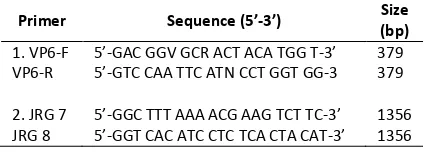
The primer pairs used in this study are shown in Table 1. ‘Primer no. 1’ included VP6-F and VP6-R for specific amplification of VP6 genes in swine and bovine rotavirus [12]. ‘Primer no. 2’ included JRG7 and JRG8 for specific amplification of full length VP6 genes [13].
Table 1. RT-PCR primers for detection of rotavirus and norovirus from swine and bovine fecal samples
Primer Sequence (5’-3’) Size
Condition of RT-PCR reactions for detection VP6 genes were reverse transcription at 42oC or 1 h, after an initial denaturation at 95oC for 5 min, 35 amplification cycles were performed with denaturation at 94oC for 30 s, annealing at 55oCfor 30 s, and extension reaction at 72oC for 45 s, followed by a final extension at 72oC for 3 min [12]. The amplification products were analyzed by 1.5% agarose gel electrophoresis and
visualized by UV light after ethidium bromide staining.
Phylogenetic Analysis
Nucleotide sequences of the VP6 genes were compared with other strains using BLAST search of the National Center for Biotechnology information (NCBI). Phylogenetic analysis based on the nucleotide alignments was constructed using the neighbor-joining method of Molecular Evolutionary Genetics Analysis (MEGA version 6.0) with a pair-wise distance comparison.
RESULT AND DISCUSSION
Distribution of Rotavirus in Swine and Bovine in Taiwan by Age
Distribution of rotavirus positive in group of swine by age was given in Table 2. The largest proportion of swine rotavirus was noted in piglet 3/30 (10%), fattening 0/30 (0%), and sow 0/30 (0%). 1 among 20 samples (5%) were positive in calf and 0% in cow. Based on the positive results, the incidence of positive rotavirus was highest among piglets and calf. Probably because pigs did not receive protective levels of maternal antibo-dy, high levels of passive antibody may tempo-rary protect pigs [17].
Table 2.prevalence of rotavirus from swine and bovine on March 2015
Species Positive Results Total Swine rotavirus in Taiwan with human strain. Circula-tion of animal rotavirus strains confirmed poten-tially zoonotic [18,19].
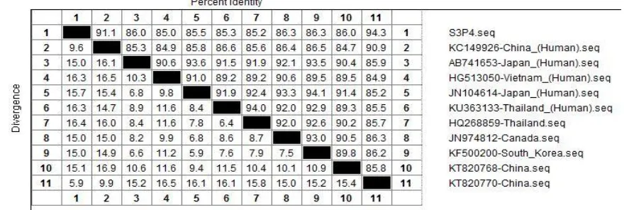
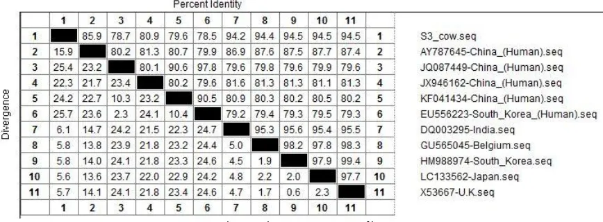
Taiwan wild strain from swine showing 85.0% - 91.1% identity relations with human strains from China (91.1%), Japan (86.0%), Vietnam (85.0%), Japan (85.5%), and Thailand (85.3%), showed on Figure 1. The wild strain of VP6 gene from bovine showing 78.7% - 85.9% identity
rela-tions with human strains from China and 78.5% from South Korea, showed on Figure 2.
Phylogenetic tree and homologous identity comparison analysis showed bovine rotavirus had lower transmission risk than swine (Fig. 3 and 4). Some study mentioned that bovine had low inci-dence and transmission risk for rotavirus [20].
Figure 1. Phylogenetic tree of swine rotavirus based on the VP6 gene (1356 bp) analyzed by MEGA version 6.0 to show the distance of identity relations. Wild strains from Taiwan: south of Taiwan (S3P4). GenBank accession number: KC149926, AB741653, HG513050, JN104614, KU363133, HQ268859, JN974812, KF500200, KT820768, KT820770.
Figure 2. Homologous identity comparison of swine rotavirus
Figure 4. Homologous identity comparison of bovine rotavirus
These results reflect 2 possible issues: the first is that VP6 rotavirus maybe transferred di-rectly from pig to human or from humans to pig, in which provides zoonotic source for swine rota-virus outbreak. Second, pigs may be co-infected with a human and a swine strain of rotavirus si-multaneously [21].
CONCLUSIONS
Evidence for zoonotic transmission of wild strain rotavirus from Taiwan both in swine and bovine showed in phylogenetic tree and homo-logous identity comparison analysis. This infor-mation will be useful in the rasionalization of genotypes for vaccines to protect Taiwan pigs and cattle. An effective vaccine may potentially reduce zoonotic transmission.
ACKNOWLEDGMENTS
This work was supported by grants from the project of center for disease control in Taiwan (Taiwan CDC) and microbiology aboratory of de-partment veterinary medicine, National Pingtung University of Science and Technology.
REFERENCES
[1]. Kim, H.H., J. Matthijnssens, H.J. Kim, H.J. Kwon, J.G. Park, K.Y. Son, E.H. Ryu, D.S. Kim, W.S. Lee, M.I. Kang, D.K. Yang, B.H. Hyun, S.I. Park, S.J. Park, K.O. Cho. 2012. Full-length genomic analysis of porcine G9P[23] and G9P[7] rotavirus strains isolated from pigs with diarrhea in South Korea. Infect.
Genet. Evol. 12. 1427–1435.
[2]. Tate, J.E., A.H. Burton, C. Boschi-Pinto, A.D. Steele, J. Duque, U.D. Parashar. 2012. 2008 estimate of worldwide rotavirus-associated mortality in children younger than 5 years before the introduction of universal
rota-virus vaccination programmes: a systematic review and meta-analysis. Lancet Infect. Dis. 12. 136–141.
[3]. Suzuki, T., A. Hasebe, A. Miyazaki, H. Tsunemitsu. 2014. Phylogenetic characteri-zation of VP6 gene (inner capsid) of porcine rotavirus C collected in Japan. Infect. Genet.
Evol. 26. 223–227.
[4]. Taiwan CDC. 2015. This year’s viral gastro -enteritis activity highest in 4 years; As long weekend approaches, public urged to pay attention to personal hygiene to ensure their health and health of others. Available at: https://www.cdc.gov.tw/english/info. [5]. Kindler, E., E. Trojnar, G. Heckel, P.H. Otto,
R. Johne. 2013. Analysis of rotavirus species diversity and evolution including the newly determined full-length genome sequences of rotavirus F and G. Infect. Genet. Evol. 14. 58–67.
[6]. Matthijnssens, J., P.H. Otto, M. Ciarlet, U. Desselberger, M. Van Ranst, R. Johne. 2012. VP6-sequence-based cutoff values as a criterion for rotavirus species demarcation.
Arch. Virol. 157. 1177–1182.
[7]. Lachapelle, V., J.S. Sohal, M.C. Lambert, J. Brassard, P. Fravalo, A. Letellier, Y. L’Homme. 2014. Genetic diversity of group A rotavirus in swine in Canada. Arch. Virol. 159. 1771–1779.
[8]. Zhirakovskaia, E.V., R.K. Aksanova, M.G. Gorbunova, A.I. Tikunov, A.M. Kuril’shchi -kov, S.N. Sokolov, S.V. Netesov, N.V. Tikunova. 2012. Genetic diversity of group A rotavirus isolates found in Western Siberia in 2007-2011. Mol. Genet. Microbiol.
Virusol. 33–41.
characterisation of group A rotavirus in asymptomatic piglets in southern Ireland.
Arch. Virol. 155, 1247–1259.
[10].Thongprachum, A., P. Khamrin, P. Saekhow, C. Pantip, S. Peerakome, H. Ushijima, N. Maneekarn. 2009. Analysis of the VP6 gene of human and porcine group A rotavirus strains with unusual subgroup specificities: Analysis of the VP6 Gene. J. Med. Virol. 81. 183–191.
[11].Zhu, J., Q. Yang, L. Cao, X. Dou, J. Zhao, W. Zhu, F. Ding, R. Bu, S. Suo, Y. Ren, et al. 2013. Development of porcine rotavirus vp6 protein based ELISA for differentiation of this virus and other viruses. Virol. J. 10(1). 1-8.
[12].Iturriza Gomara, M., C. Wong, S. Blome, U. Desselberger, J. Gray. 2002. Molecular characterization of VP6 genes of human rotavirus isolates: correlation of genogroups with subgroups and evidence of indepen-dent segregation. J. Virol. 76. 6596–6601. [13].Le, L.T., T.V. Nguyen, P.M. Nguyen, N.T.
Huong, N.T. Huong, N.T.M. Huong, T.B. Hanh, D.N. Ha, D.D. Anh, J.R. Gentsch, Y. Wang, M.D. Esona, R.I. Glass, A.D. Steele, P.E. Kilgore, N.V. Man, B. Jiang, N.D. Hien. 2009. Development and characterization of candidate rotavirus vaccine strains derived from children with diarrhoea in Vietnam.
Vaccine. 27. F130–F138.
[14].Ghosh, S., N. Urushibara, K. Taniguchi, N. Kobayashi. 2012. Whole genomic analysis reveals the porcine origin of human G9P[19] rotavirus strains Mc323 and Mc345. Infect.
Genet. Evol. 12. 471–477.
[15].Ghosh, S., V. Varghese, S. Samajdar, S.K. Bhattacharya, N. Kobayashi, T.N. Naik. 2006. Molecular characterization of a porcine Group A rotavirus strain with G12 genotype specificity. Arch. Virol. 151. 1329–1344. [16].Midgley, S.E., K. Bányai, J. Buesa, N.
Halaihel, C.K. Hjulsager, F. Jakab, J. Kaplon, L.E. Larsen, M. Monini, M. Poljšak-Prijatelj, P. Pothier, F.M. Ruggeri, A. Steyer, M. Koopmans, B. Böttiger. 2012. Diversity and zoonotic potential of rotaviruses in swine and cattle across Europe. Vet. Microbiol. 156. 238–245.
[17].Morrow, W.E.M., 2013. Rotaviral diarrhea in pigs. Swine Health. PIG 04-01-33. 1-6. [18].My, P.V.T., M.A. Rabaa, C. Donato, D.
Cowley, V.V. Phat, T.T.N. Dung, P.H. Anh, H. Vinh, J.E. Bryant, P. Kellam, G. Thwaites, M.E.J. Woolhouse, C.D. Kirkwood, S. Baker.
2014. Novel porcine-like human G26P[19] rotavirus identified in hospitalized paedia-tric diarrhoea patients in Ho Chi Minh City, Vietnam. J. Gen. Virol. 95. 2727–2733. [19].Martella, V., K. Bányai, J. Matthijnssens, C.
Buonavoglia, M. Ciarlet. 2010. Zoonotic aspects of rotaviruses. Vet. Microbiol. 140. 246–255.
[20].Mauroy, A., A. Scipioni, E. Mathijs, C. Saegerman, J. Mast, J.C. Bridger, D. Ziant, C. Thys, E. Thiry. 2009. Epidemiological study of bovine norovirus infection by RT-PCR and a VLP-based antibody ELISA. Vet. Microbiol. 137. 243–251.