DETERMINATION OF RESIDUAL OXYTETRACYCLINE
IN FISHES BY HIGH PERFORMANCE LIQUID CHROMATOGRAPHY
[Determinasi residu oxytetracycline pada ikan dengan menggunakan HPLC]
Djamartumpal F. Lumban Batu
Faculty of Fisheries and Marine Sciences, Bogor Agricultural University Jl. Agatis, Kampus IPB Dramaga 16680
Received: 8 February 2010, Accepted: 11 May 2010
ABSTRACT
A high performance liquid chromatography method (HPLC) was developed for the determination of oxytetracycline (OTC) residues in fishes. OTC was extracted from fish tissues with 5% trichloroacetic acid containing 5% disodium ethylenediaminetetraacetate (Na2EDTA). The extract was centrifuged and concentrated. The concentrated solution was
washed with n-hexane and passed through a Sep-pak C18 cartridge. The cartridge was washed with distilled water,
befo-re use. Elute OTC with methanol and evaporate to dryness. The befo-residue was dissolved in acetonitrile-distilled water (3:7), and determined by HPLC (with absorbance measurement at 360 nm) on a TOSOH TSK-GEL ODS 80 Tm Co-lumn (250 mm x 4,6 mm) with methanol-acetonitrile-0.2 M oxalic acid (1 : 1 : 4.4; pH 2,0) as a mobil phase. The ave-rage recoveries of OTC from blood, liver, muscle and kidney of rainbow trout, Oncorhynchus mykiss were 90, 72, 84, and 73%, respectively. The detection limits for muscle was 0.05 ppm, and those for liver and kidney were 0.1 ppm.
Key words: determination, fishes, HPLC, oxytetracycline, residue.
INTRODUCTION
Oxytetracycline (OTC) is a commonly
used antibiotic in commercial aquaculture of
freshwater and marine fish species. OTC is a
bacteriostatic compound with a broad
antibac-terial activity against both Gram-positive and
Gram-negative microorganisms, both aerobic and
anaerobic species (Grondel et al., 1987).
For the purpose of prevention and/or
treatment of infection fish diseases, various
anti-microbial agent have been widely used in
aqua-culture. In consequence, many drug-resistant
strains of fish pathogens have appeared in fish
frams (Watanabe et al., 1971; Aoki, 1975). The
drug may be used for prophylaxis or therapy of
bacterial infections. Several diseases are
descri-be, i.e. carp erythrodermatitis, columnaris
disea-se, edwardsiellosis, enteric redmouth diseases
and furunculosis, on which occasion OTC are
re-commended drugs for treatment (Austin, 1984).
The quantitative determination of drug in
fish is usually done by bioassay, but is well
known that this assay is lacking in sensitivity and
specifity to drug (Sporns et al., 1986). A simple
and sensitive method for determination of OTC
in fish tissue by high performance liquid
chroma-tography (HPLC) has been reported (Ueno et al.,
1989), however could not enough to find the high
recoveries of OTC, because of the influence of
the interfering substances. Therefore, it is
neces-sary to develop a HPLC method for the
determi-nation of OTC residues in fishes. The present
study was performed on the determination of
OTC residues in various tissues of yellowtail, red
sea bream, ayu, carp and rainbow trout by means
of HPLC.
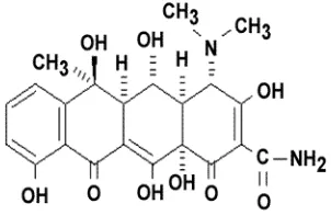
-2-napthacenecarboxamide, OTC used in this
experiment was obtained from Kyowa Hakko
Kogyo., Ltd. Japan, and a Sep-pak C18 cartridge
Other chemicals were of analytical grade or of
the grade for HPLC. The chemical structure of
OTC is shown in Fig. 1.
Fig. 1. The chemical structure of oxytetracycline
Fish
Five species, namely yellowtail, Seriola
quingueradiata; red sea bream, Pagrus major;
ayu, Plecoglossus altivelis; carp, Cyprinus
car-pio; and rainbow trout, Oncorhynchus mykiss
employed in this experiment were obtained from
fishfarms and aclimatized in aquarium al least for
one week prior to use for the experiment. The
averages body length and weight of fishes and
water temperature of aquarium are shown in
Ta-ble 1.
Tabel 1. The averages body length and weight of fishes, and water temperature
Species Body length
All the test fishes were starved for one day
before oral administration of OTC. Ninety fish of
each species were acclimatized in separated
aq-uarium (1,000 liters capacity) equipped with
flowing water system. After one week
ac-climatization, the fishes were transfered to 50
ppm MS-222 water and anaesthetized for 10 min.
Then, the diets containing adequate amount of
OTC was orally administrated to fish at one dose
of 50 mg in 3 g diet kg-1-body weight, using
Ep-pendorf Combitips. Then, the ninety fish of each
species were immediately transfered into each
150 liters aquarium, dividing into three groups,
each two aquarium in duplicate including
con-trol.
At 2, 4, 8, 12, 26, 50, 74, 98, 146, 194,
and 242 hr after OTC administration, the three
fish of each group were taken out from aquarium
and cut at the hind brain with scissors. The
blood was drawn out from cardiac puncture with
a heparinized syringe. The blood, liver, kidney,
muscle, and intestine collected from each three
fish at each interval were pooled, frozen in liquid
nitrogen and then stored in a freezer at -20oC.
Determination of OTC in fish
The extraction of OTC from fish tissues
was carried out by a modified method of Ueno et
al. (1989) (Fig. 2). The HPLC analysis of OTC
Data analysis. TOSOH Super System Controller.
RESULTS AND DISCUSSIONS
It is known that OTC antibiotic has a
ten-dency to combine with proteins and to form
al., 1986). Therefore it is difficult to extract the
muscle, kidney, and liver 74.2; 72.2; and 75.3%
(Ueno et al., 1989).
In order to increase the accuracy of OTC
recovery, in this experiment, an attemps were
made a more convenient method. Disodium
ethy-lenediaminetertaacetic acid has also been known
as one of typical chelating agents and prevents
OTC from forming complex with metal ions.
Oxytetracycline is subject to chelation and
therefore the recovery of this compound from
fish tissues is difficult. Therefore, in this
experi-ment the 5% Na2EDTA was added 3 times to
sample and homogenized, compared to the
ex-traction procedure reported by Ueno et al. (1989)
utilising 5% Na2EDTA only for once.
A mixture of methanol-acetonitrile-0.2 M
oxalic acid (aqueous, 1:1:4.4, pH 2.0) was used
for the two purposes: (1) to elute the OTC from
the Sep-pak C18 column, and (2) as a OTC
mo-bile phase. This solvent system was modified
from a method developed by Ueno et al. (1989)
using methanol-acetonitrile-0.2 M oxalic acid
(aqueous, 1:1:3.5, pH 2.0). The modification of
the extraction systems from twice (Ueno et al.,
1989) to three times and addition of 5% Na2
EDTA to solvens, following the utilisation of
methanol-aecetonitrile-0.2 M oxalic acid (1:1:4.4
pH 2.0) as a mobil phase gave the best average
recovery (79.8%) (Table 2) higher than the reco-
very of OTC from various tissues of rainbow
Tissue (muscle, 5 g; plasma, liver, muscle and kidney, 2 g each
Add 5 ml of 0.5% Na2EDTA and homogenize for 1 min. Add 25 ml of 5% TCA in 0.5 Na2EDTA. Homogenize for 1 min and centrifuge for 15 min. at 10,000 rpm
Supernatant Precipitate
Add 15 ml of 5% TCA in 5% Na2EDTA. Homogenized and centrifuged
Precipitate Add 10 ml of n-hexane, shaked and centrifuged. Repeat twice this extraction
Aqueous layer. Hexane layer.
Stand for 1 hr
Sep-pak C18
Wash the column with 20 ml of distilled water. Elute OTC with 15 ml MeOH and evaporated to dryness
Residue
Dissolved in 1 ml of acetonitrile-distilled water (3:7)
HPLC analysis
Fig. 2. Analytical procedure for oxytetracycline in fish tissues
trout reported by Ueno et al. (1989) reached an
average recovery at 73.5%. It seems this method
prevents OTC from forming complex with metal
ions, then the hexane treatment and a method
using Sep-pak C18 cartridge were a suitable
preparation but also improved effectively the
eli-mination of lipids and interfering substances
from the fish tissues. Morever, the suitable
con-dition of mobile phases was effective to find the
high recoveries of OTC compared to the previous
by Ueno et al, (1989).
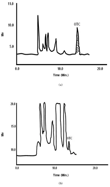
Fig. 3. Typical chromatograms of oxytetracycline from tissues (a) plasma and (b) liver of rainbow trout on HPLC
(a)
(b)
Time (Min.)
Time (Min.)
Mv
Fig. 4. Typical chromatograms of oxytetracycline from tissues (a) plasma and (b) liver of rainbow trout on HPLC
(a)
(b)
Time (Min.)
Mv
Time (Min.)
The recoveries of OTC from plasma, liver,
muscle, and kidney were 90, 72, 84, and 73%,
respectively (Table 2), was higher that of
report-ed by µeno et al., (1989) corresponding to the
average recovery at 73.5%. The detection limits
of OTC for muscle was 0.05 ppm, and those for
liver and kidney were 0.1 ppm. Fifteen percent
loss of recovery might be cause by absorption of
OTC with the gel Sep-pak C18 catridge, chemical
degradation and the irreversible binding to
prote-in durprote-ing the storage period as reported by
Murray et al., (1987). This values were in
agree-ment with the standard values provided for the
settlement of Residue Analysis (recovery > 70%)
(Norin Suisan Sho, 1980). High recovery of OTC
was observed in the order plasma (90%), muscle
(84%), kidney (73%) and liver (72%). The
re-sults of this experiment show marked differences
in the absorption levels of OTC tested in various
tissues of rainbow trout. This low recovery value
in liver may be explained that the OTC
admini-strated to fish might be biotransformed toward
the respective extractable forms by the various
reactions such as hydroxylation, epoxidation,
S-oxidation, dealkylation, reduction, hydrolysis and
conjugation (Lumban Batu, 2001a). Our data
also show that the OTC quickly released from
the kidney, resulting in the low recovery of OTC
in kidney compared with those tissues.
Oxytetracycline orally administrated was
easily absorbed and quickly distributed in issues
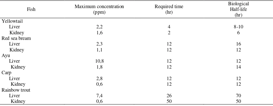
of tested fishes. Table 3 shows the maximum
concentration and its required time, and also the
biological half-life of OTC in the liver and
kid-ney of the tested fishes. The maximum
concen-tration of OTC were observed higher in the liver
of tested fishes compared with those in kidney
(Table 3). The maximum OTC concetrations in
the tissues of yellowtail, red sea bream, ayu, carp
and rainbow trout were attained respectively at
4-2 hr, 14-2 hr, 14-2 hr, 14-2 hr, and 4-26-50 hr post
dosing. The time required to attain to the
respective maximum levels and biological
half-life of OTC in the tested fishes were in the same
order with those in oxolinic acid and
thiamphenicol (Lum-ban Batu, 2001a; 2002b).
The highest maximum concentration of OTC was
found in the liver of ayu (10.8 ppm), and the
in Table 3 by both the biological half-life values.
Table 3. Maximum concentration, required time, and biological half-life of oxytetracycline in the liver and kidney of fishes after oral administration
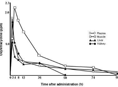
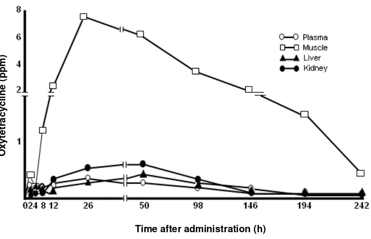
Figs. 4-9 shows the changes in the tissues
level of OTC after oral administration at dose of
50 mg/kg-body weight to yelowtail, red sea
bream, ayu, carp and rainbow trout. The
remain-ing OTC was observed in blood, liver, muscle
and kidney of yellowtail, red sea bream, ayu and
carp 98 hr after oral administration (Figs. 4-7).
Figure 8. shows the changes in the OTC
concen-tration at a dose of 50 mgkg-1 -body weight. The
intestine of rainbow trout showed the longest
du-ration of OTC among those of fishes, i.e. the
OTC in the intestines of tested fishes except
rain-bow trout disappeared after 24 hr post dosing, in
shorter periods than in their other tissues.
How-ever, the OTC in the intestine of rainbow trout
still remained at 242 hr post dosing (Fig. 8.). In
conclusion, this experiment indicate that the
di-gestive absorption of OTC administrated to the
tested fish differs from another. In our previous
paper was reported that oxolinic acid and
thiam-phenicol also remained for a long time in the
in-testine of rainbow trout compared with other
tes-ted fishes (Lumban Batu, 2002a; 2002b). Thus,
the long duration of OTC observed in the tissues
of rainbow trout might due to the slow digestive
process of the rainbow trout.
With the exception of rainbow trout which
showed a digestive singularity as mentioned
abo-ve, a good relationship was observed between the
activities of drug-metabolizing enzymes,
especi-ally aryl hydrocarbon hydroxylase and
glucu-ronyl transferase in fishes, and the duration of
OTC orally administrated to the fishes (Lumban
Batu, 2002c).
By using this developed HPLC method
has enabled to demonstrate the presence of OTC
residues in the various tissues of fish, and allows
for higher recoveries compared with the previous
paper reported by Murray et al., (1987) and Ueno
et al., (1989).
Fig. 4. Channges in the concentration of oxytetracycline in the tissues of yellowtail after oral administration at a dose of 50 mg/kg-body weight
Time after administration (h)
O
xy
tetra
cy
cl
ine
(
p
p
Fig. 5. Channges in the concentration of oxytetracycline in the tissues of red dea bream after oral administration at a dose of 50 mg/kg-body weight
Fig. 6. Channges in the concentration of oxytetracycline in the tissues of ayu after oral administration at a dose of 50 mg/kg-body weight
Time after administration (h)
O
xy
tetra
cy
cl
ine
(
p
p
m)
Time after administration (h)
O
xy
tetra
cy
cl
ine
(
p
p
Fig. 7. Channges in the concentration of oxytetracycline in the tissues of carp after oral administration at a dose of 50 mg/kg-body weight
Fig. 8. Channges in the concentration of oxytetracycline in the tissues of rainbow trout after oral administration at a dose of 50 mg/kg-body weight
Time after administration (h)
O
xy
tetra
cy
cl
ine
(
p
p
m)
Time after administration (h)
O
xy
tetra
cy
cl
ine
(
p
p
Fig. 9. Channges in the concentration of oxytetracycline in the intestine of fishes after oral administration at a dose of 50 mg/kg-body weight
ACKNOWLEDGEMENTS
The author wish to thank Prof. Dr. Kunio
Kobayashi and Dr. Junji Oshima for helpful
dis-cussions and technical assistance, and Kyowa
Hakko Kogyo., Co., Ltd, for the gift of
oxytetra-cycline.
REFERENCES
Aoki, T. 1975. Effects of chemotherapeutics on bacterial ecology in the water of ponds and the intestinal tracts of cultured fish, ayu (Plecoglossus altivelis). Jap. J. Microbiol. 19(4), 327-329.
Austin, B. 1984. The control of bacterial fish diseases by antimicrobial compounds. in Woodbine, M. Antimicrobials in Agricul-ture. pp. 255-268. Butterworths. London.
Grondel, J.L.; Nowrs, J.F.M.; De Jong, M. Schutte, A.R. & Driessens, F. 1987. Phar-macokinetics and tissue distribution of oxy-tetracycline in carp, Cyprnus carpio L., fol-lowing different routes of administration. Journal of fish Disease, 10: 153-163.
Lumban Batu, D.F. 2001a. Effects of several en-vironmental contaminants on the benzo-(a)pyrene hydroxylase enzyme activity of carp microsome. Indonesian Journal of
Aquatic Sciences and Fisheries, 8(1): 23-29.
Lumban Batu, D.F. 2002a. Determination of re-sidual Thiamphenicol in fishes by High Per-formance Liquid Chromatography. Indone-sian Journal of Aquatic Science and Fish-eries, 9 (1): 35-42.
Lumban Batu, D.F. 2002b. Comparison of tissue level of oxolinic acid in fresh and sea water fishes after oral administration. Indonesian Journal of Aquatic Sciences and Fisheries, 9(1): 53-58.
Lumban Batu, D.F. 2002c. Effect of drug-me-tabolizing enzyme activity induced by Polychlorinated Biphenyl on the duration of Oxolinic Acid in carp. Indonesian Journal of Aquatic Sciences and Fisheries, 9 (2).
Muray, J.; Mc Gill, A.S. & Hardy, R. 1987. De-velopment of a method for the determina-tion of oxytetracycline in trout. Food addi-tives and contaminations, 5 (1), 77-83.
Norin Suisan Sho. 1980. Tikusan Kyoku: Shiryo-tenka butu no Hyokakijun ni motozoku. Shiken no Tebiki. Tokyo.
Oka, K.; Uno, K; Harada, K.; Yasaka, K. & Su-zuki, M. 1985. Improvement of chemical analysis of antibiotics. VIII. Applications of prepacked C18 cartridge for the analysis of
Sporns, P.; Kwan, S. & Roth, L.A. 1986. Use of a discassay system to detect oxytetracycli-ne residues in hooxytetracycli-ney. J. Food Prot., 49: 383-388.
Ueno, R.; Uno, K.; Kubota, S.S. & Horiguchi, Y. 1989. Determination of oxytetracycline in
fish tissues by High Performance Liquid Chromatography. Nippon Suisan Gakka-ishi, 55(7): 1273 – 1276.