Research Report
Dental Journal
(Majalah Kedokteran Gigi)2016 June; 49(2): 87–92
Transforming growth factor beta 1 expression and inflammatory
cells in tooth extraction socket after X-ray irradiation
Ramadhan Hardani Putra,1 Eha Renwi Astuti,1 and Rini Devijanti2
1Department of DentomaxillofacialRadiology 2Department of Oral Biology
Faculty of Dental Medicine, Universitas Airlangga Surabaya-Indonesia
abstract
Background: Radiographic examination is often used in dentistry to evaluate tooth extraction complications. X-ray used in radiographic examination, however, has negative effects, including damage to DNA and inflammatory response during wound healing process. Purpose: This study aimed to analyze the effects of X-ray irradiation on transforming growth factor beta 1 (TGF-ß1) expression andnumber of inflammatory cells in tooth extraction sockets. Method: Thirty rats were divided into three groups, which consist of control group (with a radiation of 0 mSv), treatment group 1 (with a radiation of 0.08 mSv), and treatment group 2 (with a radiation of 0.16 mSv). These rats in each group were sacrificed on days 3 and 5 after treatment. Inflammatory cells which wereobserved in this research were PMN, macrophages, and lymphocytes. Histopathological and immunohistochemical examinations were used to calculate the number of inflammatory cells and TGF-ß1 expression. Obtained data were analyzed using SPSS 16.0 software with one way ANOVA and Tukey’s HSD tests. Result: There was no significant decrease in the number of PMN. On the other hand, there were significant decreases in the number of macrophages and lymphocytes in the sacrificed group on day-5 with the radiation of 0.16 mSv. Similarly, the most significant decreased expression of TGF-ß1 was found in the group sacrificed on day 5 with the radiation of 0.16 mSv. Conclusion: X-ray irradiation with 0.08 mSv and 0.16 mSv doses can decrease TGF-ß1 expression and number of inflammatory cells in tooth extraction sockets on day 3 and 5 post extraction.
Keywords: X-ray irradiation; inflammatory cells; TGF-ß1; tooth extraction; socket healing
Correspondence: Ramadhan Hardani Putra, Department of Dentomaxillofacial Radiology, Faculty of Dental Medicine, Universitas Airlangga. Jl. Mayjen. Prof. Dr. Moestopo no. 47 Surabaya 60132, Indonesia. E-mail: [email protected]
introduction
Radiographic examination is often conducted in the field of dentistry. Radiographic examination may assist dentists in establishing a diagnosis to determine a treatment plan and evaluation of treatment results.1 Tooth extraction often requires radiographic examination. It means that if a fracture occurs during tooth extraction, it will be evaluated with radiographic examination to see the state of the remaining teeth and to determine further treatment plan.2
Nevertheless, the use of dental X-ray to produce a radiograph has a negative impact on tooth extraction sockets since the body cannot be fully protected from the
effects of X-ray irradiation. Ionizing radiation in cells actually depends on many factors. In addition to physical factors, some cells are known to have certain characteristics which are sensitive to radiation, referred as radiosensitive. Therefore, the effects of irradiation on an organism as a whole will depend on the size and type of cells affected.
The cells, which are radiosensitive, are white blood cells or leukocytes.1,3 On tooth extraction sockets, various kinds of white blood cells will emerge as a response to the presence of injury, such as polymorphonuclear cells (PMN), lymphocytes, and macrophages that act as inflammatory cells. Growth factors also play a role in regulation of cell proliferation, differentiation, and migration, in synthesizing
Dental Journal (Majalah Kedokteran Gigi) p-ISSN: 1978-3728; e-ISSN: 2442-9740. Accredited No. 56/DIKTI/Kep./2012. Open access under CC-BY-SA license. Available at http://e-journal.unair.ac.id/index.php/MKG
extracellular matrix proteins, as well as in angiogenesis. A growth factor which plays a role and often expressed during wound healing process is transforming growth factor-β1 (TGF-β1). The role of TGF-β1 emerges on the second phase of the wound healing process, from inflammatory phase to the final phase, i.e tissue remodeling.4,5
Low dose irradiation could cause biological effects on the body since ionization process of X-ray could cause damage to DNA.6 Variation of DNA damage caused by ionization could be changes to the base, losing a nucleotide bases, breakage of hydrogen bonds between the chains, single strand fractures, double strand fractures, and cross linking in helix.7 Dental X-ray irradiation at a dose of 0.08 mSv, 0.16 mSv, and 0, 24 mSv in mice even can lead to increased apoptosis and necrosis of the oral mucosal cells.8 X-ray irradiation could also inhibit initial inflammatory response and decrease infiltration of macrophages and neutrophils, as a result, the wound healing process becomes longer.9
The effects of X-ray irradiation on inflammatory cells and TGF-ß1 expression in tooth extraction sockets are still unsolved. Thus, this research was aimed to analyze the effects of X-ray irradiation on a decrease in both inflammatory cells during the inflammatory phase of wound healing process and TGF-β1 expression during the wound healing process. As a result, the results of this research are expected to reveal the effects of X-ray irradiation with a low dose during the wound healing process of tooth extraction based on molecular biology aspect.
materialsandmethods
Thirty rats (Rattus norvegicus) aged 8-11 weeks and weighed 250-500 grams were randomly divided into three groups, which consist of control group, treatment group I, and treatment group II. Each group consisted of ten rats. All of these rats were adapted in the Laboratory of Biochemistry, Faculty of Medicine, Universitas Airlangga in Surabaya.
Tooth extraction was conducted on these thirty rats. The anterior mandibular incisor of those rats was extracted after administration of anesthesia using ketamine intramuscularly. Before the extraction, cervical preparation was carried out first using a bur with low speed. The extraction then was performed using luxation technique until fractures occurred in the crown of the teeth. After the irradiation process in each study group, the rest of the teeth were taken, the wound was stitched, and the rats were returned to the cage for adaptation.
X-ray irradiation on injured rat (tooth extraction) was performed using conventional radiographic dental instrument, Belmont Searcher model Dx-068 70 kVp 8 mA. Before the X-ray irradiation, those rats were fixed with a wire mesh so that the rats would not move around when exposed to radiation. The control group was not given X-ray irradiation. Treatment group I was given radiation
at a dose of 0.08 mSv or one X-ray irradiation exposure. Meanwhile, treatment group II was given radiation at a dose of 0.16 mSv or twice the X-ray irradiation exposure. The rats in each group then would be sacrificed on days 3 and 5 after the extraction process.
Retrieval and processing of tissues were started by cutting the mandibular tissue of the rats under anesthesia with 10% ether on day 3 and day 5. Fixation of mandibular tissue then was performed using 10% neutral buffered formalin (NBF) and decalcified using 10% EDTA. After the bone tissues become soft, dehydration, clearing, impregnation, and embedding processes were performed on the tissues. The paraffin blocks then were cut. Next, the results were embedded in solid paraffin. The resultswhich obtained in this phase were preparation slides.
Hematoxylin eosin stains was conducted to observe the number of inflammatory cells. Meanwhile, immunohistochemical method with monoclonal
anti-TGF-β1 (T0438; Sigma-Aldrich) was used to observe TGF-β1 expression. Inflammatory cells and TGF-ß1 expressions on the mandibular preparations then were observed using HE staining under a light microscopy, a Nikon H600L digital camera equipped with 300 megapixel DS Fi2. After that, observations were made on the healing area, one-third of the apical incisor sockets.
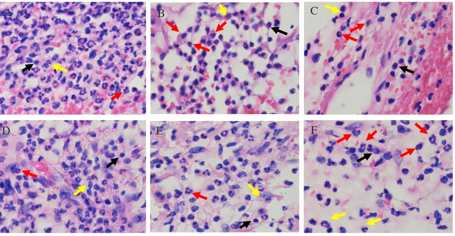
Inflammatory cells observed in this research were PMN cells, macrophages, and lymphocytes. The mean number of the inflammatory cells was calculated by using a light microscope with a magnification of 1000x on five fields of view. PMN cells have segmented cell nucleus with 2-4 purple cores. Meanwhile, themacrophage cells have oval nucleus located eccentrically, and the lymphocytes have a round and dark nucleus which almost fills the entire cell with little cytoplasm.
TGF-β1 expressions were calculated by counting the number of cells expressing TGF-β1. The mean positive expressions of TGF-β1 were observed by counting the number of macrophages expressing TGF-β1 which characterized by a brownish color in the cytoplasm counted under a light microscope with a magnification of 400 times on five field of view. Data obtained in this research were analyzed using SPSS 16.0 software and statistical tests, namely one way Anova test followed by post hoc Tukey’s HSD test.
results
Based on the calculation results, the mean expressions of TGF-β1, PMN, macrophages, and lymphocytes in each sample group were presented in Table 1 and Figure 1. The results of histopathologic examination with IHC staining on TGF-β1 expression were presented in Figure 2. Meanwhile, the results of histopathologic examination with HE staining on PMN cells, macrophages, and lymphocytes were presented in Figure 3.
Dental Journal (Majalah Kedokteran Gigi) p-ISSN: 1978-3728; e-ISSN: 2442-9740. Accredited No. 56/DIKTI/Kep./2012. Open access under CC-BY-SA license. Available at http://e-journal.unair.ac.id/index.php/MKG
Table 1. The mean and standard deviation of TGF-ß1 expression and inflammatory cells on days 3 and 5
Control group Treatment group I Treatment group II
TGF-ß1 Day 3 7.4 ± 1.14 5.8 ± 1.64 3.0 ± 0.70
Day 5 7.6 ± 1.14 6.0 ± 1.00 2.2 ± 0.83
PMN Day 3 271.4 ± 75.25 269.6 ± 63.89 235.2 ± 67.69
Day 5 156.8 ± 64.91 152.4 ± 41.22 124.2 ± 48.47
Macrophages Day 3 64.6 ± 25.98 51.6 ± 21.98 25.4 ± 9.91
Day 5 69.0 ± 26.63 57.6 ± 31.43 21.8 ± 6.76
Lymphocytes Day 3 37.2 ± 11.73 20.8 ± 1.92 19 ± 6.32
Day 5 51.8 ± 21.54 26.6 ± 7.40 18.0 ± 7.58
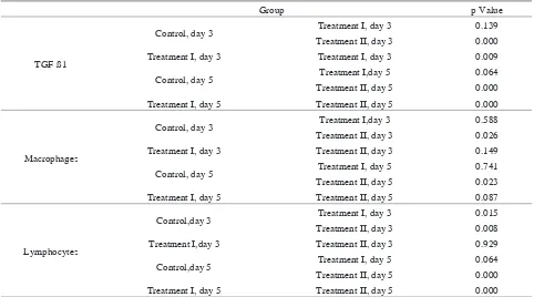
Table 2. The results of Post-hoc Tukey’s HSD test on TGF-ß1 expression and inflammatory cells
Group p Value
TGF-ß1
Control, day 3 Treatment I, day 3 0.139
Treatment II, day 3 0.000
Treatment I, day 3 Treatment I, day 3 0.009
Control, day 5 Treatment I,day 5 0.064
Treatment II, day 5 0.000
Treatment I, day 5 Treatment II, day 5 0.000
Macrophages
Control, day 3 Treatment I,day 3 0.588
Treatment II, day 3 0.026
Treatment I, day 3 Treatment II, day 3 0.149
Control, day 5 Treatment I, day 5 0.741
Treatment II, day 5 0.023
Treatment I, day 5 Treatment II, day 5 0.087
Lymphocytes
Control,day 3 Treatment I, day 3 0.015
Treatment II, day 3 0.008
Treatment I,day 3 Treatment II, day 3 0.929
Control,day 5 Treatment I, day 5 0.064
Treatment II, day 5 0.000
Treatment I, day 5 Treatment II, day 5 0.000
Note: p Value<0.05 indicating a significant difference
Figure 1 Revisi
Figure 1. The mean expression of TGF-ß1 and the mean number of inflammatory cells.
Dental Journal (Majalah Kedokteran Gigi) p-ISSN: 1978-3728; e-ISSN: 2442-9740. Accredited No. 56/DIKTI/Kep./2012. Open access under CC-BY-SA license. Available at http://e-journal.unair.ac.id/index.php/MKG
10
C A
B
10
C
D
E A
B
F
10
C B
10
C
10
C
E B
F
10
C F
Figure 2. The positive expression of TGF-ß1 observed under light microscope at 400x magnification. (A) control group on day 3; (B) treatment group I on day 3; (C) treatment group II on day 3; (D) control group on day 5; (E) treatment group I on day 5; and (F) treatment group II on day 5. The lowest number of cells expressing TGF-ß1 was found in the Treatment Group II both on day 3 and day 5 compared to the control group and the treatment group I (arrows indicating cells expressing TGF-ß1).
11
A
11
A D
11
11
11
11
Figure 3. The HPA on PMN (red arrows), macrophages (yellow arrows), and lymphocytes (black arrows) with hematoxylin-eosin staining technique observed under a microscope at 1000x magnification. (A) control group on day 3; (B) treatment group I on day 3; (C) treatment group II on day 3; (D) control group on day 5; (E) treatment group I on day 5; and (F) treatment group II on day 5.
A
B
C
D
E
F
Dental Journal (Majalah Kedokteran Gigi) p-ISSN: 1978-3728; e-ISSN: 2442-9740. Accredited No. 56/DIKTI/Kep./2012. Open access under CC-BY-SA license. Available at http://e-journal.unair.ac.id/index.php/MKG
According to the results of Kolmogorov-Smirnov and levene tests, the expression of TGF-ß1 on days 3 and 5 had a normal and homogeneous distribution. Next, according to the results of one way Anova test results, there was a significant difference in TGF-β1 expression between the research groups on days 3 and 5 since a value of P was less than 0.05. Similarly, the results of post hoc Tukey’s HSD test showed that there were significant differences (p<0.05) in TGF-β1 expression between the control group and the treatment group II as well as between the treatment group I and the treatment group II both on day 3 and day 5 as seen in Table 2. Nevertheless, there was no significant difference (p>0.05) in TGF-β1 expression between the control group and the treatment group I.
In addition, according to the results of Kolmogorov-Smirnov and Levene tests, the number of PMN, macrophages, and lymphocytes on day 3 and 5 had a normal and homogeneous distribution. Thus, one way Anova test then was performed. The results of one way Anova test showed that there was no significant difference in the number of PMN between those research groups on days 3 and 5 (p>0.05). However, there were significant differences in the number of macrophages and lymphocytes between the research groups (p<0.05).
Post-hoc Tukey’s HSD test was conducted to find out which groups that differed in the number of macrophages and lymphocytes. The results of post-hoc Tukey’s HSD test showed that there was a significant difference (p<0.005) in the number of macrophage cells between the control group (without X-ray radiation) and the treatment group II (with X-ray radiation at a dose of 0.16 mSv), either on day 3 or on day 5 as seen in Table 2. Similarly, there was also a significant difference (p<0.005) in the number of lymphocytes between the control group, the treatment group I (with X-ray radiation at a dose of 0.08 mSv), and the treatment group II, either on day 3 or on day 5.
discussion
X-ray irradiation is a type of ionizing radiation which could cause ionization process in the media path, including
human body. The radiation dose for dental X-ray is
categorized as low dose in the range of 0.01-10 mSv.10
However, ionizing radiation has been known to cause a
varied effects associated with the occurrence of changes or damage in cells as a result of the consequences. Sometimes, cell damage caused by interaction with the radiation can
be recovered through the process of cell repair which
possessed by every individual living cell, but it depends on
the cell type and the radiation exposure dose.6
DNA damage caused by X-ray irradiation could be
either direct or indirect. Irradiation can damage DNA
directly or through the mechanism of free radical formation.
The damage to DNA,which cannot be repaired,would
activate apoptosis, which in this case, is the pathological apoptosis. Effect of X-ray irradiation on cells of the body
could be affected by the amount of the received dose and the type of cell. Apotosis due to irradiation could occur to leukocytes since leukocytes are one of the radiation-sensitive cells.3 Leukocytes or white blood cells have a very important role in the inflammatory phase during wound healing revocation, which act as both acute and chronic inflammation cells.
The results of this research showed that there were significant differences in TGF-β1 expression between the control group and the treatment group I and the treatment group II, either on day 3 and day 5. Decreased expression of TGF-β1 might be caused by a decrease in the number of macrophages due to X-ray irradiation. TGF-β1 is secreted by macrophages, platelets, and keratinosit.11 In this research, platelets and keratinocytes were not observed, but a significant decrease in the number of macrophages occurred in the treatment group II, both on day 3 and day 5.
Decreased expression of TGF-β1, may disrupt the healing process of tooth extraction. TGF-β1 has a broad role in wound healing which plays an important role in inflammatory phase and formation of tissue granulation in proliferation phase.12 In addition, TGF-β1 also plays a role in angiogenesis, extracellular matrix formation, and bone formation in maturation phase.13 In formation bone, TGF-β1 has a role as chemoattractor and stimulates the proliferation and differentiation of osteoblast precursors. TGF-β1 may also increase bone formation by recruiting progenitor of osteoblasts and stimulating proliferation of osteoblasts.14
In this research, the studied inflammatory cells were PMN, macrophages, and lymphocytes. The inflammatory cells play an important role in wound healing, which can kill bacteria and prevent infection in a wound.15 PMN are cells which were very dominant in acute inflammatory phase. In this research, there was no significant difference in the number of PMN between the control group and the treatment groups. But, the number of PMN were the most in the control group, while the least number was found in the treatment group II, either on day 3 and day 5. This indicates that the greater the radiation is given to the injured tooth extraction, the higher the number of PMN will decrease although not significant. This could happen because the given radiation dose can be categorized as low so that a decrease in the number of PMN did not occur significantly. Damage to DNA in the cell nucleus as a result PMN X-ray irradiation could actually be repaired by the body so that the occuring decrease was not significant.
In addition, the results of this research also showed that there was a significant difference in the number of macrophages between the control group and the treatment group II, either on day 3 or day 5. It has similarities with a research conducted by Liu X et al.9 showing that the effects of X-ray radiation on wound healing incision in the skin of mice could reduce macrophage infiltration. Macrophages are derived from monocytes that circulate in the blood to the tissues. X-ray irradiation on wound
Dental Journal (Majalah Kedokteran Gigi) p-ISSN: 1978-3728; e-ISSN: 2442-9740. Accredited No. 56/DIKTI/Kep./2012. Open access under CC-BY-SA license. Available at http://e-journal.unair.ac.id/index.php/MKG
healing can form reactive oxygen species (ROS) that could cause oxidative damage to DNA monocytes. Monocytes are blood cells that are particularly sensitive to X-ray irradiation in which the expression of proteins playing a role in DNA repair would be disturbed, thus influencing DNA repair. Monocytes which cannot be repaired by proteins of DNA repair will activate caspase 8, caspase 3, and caspase 7, which can cause apoptosis of monocyte cells.3 The number of monocytes will be indirectly decreased as a result of apoptosis. In wound healing, monocyte will differentiate into macrophages. The decrease in the number of monocytes, consequently, will decrease the number of macrophages. Although there was no significant difference between the control group and the treatment group I, but the number of macrophages was still decreased due to X-ray irradiation.
In lymphocytes, moreover, there was also a significant difference between the control group and the treatment group I and the treatment group II, either on day 3 or day 5. The decrease in the number of lymphocytes could occur because of the rapid mechanism of apoptosis after experiencing Double Strand break in DNA before the DNA is repaired.16 A research conducted by Faraj et al. showed that the percentage of apoptosis in lymphocytes increases as the given dose of X-ray irradiation increases as well. Apoptosis in lymphocyte cells could be detected by the activated caspase 3 since caspase 3 is a protease which is often activated in apotosis mechanism.17
In the treatment group II (with a radiation dose of 0.16 mSv), the number of macrophages and lymphocytes decreased from day 3 to day 5. This was different from what occured in the control group and the treatment group I in which there was an increase in the number of macrophages and lymphocytes on day 5. The decrease in the number of macrophages and lymphocytes from day 3 to day 5 might indicate a delay in the acute inflammatory phase because in normal wound healing, an increase in the number of macrophages and lymphocytes should occur on day 5. Long inflammatory phase would inhibit the healing process since components in the inflammatory reaction that destroy and eliminate the microorganisms or tissue injury may also damage normal tissue.15
In the treatment group II (with a radiation dose of 0.16 mSv), the number of TGF-β1 expression also decreased from day 3 to day 5. This was different from what happened in the control group and the treatment group I in which there was an increase in the number of TGF-β1 on day 5. Besides, there was also a significant decrease in the number of macrophages and lymphocytes in the treatment group II (with a X-ray irradiation dose of 0.16 mSv). These results can be taken into consideration before taking periapical radiograph. Although there is no clinical evidence, a dentist or radiographer should be more cautious in the making of periapical radiograph in patients with fractures which caused by tooth extraction in order to avoid both a failure in radiograph and unnecessary repetition of X-ray irradiation
exposure since the repeated process of making periapical radiograph on the wound of the tooth extraction may have an impact on the molecular aspects of inflammatory cells, especially macrophages and lymphocytes.
Finally, it can be concluded that the X-ray irradiation at a dose of 0.08 mSv and 0.16 mSv can disrupt the wound healing process of tooth extraction caused by a decrease in TGF-β1 expression and number of inflammatory cells in the tooth extraction sockets on day 3 and day 5. Nevertheless, further researches on the effects of X-ray irradiation on cells or growth factors affecting the wound healing process still need to be conducted.
references
1. White SC, Pharoah MJ. Oral radiology: principles and interpretation. 6th edition. Missouri: Mosby; 2008. p. 175.
2. Miloro M. Peterson’s principle of oral and maxillofacial surgery. 2nd ed. London: BC Decker Inc; 2003. p. 151.
3. Bauer M, Goldstein M, Christmann M, Becker H, Heylmann D, Kaina B. Human monocytes are secverely impaired in base dan dna double strand break repair that renders them vulnerable to oxidative stress. Proc Natl Acad Sci U S A 2011; 108(52): 21105-10. 4. Barrientos S, Stojadinovic O, Golinko MS, Brem H, Tomic-Canic
M. Growth factors and cytokines in wound healing. Wound Repair Regen 2008; 16(5): 585-601.
5. Reed MJ, Koike T, Puolakkainen P. Wound repair in aging: a review. Methods in Molecular Medicine 2003:78: 217–37.
6. Alatas Z. Efek kesehatan pajanan radiasi dosis rendah. Cermin Dunia Kedokteran 2007; 154: 27-39.
7. Minicucci EM, Kowalski LP, Maia MA, Pereira A, Ribeiro LR, de Camargo JL. Cytogenetic damage in circulating lymphocytes and buccal mucosa cells of head and neck cancer patiens undergoing radiotherapy. J Radiat Res 2005; 46: 135-42.
8. Saputra D. Apoptosis dan nekrosis sel mukosa rongga mulut akibat radiasi sinar-x dental radiografik. Surabaya: Universitas Airlangga; 2012. p. 61-78.
9. Liu X, Liu JZ, Zhang E, Li P, Zhou P, Cheng TM, Zhou YG. Impaired wound healing after local soft X-ray irradiation in rat skin: time course study of pathology, proliferation, cell cycle, and apoptosis. J Trauma 2005; 59(3): 682-90.
10. Whaites E. Essentials of dental radiography and radiology. 4th ed.
Philladelphia: Churchill Livingstone; 2007. p. 187.
11. Rolfe KJ, Richardson J, Vigor C, Irvine LM, Grobbelaar AO, Linge C. A role for TGF-beta1-induced cellular responses during wound healing of the non-scarring early human fetus. J Invest Dermatol 2007; 127(11): 2656–67.
12. Mohammadreza P, Ali F, Mohsen KM, Aziz G. Critical role of transforming growth factor in different phases of wound healing. J Adv Wound Care (New Rochelle) 2013; 2(5): 215–24.
13. Richard WDG, Matthew KV, Alicia MVP. Signalling by transforming growth factor beta isoforms in wound healing and tissue regeneration. J Dev Biol 2016; 4(2): 21.
14. Maeda SM, Hayashi M, Komiya S, Imamura T, Miyazono K. Endogemous TGF-ß1 signalling suppreses maturation of steoblastic mesenchymal cells. EMBO J 2004; 23(3): 552-63.
15. Larjava H. Oral wound healing: an overview of biological science. Endodontic Topics 2012; 24(1): 1-3.
16. Fujikawa K, Hasegawa Y, Matsuzawa S, Fukunaga A, Itoh T, Kondo S. Dose and dose-rate effects of X-ray and fission neutrons on lymphocyte apoptosis in p53(+/+) and p53(-/-) mice. J Radiat Res 2000; 41(2): 113-27.
17. Faraj KA, Elias MM, Baaout S. Effect of x and gamma rays on human lymphocytes. Romanian J Biophys2010; 20(4): 355-67.
Dental Journal (Majalah Kedokteran Gigi) p-ISSN: 1978-3728; e-ISSN: 2442-9740. Accredited No. 56/DIKTI/Kep./2012. Open access under CC-BY-SA license. Available at http://e-journal.unair.ac.id/index.php/MKG