A
SIAN
J
OURNAL OF
C
HEMISTRY
A
SIAN
J
OURNAL OF
C
HEMISTRY
INTRODUCTION
Neuraminidase (NA) plays an important role in virus proliferation and infectivity1. Therefore, blocking its activity
generates antiviral effects and neuraminidase is considered as a highly valid target for the design and development of anti-influenza drugs because the active site of viral neuraminidase of both virus A and B is highly conserved in amino acid sequence variation2.
At the recent time, many Neu5Ac2en-based compounds have been synthesized and tested for their influenza virus sialidase inhibitory potential, there are at least 268 compounds of Neu5Ac2en derivatives have been created by many rese-archer (www.bindingDB.org). When a set of active ligands is available, it is possible to compute their shared pharma-cophore. After detected of the chemical feature in training set of active ligands, a pharmacophore can serve as an important model for virtual screening; Pharmacophore-based screening has ability to detect a diverse set of presumed active compounds with totally different chemical scaffolds, even this increase the chances that some of the detected compounds will pass all
Evidence of Combining Pharmacophore Modeling-Docking Simulation for
Screening on Neuraminidase Inhibitors Activity of Natural Product Compounds
MUCHTARIDI MUCHTARIDI1,*, CHOI SY BING2, AISYAH S. ABDURRAHIM3 and HABIBAH A. WAHAB2
1Department of Pharmaceutical Analysis and Medicinal Chemistry, Faculty of Pharmacy, Universitas Padjadjaran, Indonesia 2Malaysian Institute of Pharmaceuticals and Nutraceuticals, Ministry of Science, Technology and Innovation, Penang, Malaysia 3School of Pharmaceutical Sciences, University Sains Malasysia, 11800, Penang, Malaysia
*Corresponding author: E-mail: [email protected]
Published online: 24 December 2014; AJC-16438
In this study, chemical features of 3D-pharmacophore were built to screen of neuraminidase inhibitors from natural products. The pharmacophore hypothesis selected had five features [one hydrogen bond donor, one negative ionizable (N), one positive ionizable (P) and two hydrophobic moiety (Hy)], also included two excluded volume. The best pharmacophore was created by hypogen in DS 2.5 which has a lowest total cost value (92.055), the highest cost difference (107.807), the lowest RMSD (1.197) and the best correlation coefficient (0.944651). The screening of NADI database natural compounds shows that 167 compounds of 3000 NADI compounds were mapped by using four feature at this screening by optimizing the minimum predicted activity to 0.41. In a docking study, good bonding interaction has been shown by coumarilquinic acid derivatives of NADI compounds mapped against neuraminidase (1FD8B), but only MSC927 has a free energy docking score smaller than DANA-1F8B (-9.56 kcal/mol). We have proved both these methods pharmacophore and docking with the statistic and assay experiment methods. Receiver operating under curve in statistic methods have high value (0.907), whereas MUNANA assay experiment resulted that MSC927 has IC50 smaller than others as mention in silico resulted.
Keywords: Neuraminidase, Pharmacophore, Docking, Virtual screening, Nature based drug discovery.
the stages of drug development). In this study, the combining molecular docking-pharmacophore based were the strength this methods for screening when compared to other ligand similarity screening approaches lies3.
Both models were employed to screen for neuraminidase inhibitors from natural product compounds. NADI (Nature Based Drug Discovery)4 is a database of Malaysian medicinal
plants which aims to be a one-stop centre for in silico drug discovery from natural products. It provides structural mation on 3500 different compounds along with the infor-mation of the botanical sources of plants species that could be used in virtual screening.
EXPERIMENTAL
Database of bioactive compounds in nature based drug discovery (NADI): The bioactive compounds of diversity laboratories collection was filtered by NADI of USM properties ligands dataset available from http://phd.usm.my/NADI. These databases contain 3000 compounds of Malaysia natural product.
Pharmachopore modeling
Generation of conformation library of bioactive com-pounds: For the training and test sets molecules, conforma-tional models representing their available conformaconforma-tional space were calculated. All molecules were built using the 2D and 3D sketcher of Hyperchem 7.0 and optimized using MM2 in Hyperchem 7.0. A conformational set was generated for each molecule using the poling algorithm and the best energy option, based on CHARMm force field from Discovery studio 2.55.
The molecules associated with their conformational models were mapped onto the pharmacophore model using the "best fit" option to obtain the bioactive conformation of each mole-cule.
Pharmacophore models: Two hundred fifty five confor-mers was chosen to be minimized as best conformation and 20 kcal/mol was set as energy threshold as global energy mini-mum for conformation searching6, this protocol is available
in DS 2.5 packages. A good pharmacophore model should have a high correlation coefficient, lowest total cost and RMSD values and the total cost should be close to the fixed cost and away from the null cost. The best pharmacophore model was further validated by test set method, receiver operating under curve, fischer's randomization test7 and MUNANA assay
experiment.
Pharmacophore-based virtual screening of nature base drug discovery (NADI) database: Out of the 3000 molecules contained in NADI database, 2350 molecules were filtered as drug-like molecules which were then converted into separate catalyst libraries. Using the ligand pharmacophore mapping protocol, the 'best mapping' was performed with the 'rigid fitting method' and maximum omitted features were set to zero and two7.
Molecular docking: The neuraminidase protein of subtype N1 binding with DANA complex (PDB code : 1F8B)8
was used as the target. Docking simulations were performed with Auto dock9. The auto dock tools (ADT) script was used
to convert the ligand PDB to the pdbq format by adding Gasteiger charges, checking polar hydrogens and assigning ligand flexibility. In addition, the auto dock tools was also performed to prepare the protein targets for the simulations. Using auto dock tools interface, the Kollman charges were added for the macromolecule and a grid box of 60 × 60 × 60 points, with a spacing of 0.375 Å, centered on the binding site for the co-crystallized ligand (26.507; 17.972; 57.828) was setup for auto grid and auto dock calculations.
MUNANA assay: Chlorogenic acid and caffeic acid from coffee seeds and quinic acid (Acros Organic®) was utilized in assay as neuraminidase inhibitors. The assays were carried out on the bacterial neuraminidase. The procedure of assay was referred to Hurt10.
RESULTS AND DISCUSSION
We have collected a total 126 compounds from different literature11-17. Based on principles structural diversity and
activity range of the training set compounds, we have chosen strictly for the 26 compounds used as training set.
3D-QSAR Pharmacophore model: The 10 hypotheses were produced using hypo refine run in 3D-QSAR
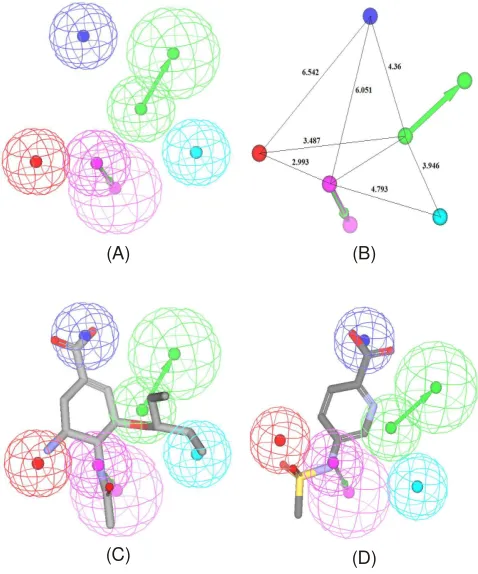
pharma-cophore DS 2.5 packages The best hypothesis hypo 2 (Fig. 1a) was employed in virtual screening tools, because this model has requirement validity (statistical, test set and receiver opera-ting under curve analysis), is characterized by the lowest total cost value (92.055), the highest cost difference (84.395), the lowest RMSD (1.197) and the best correlation coefficient (0.944651). The fixed cost and null cost are 97.2168 and 197.498 bits, respectively. Hypo2 contains five features: one hydrogen-bond donor (D), two hydrophobic aliphatic moiety (Hy), one negatively ionizable (N) and one positive ionizable (P). Two excluded volumes are also involved in hypo2. The 3D space and distance constraints of these pharmacophore features are shown in Fig. 1b.
(A) (B)
(C) (D)
Fig. 1. Feature of Best Pharmacophore with Validation by Hyporefine Run in DS 2.5. (A) The best HypoRefine pharmacophore model, Hypo2. (B) 3D spatial relationship and geometric parameters of Hypo2. (C) Hypo2 aligned with the most-active compound 1 (IC50: 0.5 nM). (D) Hypo2 aligned with the least active compound 24 (IC50: 128825 nM). Pharmacophore features are color coded; magenta: hydrogen-bond donor (HBD), blue - hydrophobic feature (Hy), dark blue - negative ionizable(N), and red - positive ionizable (P)
Validation of pharmacophore model
Fischer randomization test: Fischer randomization is provided in the DS 2.1 package to evaluate models of best pharmacophore. The program used is CatScramble module within catalyst. This statistical program in CatScramble mixes up activity values of all training set compounds to check whether there are strong correlation between the structure and activity. The confidence level was set to 95 % to produce a total 19 random spread sheets were built. The results are shown in Fig. 3. Fig. 2a showed that the correlation (r2) of all pharma-cophore models generated using the 19 random spreadsheet are much lowest than the correlation of corresponding original pharmacophore models and Fig. 2b showed that total costs of
19 random spread sheet are much higher than total costs of best pharmacophore. These results provide confidence on our pharmacophore.
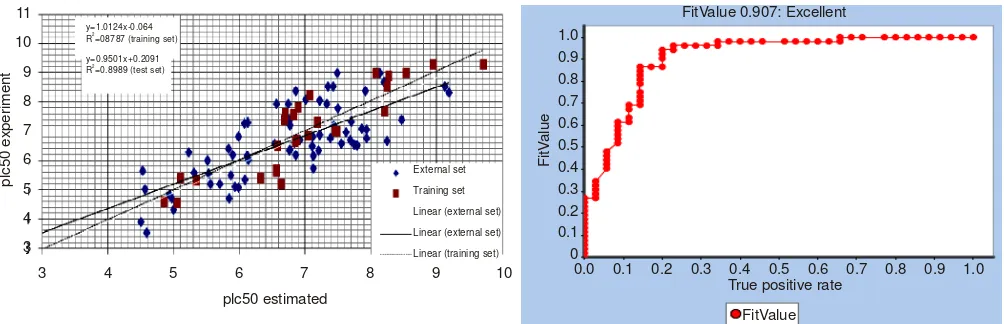
Test set and receiver operating under curve analysis methods: All the test set molecules were prepared by the same way as that for the training set molecules. Hypo2 was applied against the 96 test set compounds which gave a correlation coefficient of between experimental and estimated activities as shown in Fig. 3a.
Model hypo2 illustrated good overall performance with an area under curve value of 0.907 (Fig. 3b), respectively. In general, the greater the area under curve, the more effective the virtual screening workflow in discriminating active from inactive compounds. In terms of probabilities, an area under curve of 0.9 means that a randomly selected active molecule has a higher score than a randomly selected inactive 9 times out18 of 10.
Virtual screening of NADI based on feature pharma-cophore model: The validated Hypo2 of best model were applied as a 3D structural query for screening potent compounds from NADI database. A total of 3500 compounds were screened from the first screening. The hit compounds were further screened by using Lipinski's rule of five to make them more drug-like19
and a total of 2350 molecules passed this filtration. Finally, 167 compounds were mapped at this screening by optimizing the minimum predicted activity to 0.41 mM (MSC2273) with two missing features (Maximum Omitted: 2).
According to the results, there are no compounds of NADI which has high affinity to the fifth feature into one hydrogen bond donor, NI and PI and two Hy by set maximum omitted zero.
All compounds mapped have carboxylic acid and hydroxyl groups. Such as sialic acid, carboxylic group of NADI compounds was mapped into negative ionizable and hydroxyl group was mapped onto hydrogen bond donor feature. Actu-ally, HBA mapped into carbonyl or enol groups of compounds NADI and aromatic moiety and alkyl chain mapped into hydrophobic feature.
Molecular docking simulation study: Based on hits of screening of 3D structural query pharmacophore, the 167 compounds were docked into the inhibitor binding site of neuraminidase by using within Autodock 3.0.5. The crystal structure of neuraminidase complex-DANA (PDB entry: 1F8B)8 was taken from the RSCB protein data bank. Free
energy scoring function of Autodock was used as the ranking function since it performed better than others in a pre-evaluating process. Docking simulation of NADI compounds on N1 showed inverse orientation relative to DANA.
MSC927 was found that binding interaction into N1 (1F8B) have similarity with DANA X-ray, although have fit value (mapping) and free energy docking less than better if be compared with Oseltamivir (2HU4)20 and DANA X-ray
(1F8B). MSC927 also have mapping similar with OSTM, but
1.0
(a) Correlation (r2) of Hypo2 (b) Total cost of Hypo2
Fig. 2. Difference in total cost (b) and correlation (a) of hypotheses between the initial spreadsheet and 19 random spreadsheets after CatScramble run
p R =08787 (training set)
y=0.9501x+0.2091 R =0.8989 (test set)
2
MSC927 doesn't has positive ionizable feature. Fig. 4a and 4b show MSC927 and OSTM interaction at N1. Based on chemical structure, MSC927 or 3-O-Caffeoylquinic acid is ester formed between coffee acid and quinic acid in secondary metabolites of plants21. MSC927 have three isomer position
at 3, 4 and 5 which attack -OH of quinic acid22.
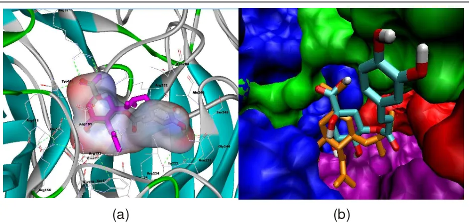
In Fig. 4a, cyclopentenone ring of DANA (1F8B) (pink) coincide well with cyclopentane of MSC927, whereas in Fig. 4b explained that -COOH at oseltamivir (orange) and MSC927 (colourful) given same interaction with Arg371, Arg118 and Arg292 and N-acetyl of OSTM and OH-C4 of MSC927 interact into Asp151 and Glu227 residue, while OH-C5 in MSC927 has same interaction with NH3+-OSTM into Glu119
and Asp151. Oseltamivir at isopentyl groups and moiety of MSC927 given strong hydrophobic interaction into Glu 276, Glu 277, Ala 246 and Arg 224, whereas catechol moiety of MSC927 interacts into most broadly of hydrophobic residues such as Ser245, Gly224 and Asn221. Fig. 4b explained that MSC927 doesn't have positive ionizable pocket (purple), but provides a large hydrophobic pocket (red). Aromatic moiety leads to hydrophobic pocket (red) and -dihydroxy of catechol interact with Ala246
Bioassay for verification simulation models: In analysis of combining results pharmacophore-docking screening, the most interesting active hits are MSC1713 and MSC927. Both compounds have high fit value into model pharmacophore. Molecular docking MSC1713 and MSC927 are almost same, since the compounds gave same interaction into neuramini-dase. In this study, the glycosides of these compounds play an important role in the affinity fitting of features models. Carbo-xylate derivative of glycoside was captured by negative ionizable feature of the both models, hydroxyl of glycoside attached into hydrogen bond donor or HBA features. Aromatic ring of catechol moiety fitted into hydrophobic feature of model, while alkyl groups of catechol side fitted into hydro-phobic feature of model even position of catechol moiety as hydrophobic feature is similar with isopentyl of OST (GS4071).
As described by Luo et al.23, carboxylate acid of MSC927 binds well with triad arginine of neuraminidase as well as sialic acid. Luo et al.23 also explains that chlorogenic acid or MSC927 have potent for NAI of H5N1 (2HUO) because FEB of this compound lower than OST. However in this study, FEB of OST (-10.99 kcal/mol ) is less than MSC927 (-9.88 kcal/mol). Cation-piinteraction (orange line) might occur between ring aromatic of catechol moiety with positive charges of Arg152. Hydrophobic interaction was seemed between chain of caffeic acid moiety and Ser246, Ile222 and Arg224 of neuraminidase binding site, as well as the isopentyl of OSTM with same residue of neuraminidase.
According to the results above, DANA, chlorogenic acid (MSC927), caffeic acid (MSC94) and quinic acid (MSC928) were purchased from ACROS®, asiatic acid (MSC517)
(SIGMA®) and kuguacin R (Tianjin) to verify our models that
these compounds have role in inhibiting of neuraminidase activity. Table-1 showed that all compounds have capability to inhibit neuraminidase activity of C. perfringens.
ACKNOWLEDGEMENTS
This work was supported by Universiti Sains Malaysia Post Graduate Research Student Grant (1001/PFARMASI/ 844111), MOSTI through Nutraceuticals R&D Initiative Grant (Grant no: 09-05-IFN-MEB 004), and Seeds Grant of Faculty of Pharmacy, Universitas Padjadjaran (05/UPPF-FFUP/V/ 2014).
REFERENCES
1. G.M. Air and W.G. Laver, Proteins, 6, 341 (1989).
2. M. von Itzstein, J.C. Dyason, S.W. Oliver, H.F. White, W.-Y. Wu, G.B. Kok and M.S. Pegg, J. Med. Chem., 39, 388 (1996).
3. O. Dror, D. Schneidman-Duhovny, Y. Inbar, R. Nussinov and H.J. Wolfson, J. Chem. Inf. Model., 49, 2333 (2009).
4. H.A. Wahab, R.M. Asarudin, S. Ahmad et al., Nature Based Drug Dis-covery (NADI) & Its Application to Novel Neuraminidase Inhibitors Identification by Virtual Screening, Pharmacophore Modelling and Mapping of Malaysian Medicinal Plants.
(a)
(b)
Fig. 4. (a) Conformation of MSC927 (green) and DANA X-ray (pink), bonded N1(1F8B), surface visualizased with DS 2.5. (b) Pocket area of site active of neuraminidase A (green : NI, red : strong hydrophobic, purple : PI, blue : weak hydrophobic) with visualizased VMD 1.8.5 by Linux
5. B.R. Brooks, R.E. Bruccoleri, B.D. Olafson, D.J. States, S. Swaminathan and M. Karplus, J. Comput. Chem., 4, 187 (1983). 6. H.Y. Wang, Z.X. Cao, L.L. Li, P.-D. Jiang, Y.-L. Zhao, S.-D. Luo, L.
Yang, Y.-Q. Wei and S.-Y. Yang, Bioorg. Med. Chem. Lett., 18, 4972 (2008).
7. X.Q. Deng, H.Y. Wang, Y.L. Zhao, M.-L. Xiang, P.-D. Jiang, Z.-X. Cao, Y.-Z. Zheng, S.-D. Luo, L.-T. Yu, Y.-Q. Wei and S.-Y. Yang, Chem. Biol. Drug Des., 71, 533 (2008).
8. B.J. Smith, P.M. Colman, M. Von Itzstein, B. Danylec and J.N. Varghese, Protein Sci., 10, 689 (2001).
9. G.M. Morris, R. Huey and A.J. Olson, Using AutoDock for Ligand-Receptor Docking, Current Protocols in Bioinformatics, Chapter 8, Unit 8.14 (2008).
10. A. Hurt, Fluorometric Neuraminidase Inhibition Assay, WHO Collabo-rating Centre for Reference and Research on Influenza, pp. 1-10 (2007). 11. V.R. Atigadda, W.J. Brouillette, F. Duarte, Y.S. Babu, S. Bantia, P. Chand, N. Chu, J.A. Montgomery, D.A. Walsh, E. Sudbeck, J. Finley, G.M. Air, M. Luo and G.W. Laver, Bioorg. Med. Chem., 7, 2487 (1999). 12. P. Chand, S. Bantia, P.L. Kotian, Y. El-Kattan, T.-H. Lin and Y.S. Babu,
Bioorg. Med. Chem., 13, 4071 (2005).
13. P. Chand, P.L. Kotian, A. Dehghani, Y. El-Kattan, T.-H. Lin, T.L. Hutchison, Y.S. Babu, S. Bantia, A.J. Elliott and J.A. Montgomery, J. Med. Chem., 44, 4379 (2001).
14. P. Chand, P.L. Kotian, P.E. Morris, S. Bantia, D.A. Walsh and Y.S. Babu, Bioorg. Med. Chem., 13, 2665 (2005).
15. C.U. Kim, W. Lew, M.A. Williams, H. Wu, L. Zhang, X. Chen, P.A. Escarpe, D.B. Mendel, W.G. Laver and R.C. Stevens, J. Med. Chem., 41, 2451 (1998).
16. W. Lew, P.A. Escarpe, D.B. Mendel, D.J. Sweeny and C.U. Kim, Bioorg. Med. Chem. Lett., 9, 2811 (1999).
17. W. Lew, H. Wu, D.B. Mendel, P.A. Escarpe, X. Chen, W.G. Laver, B.J. Graves and C.U. Kim, Bioorg. Med. Chem. Lett., 8, 3321 (1998). 18. N. Triballeau, F. Acher, I. Brabet, J.-P. Pin and H.-O. Bertrand, J. Med.
Chem., 48, 2534 (2005).
19. C.A. Lipinski, F. Lombardo, B.W. Dominy and P.J. Feeney, Adv. Drug Deliv. Rev., 46, 3 (2001).
20. R.J. Russell, L.F. Haire, D.J. Stevens, P.J. Collins, Y.P. Lin, G.M. Blackburn, A.J. Hay, S.J. Gamblin and J.J. Skehel, Nature, 443, 45 (2006).
21. M. Sefkow, A. Kelling and U. Schilde, Eur. J. Org. Chem., 2735 (2001). 22. M.N. Clifford, S. Marks, S. Knight and N. Kuhnert, J. Agric. Food
Chem., 54, 4095 (2006).
23. H.-J. Luo, J.-Z. Wang, J.-F. Chen and K. Zou, Med. Chem. Res., 20, 554 (2011).
24. I. Koji, Y. Makoto and N. Takehiko, Neuraminidase Inhibitory Component, N. A. F. R. Organization, Japan (2011).
TABLE-1
COMPARISON OF IC50 VALUE BETWEEN EXPERIMENT AND in silicoPREDICTION
No. Compounds2 IC50 (Virus-N1)
experiments (µM)
IC50 predicted
(T2S202) (µM)1 Fit value
1 AD (3B7E) FEB2
1 DANAB 12.18 9.3012 7.80440 -9.21
2 MSC 517 16.4 15.1823 7.59166 -11.44
3 NSC 99660 2.9 17.85 7.52145 -8.49
4 NSC 5069 51.288 19.45 7.48408 -14.94
5 NSC 45527 3.2854 25.85 7.36054 -8.29
6 MSC 927 32.69 27.36 7.67081 -9.27
7 NSC 609699 270 35.04 7.2284 -9.84
8 NSC 112257 350 35.51 7.22271 -11.71
9 NSC 10458 190 37.96 7.19372 -11.7
10 NSC 65689 760 60.78 6.98924 -10.81
11 MSC 94 109.8 108.84 6.58919 -9.19
12 NSC 43413 650 152.00 6.58981 -11.93
13 NSC 80997 2541.3 153.00 6.58956 -12.89
14 NSC 16087 540 153.00 6.58812 -11.40
15 MSC 2307 1,880 447.05 6.12264 -11.26
16 NSC 202386 1115.9 892 5.82271 -12.01
17 NSC 45384 1613.9 1060 5.74948 -9.93
18 NSC 146771 2953.1 1400 5.6254 -9.35
19 MSC 928 208.48 108.41 7.51884 -7.77