Isolation of Potential Probiotic
Lactobacillus rhamnosus
Strains
from Traditional Fermented Mare Milk Produced
in Sumbawa Island of Indonesia
Tala S
HI,
1Keita N
ISHIYAMA,
2Koichi N
AKAMATA,
2Ni Putu Desy A
RYANTINI,
1Dai M
IKUMO,
3Yuji O
DA,
3Yuji Y
AMAMOTO,
2Takao M
UKAI,
2I Nengah S
UJAYA,
4Tadasu U
RASHIMA,
1and Kenji F
UKUDA1;y1
Department of Animal and Food Hygiene, Obihiro University of Agriculture and Veterinary Medicine,
Inada-cho, Obihiro, Hokkaido 080-8555, Japan
2
School of Veterinary Medicine and Animal Sciences, Kitasato University, Towada, Aomori 034-8628, Japan
3
Department of Food Science, Obihiro University of Agriculture and Veterinary Medicine,
Inada-cho, Obihiro, Hokkaido 080-8555, Japan
4
Integrated Laboratory for Bioscience and Biotechnology, Udayana University,
Bukit Jimbaran Campus, Badung, Bali, Indonesia
Received May 14, 2012; Accepted July 4, 2012; Online Publication, October 7, 2012 [doi:10.1271/bbb.120385]
To explore potential probiotics in the traditional
foods of Indonesia, fermented mare milk produced in
Sumbawa Island was investigated in this study. Gram
stain, catalase activity, gas production, cell morphology,
carbohydrate utilization pattern, and 16S rDNA
se-quencing were performed to identify isolated lactic acid
bacteria. To assess their probiotic ability, tolerance of
low pH, bile salts, artificial gastrointestinal fluids, and
adhesion properties to extracellular matrices, were
examined. In total 27 strains, 25
Lactobacillus
rhamno-sus
and two
Lactobacillus fermentum
, were obtained.
Among the isolated lactobacilli, three
Lb. rhamnosus
strains, FSMM15, FSMM22, and FSMM26, were
se-lected as candidates for probiotics, using
Lb. rhamnosus
GG as index.
In vitro
binding assay of the three strains
against several extracellular matrix proteins revealed
that FSMM15 and FSMM26 gave greater binding ratios
of mucin/bovine serum albumin (BSA) and significantly
higher adhesive abilities to fibronectin than
Lb.
rham-nosus
GG. FSMM22 showed significantly higher
adhe-sion to laminin than
Lb. rhamnosus
GG.
Key words:
Lactobacillus rhamnosus
; laminin adhesive
ability;
probiotic
properties;
Sumbawa
mare; traditional fermented milk
Probiotics are defined as live microorganisms which
when administered in adequate amounts confer a health
benefit on the host.
1)They can reach the gastrointestinal
(GI) tract alive, and exhibit their health promoting effects
in the host, even though they colonize the GI tract only
temporarily. Hence their ability to adhere constituents of
the GI mucosal layer such as mucin
2)and also to
extracellular matrix (ECM) components including
fibro-nectin,
3)laminin,
4)and collagen
4)is a key function of
probiotics in promoting beneficial health effects, as well as
their antibacterial
5)and immunomodulatory
6)activities.
Probiotics can protect the host defensive mechanism
against pathogenic infection in the gut lumen. Rapid
formation of microbial communities is considered to
reduce pH and to compete with pathogenic bacteria for
adhesion sites, resulting in prevention of pathogenic
colonization.
7)Secretion of antibacterial substances,
e.g.
, acetate
8)and bacteriocin,
9)also prevents the growth
of pathogens. Some lactobacilli
10)and probiotic
Esche-richia coli
11)stimulate human intestinal barrier functions
through induction of epithelial
-defensin.
Several glycoproteins are localized on the surface of
the basement membrane (BM), a thin layer surrounding
epithelial tissues, nerves, fat cells, and muscles.
12,13)These including laminin, type IV collagen, perlecan,
and entactin/nidogen which assemble into fibrils or
other complex macromolecular arrays. Their bind ability
to adhesion receptors enables a tight association with the
cell surface.
14)They are frequently targeted by
patho-genic bacteria that express surface proteins with affinity
for ECM proteins.
15–17)In this context, the ability to
adhere ECM proteins expressed on the surface of BM is
one of the most important criteria in selecting probiotics,
which potentially interfere with infection pathogenic
bacteria in the GI tract.
18)Fermented dairy products are believed to be
promis-ing sources of probiotics because of their history in the
human diet and functionality. From
dadih
, a traditional
Indonesian fermented buffalo milk,
Lactococcus lactis
IS-16183 and
Lb. rhamnosus
IS-7257 were isolated as
potential probiotics inhibiting the adhesion of
E. coli
O157:H7 to human mucin
in vitro
.
19)Lb. casei
Zhang,
Lb. helveticus
ZL12-1, and
Lb. plantarum
BX6-6 were
isolated from
koumiss
, a traditional fermented alcoholic
beverage prepared from mare milk in Inner Mongolia, as
showing antimicrobial activities.
20)Lb. salivarius
,
Lb.
buchneri
, and
Lb. plantrum
I were also found in
koumiss
by Danova
et al.
21)Lb. paracasei
UI14 and
Weissella
y To whom correspondence should be addressed. Tel: +81-15-549-5564; Fax: +81-15-549-5577; E-mail: [email protected]
confusa
UI7 were isolated from whey and cheese
respectively from Nigerian cow’s milk.
22)Furthermore,
Lb. acidophilus
E2 and
Lb. casei
G12 were selected
as potential probiotics from traditional fermented yak
milk.
23)Mare milk is traditionally utilized as a dairy product
in Central Asia, Mongolia, and the former Soviet Union,
where it provides a critical nutritional source.
24)In
addition, Sumbawa Island, located in the middle of the
Lesser Sunda Islands of Indonesia, is a production area
for mare milk and fermented products of it, which are
believed to possess beneficial functionalities. Literature
on Sumbawa mare milk and its fermented products is
scarce, although it is an attractive resource for exploring
novel beneficial compounds and microorganisms. For
example, Sujaya
et al.
isolated
Lb. rhamnosus
SKG34
and SKG49, which showed tolerance of acidic
con-ditions at pH 2 and 3, from raw milk of the Sumbawa
mare,
25)but nothing has been reported to date on the
isolation of lactobacilli from fermented Sumbawa mare
milk (FSMM). In this study, we performed screening of
potential probiotics from FSMM, focusing mainly on
lactobacilli, in relation to their ability to adhere the
colonic mucin and two ECM proteins, including
fibronectin and laminin. To our knowledge, this is the
first report of the isolation of potential probiotic
Lb.
rhamnosus
strains from FSMM produced in Indonesia.
Materials and Methods
Sample collection.Fermented mare milk (approximately 300 mL) was donated by a farmer on Sumbawa Island of Indonesia. Mature mare milk was collected manually in plastic containers in December 2010, and then kept at ambient temperature for 1 week at the farm, which allowed spontaneous fermentation by microorganisms that persisted from previous fermentation in the same container, without any additives. After fermentation, the fermented milk was transported at ambient temperature to Udayana University in Bali and immediately subjected to procedures to isolate lactobacilli.
Isolation of lactobacilli from FSMM.A 100mL-aliquot of FSMM were mixed with 5 mL of de Man-Rogosa-Sharpe (MRS; Oxoid, Basingstoke, UK) broth and incubated statically at 37C for 48 h under
aerobic conditions. A 10-fold dilution series (102–103) of the mixtures
was performed to give 30–300 of colony forming units (CFUs), and each diluted mixture was spread on MRS agar plates supplemented with 1.1 mMbromocresol purple, and was incubated anaerobically at 37C for up to 48 h.26)Single yellow colonies were randomly selected from the MRS agar plates, transferred into test tubes containing 5 mL of MRS broth, and incubated statically at 37C for 24 h under aerobic
conditions. The culture broth was again subjected to dilution and was streaked onto MRS agar plates for purification. Single colony isolation was performed, and the resulting pure isolates were stored at 4C in
stab agar or at80C in 30% glycerol for further investigation. Gram
staining properties, catalase activity, gas production with glucose as carbon source, and cell morphology were confirmed for initial characterization of all isolates.27,28)
Identification of the isolated lactobacilli.The isolated lactobacilli were characterized by carbohydrate utilization test using an API 50 CH kit (bioMe´rieux, Marcy I’Etoile, France) following the manufacturer’s instructions, and by 16S rDNA sequence analysis.29)Two type strains,
Lb. fermentumAmerican Type Culture Collection (ATCC) 14931Tand Lb. rhamnosusJapan Collection of Microorganisms (JCM) 1136T, and Lb. rhamnosusGG ATCC53103 (LGG), one of the most intensively investigated probiotics, isolated from the GI tract of healthy humans,30) were used as controls. The isolates were grown in 5 mL of MRS broth at 30C or 37C for 24 h. One mL of the broth culture was centrifuged at
12;000gfor 5 min at 4C, and then the cells mass was washed twice
with phosphate-buffered saline (PBS). The cell mass was resuspended in PBS, and then an aliquot of the culture broth was mixed with API CHL medium to give the equivalent of 2 McFarland turbidity standard. Inoculated medium (100mL) was applied to the API strips and covered with mineral oil. Fermentation was observed after incubation for 24 and 48 h anaerobically at 30C and 37C. Carbohydrates fermentation
profiles were analyzed by Apiweb (https://apiweb.biomerieux.com/ servlet/Authenticate?action=prepareLogin).
Genetic identification was done by sequencing of the variable area, V1–V3, of the 16S rDNA.31)The isolates were amplified by colony-polymerase chain reaction (PCR) using 16S 27F (50
-AGAGTTTGA-TCCTGGCTCAG-30) and 16S 520R (50-ACCGCGGCTGCTGGC-30)
as universal primers.29)ExTaq DNA polymerase (Takara Bio, Ohtsu, Japan) was used following the manufacturer’s instructions. PCR reaction was performed as follows: first denaturation at 95C for
2 min, 35 cycles of 95C for 30 s (denaturation), 55C for 30 s
(annealing), 72C for 30 s (extension), and the final extension at
72C for 7 min. After colony-PCR amplification, the PCR products
were purified using a GenElute PCR clean-up kit (Sigma-Aldrich, St. Louis, MO) following the manufacturer’s instructions. Sequencing reaction was performed using approximately 40 ng of the amplicon as template, 16S 27F as primer, and a BigDye terminator v1.1 cycle sequencing ready reaction mix kit (Life Technologies, Carlsbad, CA) following the manufacturer’s instructions. The reaction products were purified by ethanol precipitation and analyzed using an automated 310 DNA sequencer (Life Technologies). Identification to the species level was defined as 16S rDNA sequence similarity with the prototype strain sequences in the DNA Data Bank of Japan (DDBJ) and the National Center for Biotechnology Information. When the two databases gave different results, identification was done by the criteria of Dobsonet al.32)
Tolerance of low pH and bile salts of the isolated lactobacilli.The isolated strains were tested for their ability to resist low pH and bile salts. LGG was used as control. Because the pH value of gastric acid varies in a range of about 1.5–4.5 over a period of 2 h depending on a food’s entering time and the gastric contents,33)the pH value of the MRS broth was adjusted to 2.0, 3.0, and 4.0 by 1MHCl. Cells were pre-cultured in 5 mL of MRS broth at 37C for 18 h, and then a 1-mL
aliquot of the culture broth was harvested by centrifugation at
15;000gfor 5 min and washed twice with PBS. The cells were
suspended in 100mL of PBS and incubated in 5 mL of fresh MRS broth
at various pHs at 37C for 3 h. After incubation, 50
mL of the culture
broth was appropriately diluted with PBS and then streaked on MRS agar plates. Viable cell numbers were counted after anaerobic incubation at 37C for 36 h. Tolerance of bile salts was verified by
inoculating 100mL of cells pre-cultured for 18 h into 5 mL of MRS
broth containing 0.3, 0.5, and 1% bile salts (Oxoid). After 4 h of incubation at 37C, viable cells were counted as described above.
Transit tolerance of the isolated lactobacilli as against artificial gastric and intestinal fluids.The transit tolerance of the isolated strains as against simulated gastric and intestinal fluids was tested as described by Ferna´ndezet al.,34)with minor modifications. LGG was used as control. One mL of 18-h culture-broth was harvested by centrifugation at 15;000g for 5 min at 4C, washed with sterilized PBS, and
suspended in 100mL of PBS. The cell suspension was added to 900mL of artificial gastric fluid (125 mMNaCl, 7 mMKCl, 45 mMNaHCO3,
3 g/L pepsin, pH 2 and 3 adjusted with 1M HCl). The bacterial suspensions were incubated at 37C for up to 180 min with agitation
(160 rpm). Aliquots of the mixture (50mL) were taken at 0 and 180 min of incubation, an appropriate dilution of the aliquot was streaked on MRS agar plates, and then this was incubated at 37C for 36 h under
anaerobic conditions, followed by counting of viable cells. The resistance of the strains to intestinal fluids was determined as follows: The resting cell suspension, exposed to artificial gastric fluid for 180 min, was centrifuged at 15;000gfor 5 min at 4C. The cells
were washed using PBS buffer and then resuspended into 850mL of
simulated intestinal fluid (0.1% pancreatin, Sigma-Aldrich, 0.15% bile salts, pH 8.0 adjusted with 1MNaOH). The suspension was incubated anaerobically at 37C with agitation (160 rpm) for 180 min. After
incubation, a 50-mL aliquot was subjected to counting of viable cells as
Adhesion properties of the isolated lactobacilli to two ECM proteins. Out of 27 isolated lactobacilli, 14 strains which were reculturable from the stab agar transferred from Indonesia to Japan were checked for their adherence to several ECM proteins. LGG was used as control. Porcine colonic mucin was purified from the porcine large intestine by gel filtration and density-gradient centrifugation by the method of Kodairaet al.35)Other ECM proteins, fibronectin and laminin, were purchased from Collaborative Biomedical Products (Bedford, MA) and Upstate Biotechnology (Lake Placid, NY) respectively. The ECM proteins with bovine serum albumin (BSA, Sigma-Aldrich) as negative control were immobilized on a 96-well plate as follows: Each 1 mg/mL of mucin, 100 nMfibronectin, 100 nM laminin, and 1 mg/mL BSA in 50 mMphosphate buffer (pH 7.5) was incubated at 4C for 12 h, and then blocking was done with 1.0% BSA
in PBS at ambient temperature for 1 h. Each of the isolated strains and LGG grown in MRS broth at 37C until the absorbance at 600 nm
reached 1.0 was washed twice with PBS, and then 100mL of bacterial
suspension (1109CFU/mL) was applied to the ECMs-coated
96-well plate. After incubation at 37C for 1 h, these were washed 3 times
with 0.1% BSA in PBS. Attached cells were collected with 100mL of 0.01% Triton X-100 in PBS. The suspensions were plated on MRS agar to determine numbers of adhering bacteria by colony plate counting.
Statistical analysis.Results were expressed as means and standard deviation. Data for five independent replicates were subjected to one-way analysis of variance (ANOVA) with Dunnett’s multiple compar-ison of means test, using XLSTAT 2011 and Statcel2. Apvalue of less than 0.05 was regarded as indicating a significant difference.
Results and Discussion
Isolation, characterization, and identification of
lac-tobacilli in FSMM
In total of 27 pure isolates were obtained from the
FSMM following the procedures described above. All
the isolates were primarily assigned lactobacilli, since
they appeared as Gram-positive rods without catalase
activity (data not shown). Of the 27 isolates, 24
homofermenter rods and one gas producing isolate were
tentatively identified as
Lb. rhamnosus
and
Lb.
fermen-tum
respectively, and two were not clearly assigned to
the species level based on their carbohydrate utilization
profiles (Tables 1 and 2). For further identification, a
homology search using the 16S rDNA sequence was
conducted. The sequence data were deposited at DDBJ
under
consecutive
accession
nos.
AB703579
to
AB703605 (Table 2). All the strains, except for the
two strains identified as
Lb. fermentum
, were highly
similar (97–100% homology) to
Lb. rhamnosus
by
database search (Table 2). FSMM9 was
Lb. fermentum
based on high 16S rDNA sequence homology (99%) to
Lb. fermentum
strain VB1 (accession no. JQ073735),
although their carbohydrate metabolism patterns were
not coincident.
Among traditional fermented milks,
Lb. rhamnosus
has to date been found in
kule naoto
(made from zebu
cow’s milk in Kenya)
36)and
gariss
(made from camel’s
milk in Sudan)
37)as minor species, and in
dadih
(made
from buffalo’s milk in Indonesia)
19,38)as major species
together with
Leuconostoc paramesenteroides
and
Lc.
lactis
ssp.
lactis
. In the case of FSMM, most of the
isolated lactobacilli were
Lb. rhamnosus
under our
experimental conditions. These results clearly indicate
that mare milk and fermented products of it are not the
sole source of
Lb. rhamnosus
among livestock animals.
On the other hand,
Lb. rhamnosus
was not isolated from
any fermented milks of cow, yak, goat, or mare in
Mongolia
39)or from
koumiss
, a low alcohol beverage
made from mare’s or camel’s milk in Central Asia.
21)Therefore, the presence or predominance of
Lb.
rham-nosus
strains in the traditional fermented dairy products
is likely to stem from environmental factors such as
temperature favorable to growth. Most of the lactobacilli
isolated from FSMM metabolize neither sucrose nor
melibiose (Table 1). The latter can be obtained only by
invertase-catalyzed hydrolysis of raffinose, which
dis-tributed widely in the plant world,
40)and hence most is
lactobacilli in FSMM are not likely to have originated in
plants.
Probiotic properties of lactobacilli isolated from
FSMM
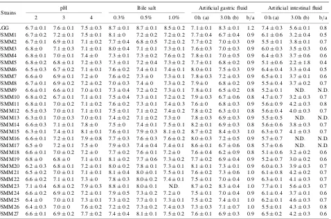
The tolerance of all the lactobacilli isolates of an
acidic environment was found to be similar to that of
LGG. A continuous decrease in cell viability was
observed for all the strains at lower pH, but they
maintained more than
10
6CFU/mL at pH 2 (Table 3).
In the bile test, all the isolates except for FSMM23
showed resistance at varied concentrations (0.3%–1.0%)
of bile salts. In general, the survival rate was constant
with increasing concentrations of bile salts, but FSMM1,
FSMM19, and FSMM20 showed a tendency to decrease
in viable cell numbers (Table 3). LGG proliferated for
3 h in the artificial gastric fluid at pH 3. In contrast, none
of the FSMM isolates grew under the same conditions,
although they maintained high cell viability. FSMM
strains showing values not greater than 0.8 of the CFU
mean ratio (b/a in Table 3) of the artificial gastric fluid
treatment were eliminated from selection. Subsequent
treatment with artificial intestinal fluid damaged LGG,
giving a CFU ratio of 0.8. FSMM11 and FSMM26
exhibited cell survival rates comparable to LGG, but
most of the FSMM strains were severely damaged by
this treatment. In selection, strains giving the CFU ratio
of less than 0.7 were eliminated. Consequently,
FSMM2, FSMM8, FSMM11, FSMM15, FSMM21,
FSMM22, FSMM25, and FSMM26 were selected as
probiotic candidates in terms of cell viability in the
artificial GI fluids. It was confirmed that no lactobacilli
strains, including LGG, proliferated after exposure to the
artificial gastric and intestinal fluids at pH 2 by
measurement of the absorbance of the culture media at
a wavelength of 660 nm (data not shown).
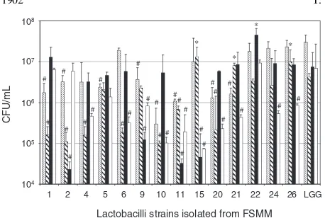
and FSMM22 showed highest adhesion, significantly
higher than LGG.
Finally, three strains, FSMM15, FSMM22, and
FSMM26, were selected as comparable to LGG in
respect to their abilities
in vitro
to survive under
artificial GI conditions and to adhere to colonic mucin
(Fig. 1 and Table 3). Among these, FSMM15 and
FSMM26 are likely to be advantageous for colonization
of the intestinal tract, because they showed higher
binding ratios of mucin/BSA than LGG (Fig. 1).
Pro-biotics that can adhere to ECM proteins are assumed to
interfere competitively with infection by enteric
patho-gens in that the pathopatho-gens also bind to ECM proteins.
18)We found that FSMM15/26 and FSMM22 show
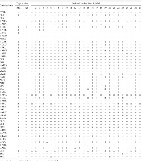
Table 1. Carbohydrate Utilization Patterns of Two LAB Type Strains and Strains Isolated from FSMM
Carbohydrates Type strains Isolated strains from FSMM
Rha Fer 1 2 3 4 5 6 7 8 9 10 11 12 13 14 15 16 17 18 19 20 21 22 23 24 25 26 27
CTRL
GLY þ þ
ERY
D-ARA þ þ þ þ
L-ARA
D-RIB þ þ þ þ þ þ þ þ þ þ þ þ þ þ þ þ þ þ þ þ þ þ þ þ þ þ þ þ þ
D-XYL
L-XYL
D-ADO
MDX
D-GAL þ þ þ þ þ þ þ þ þ þ þ þ þ þ þ þ þ þ þ þ þ þ þ þ þ þ þ þ þ
D-GLU þ þ þ þ þ þ þ þ þ þ þ þ þ þ þ þ þ þ þ þ þ þ þ þ þ þ þ þ þ
D-FRU þ þ þ þ þ þ þ þ þ þ þ þ þ þ þ þ þ þ þ þ þ þ þ þ þ þ þ
D-MNE þ þ þ þ þ þ þ þ þ þ þ þ þ þ þ þ þ þ þ þ þ þ þ þ þ
L-SBE þ
L-RHA þ þ þ þ þ þ þ þ þ þ þ þ þ þ þ þ þ þ þ þ þ þ þ þ þ þ þ
DUL þ þ þ þ þ þ þ þ þ þ þ þ þ þ þ þ þ þ þ þ þ þ þ þ
INO
D-MAN þ þ þ þ þ þ þ þ þ þ þ þ þ þ þ þ þ þ þ þ þ þ þ þ þ
D-SOR þ þ þ þ þ þ þ þ þ þ þ þ þ þ þ þ þ þ þ þ þ þ þ þ þ
MDM
MDG þ
NAG þ þ þ þ þ þ þ þ þ þ þ þ þ þ þ þ þ þ þ þ þ þ þ þ þ þ þ
AMY þ þ þ þ þ þ þ þ þ þ þ þ þ þ þ þ þ þ þ þ þ þ þ þ þ
ARB þ þ þ þ þ þ þ þ þ þ þ þ þ þ þ þ þ þ þ þ þ þ þ þ þ
ESC þ þ þ þ þ þ þ þ þ þ þ þ þ þ þ þ þ þ þ þ þ þ þ þ þ þ þ
SAL þ þ þ þ þ þ þ þ þ þ þ þ þ þ þ þ þ þ þ þ þ þ þ þ þ
D-CEL þ þ þ þ þ þ þ þ þ þ þ þ þ þ þ þ þ þ þ þ þ þ þ þ þ
D-MAL þ þ þ
D-LAC þ þ þ þ þ þ þ þ þ þ þ þ þ þ þ þ þ þ þ þ þ þ þ þ þ þ
D-MEL þ þ
D-SUC þ þ
D-TRE þ þ þ þ þ þ þ þ þ þ þ þ þ þ þ þ þ þ þ þ þ þ þ þ þ þ
INU
D-MLZ þ þ þ þ þ þ þ þ þ þ þ þ þ þ þ þ þ þ þ þ þ þ þ þ þ
D-RAF þ
Starch
GLG
XLT
GEN þ þ þ þ þ þ þ þ þ þ þ þ þ þ þ þ þ þ þ þ þ þ þ þ þ
D-TUR þ
D-LYX
D-TAG þ þ þ þ þ þ þ þ þ þ þ þ þ þ þ þ þ þ þ þ þ þ þ þ þ
D-FUC
L-FUC þ þ þ þ þ þ þ þ þ þ þ þ þ þ þ þ þ þ þ þ þ þ þ þ þ
D-ARL
L-ARL þ þ þ þ þ þ þ þ þ þ þ þ þ þ þ þ þ þ þ þ þ þ
GNT þ þ þ þ þ þ þ þ þ þ þ þ
2KG
5KG
Rha,Lb. rhamnosusJCM1136T; Fer,Lb. fermentumATCC14931T.
CTRL, control; GLY, glycerol; ERY, erythritol;D-ARA,D-arabinose;L-ARA,L-arabinose;D-RIB,D-ribose;D-XYL,D-xylose;L-XYL,L-xylose;D-ADO,D-adonitol; MDX, methyl--D-xylopyranoside;D-GAL,D-galactose;D-GLU,D-glucose;D-FRU,D-fructose;D-MNE,D-mannose;L-SBE,L-sorbose;L-RHA,L-rhamnose; DUL, dulcitol; INO, inositol; D-MAN, D-mannitol; D-SOR, D-sorbitol; MDM, methyl--D-mannopyranoside; MDG, methyl--D-glucopyranoside; NAG,N-acetyl glucosamine; AMY, amygdalin; ARB, arbutin; ESC, esculin ferric citrate; SAL, salicin;D-CEL,D-cellobiose;D-MAL,D-maltose;D-LAC,D-lactose;D-MEL, D-melibiose;D-SUC,D-sucrose;D-TRE,D-trehalose; INU, inulin;D-MLZ,D-melezitose;D-RAF,D-raffinose; GLG, glycogen; XLT, xylitol; GEN, gentiobiose;D-TUR, D-turanose; D-LYX, D-lyxose; D-TAG,D-tagatose;D-FUC, D-fucose; L-FUC, L-fucose; D-ARL, D-arabitol; L-ARL, L-arabitol; GNT, gluconate; 2KG, 2-keto gluconate; 5KG, 5-keto gluconate.
Table 2. Identification of Lactobacilli Strains Isolated from FSMM
Strains (accession no.)
API identification
16S rDNA identification
Sequence lengtha)(bp)
Homologyb)
(%)
Closely related strains (accession no.) FSMM1 (AB703579) Lb. rhamnosus Lb. rhamnosus 416 99 Lb. rhamnosusGYB9 (AF375918) FSMM2 (AB703580) Lb. rhamnosus Lb. rhamnosus 413 98 Lb. rhamnosusJSW10 (AF375896) FSMM3 (AB703581) Lb. rhamnosus Lb. rhamnosus 370 98 Lb. rhamnosusATCC8530 (CP003094) FSMM4 (AB703582) Lb. rhamnosus Lb. rhamnosus 450 99 Lb. rhamnosusMAB22 (AF375897) FSMM5 (AB703583) Lb. rhamnosus Lb. rhamnosus 442 97 Lb. rhamnosusATCC8530 (CP003094) FSMM6 (AB703584) Lb. rhamnosus Lb. rhamnosus 445 100 Lb. rhamnosusNBRC14710 (AB680649) FSMM7 (AB703585) Lb. rhamnosus Lb. rhamnosus 371 98 Lb. rhamnosusATCC7469 (AB008211) FSMM8 (AB703586) Lb. rhamnosus Lb. rhamnosus 462 100 Lb. rhamnosusATCC8530 (CP003094) FSMM9 (AB703587) Lb. rhamnosus Lb. fermentum 449 99 Lb. fermentumVB1 (JQ073735) FSMM10 (AB703588) Lb. rhamnosus Lb. rhamnosus 373 99 Lb. rhamnosusLrJ3 (HQ418482) FSMM11 (AB703589) Lb. rhamnosus Lb. rhamnosus 455 100 Lb. rhamnosusRB4 (AF375898) FSMM12 (AB703590) Lb. rhamnosus Lb. rhamnosus 443 100 Lb. rhamnosusATCC8530 (CP003094) FSMM13 (AB703591) Lb. rhamnosus Lb. rhamnosus 454 99 Lb. rhamnosusX211 (JN415185.1) FSMM14 (AB703592) Lb. rhamnosus Lb. rhamnosus 419 100 Lb. rhamnosusX211 (JN415185.1) FSMM15 (AB703593) N.I. Lb. rhamnosus 331 99 Lb. rhamnosusLrJ3 (HQ418482) FSMM16 (AB703594) Lb. rhamnosus Lb. rhamnosus 413 99 Lb. rhamnosusATCC8530 (CP003094) FSMM17 (AB703595) Lb. rhamnosus Lb. rhamnosus 403 99 Lb. rhamnosusATCC8530 (CP003094) FSMM18 (AB703596) Lb. rhamnosus Lb. rhamnosus 445 97 Lb. rhamnosusLrJ3 (HQ384288) FSMM19 (AB703597) N.I. Lb. rhamnosus 419 97 Lb. rhamnosusLr18 (HQ418480) FSMM20 (AB703598) Lb. rhamnosus Lb. rhamnosus 480 99 Lb. rhamnosusATCC8530 (CP003094) FSMM21 (AB703599) Lb. rhamnosus Lb. rhamnosus 327 98 Lb. rhamnosusChPR-II-str56 (HM462427) FSMM22 (AB703600) Lb. rhamnosus Lb. rhamnosus 492 99 Lb. rhamnosusMAB22 (AF375897) FSMM23 (AB703601) Lb. fermentum Lb. fermentum 441 100 Lb. fermentumNS9 (JQ013298) FSMM24 (AB703602) Lb. rhamnosus Lb. rhamnosus 378 97 Lb. rhamnosusMAB22 (AF375897) FSMM25 (AB703603) Lb. rhamnosus Lb. rhamnosus 451 100 Lb. rhamnosusMAB22 (AF375897) FSMM26 (AB703604) Lb. rhamnosus Lb. rhamnosus 377 100 Lb. rhamnosusATCC8530 (CP003094) FSMM27 (AB703605) Lb. rhamnosus Lb. rhamnosus 495 99 Lb. rhamnosus38-180a (HQ697635)
N.I., not identified.
a)Sequence length of amplicon for 16S rDNA identification.
b)Sequence homology of the strain in the leftmost column against the closely related strain rightmost.
Table 3. Resistance of Isolated Lactobacilli Strains to Low pH, Bile Salt, and Artificial Gastric and Intestinal Fluids
Strains pH Bile salt Artificial gastric fluid Artificial intestinal fluid 2 3 4 0.3% 0.5% 1.0% 0 h (a) 3.0 h (b) b/a 0 h (a) 3.0 h (b) b/a LGG 6:70:1 7:60:1 7:50:3 8:70:1 8:70:1 8:50:2 7:10:1 8:30:1 1.2 7:40:3 5:60:1 0.8 FSMM1 6:70:2 7:20:1 7:50:1 8:10 7:20:2 7:20:2 7:70:4 6:70:4 0.9 6:10:6 3:20:4 0.5 FSMM2 6:70:1 6:90:1 7:10:2 7:70:4 6:80:5 7:20:2 7:70:2 7:00:3 0.9 5:50:1 3:80:1 0.7 FSMM3 6:80 7:10:3 7:10:1 8:00:4 7:10:1 7:30:1 7:60:3 7:00:3 0.9 6:00:3 3:50:3 0.6 FSMM4 6:80:1 7:00:1 7:40 7:30:1 7:30:2 7:60:2 7:80:1 7:00:5 0.9 6:40:3 3:70:6 0.6 FSMM5 6:80:2 6:80:1 7:20:3 7:30:1 7:20:4 7:30:2 7:70:1 6:80:2 0.9 5:10:6 2:21:8 0.4 FSMM6 6:50:3 6:70:2 7:10:1 7:60:2 7:40:1 7:40:1 8:00:1 7:50:3 0.9 6:40:4 3:30:4 0.5 FSMM7 6:60 6:90:1 7:20 7:60:2 7:30 7:30:1 7:80:3 7:20:3 0.9 6:50:1 3:70:1 0.6 FSMM8 6:70:1 6:90:2 7:20:2 7:00:3 7:40 7:30:2 7:90 6:80:2 0.9 5:50:4 3:70:2 0.7 FSMM9 6:60:1 6:60:1 7:00:1 7:30:4 7:20:2 7:30:1 7:80:1 6:50:2 0.8 5:20:1 N.D. N.D. FSMM10 6:80:2 6:70:1 7:10:1 7:50:4 7:30:1 7:20:2 7:90:3 6:70:6 0.8 4:70:7 3:20:3 0.7 FSMM11 6:80:1 7:00:2 7:10:2 7:60:2 7:30:1 7:40:3 7:60 6:80:3 0.9 5:60:9 4:20:3 0.8 FSMM12 6:50:3 7:00:1 7:10:1 7:50:1 7:10:2 7:40:2 7:80:2 6:30:1 0.8 5:60:4 4:00:3 0.7 FSMM13 6:30:1 7:00:3 7:00:1 7:40:2 7:10:2 7:30 7:80:3 6:90:3 0.9 5:50:5 N.D. N.D. FSMM14 6:60:3 7:10:1 7:80 7:50 7:40:1 7:50:1 8:20:1 6:90:3 0.8 5:60:6 3:80:3 0.7 FSMM15 6:30:1 7:40:1 8:10:1 7:60:1 7:90:3 8:10:2 8:70:2 8:40:3 1.0 6:30:7 4:10:3 0.7 FSMM16 6:60:1 7:20:1 7:90:8 7:70:3 7:60:3 7:60:2 8:00:3 7:20:5 0.9 5:70:7 N.D. N.D. FSMM17 6:50 7:20:1 7:50 7:90:3 7:40:4 7:40:1 8:60:1 6:70:6 0.8 5:70:6 N.D. N.D. FSMM18 6:60:1 7:00:2 7:20 7:70:2 7:60:1 7:20 7:60:4 6:20:9 0.8 5:10:6 3:20:2 0.6 FSMM19 6:80 6:80 7:10:1 8:10:2 7:70:6 7:30:2 7:70:2 6:90:4 0.9 5:20:7 3:00:2 0.6 FSMM20 6:20:3 6:80:1 7:20:1 8:00:2 7:80:1 7:30:1 8:10:1 7:30:1 0.9 6:00:3 3:90:3 0.7 FSMM21 6:50:2 7:00:1 7:10:1 8:10:4 8:00:1 7:50:1 7:60:2 7:30:6 1.0 6:10:8 4:20:2 0.7 FSMM22 6:60:2 7:10:1 7:30 7:80:3 8:00:2 7:40:1 7:50:1 7:00:4 0.9 6:30:1 4:10:3 0.7 FSMM23 7:10:4 6:80:2 7:90:3 8:80:1 8:00:1 N.D. 8:70:2 8:30:4 1.0 7:70:1 5:60:3 0.7 FSMM24 6:60:2 6:90:2 7:20:1 7:90:5 7:30:2 7:20 7:50:1 7:00:4 0.9 6:10:4 3:70:1 0.6 FSMM25 6:40 7:00:1 7:30:1 7:30:2 7:70:1 7:30:1 7:50:2 7:40:1 1.0 6:20:1 4:60:3 0.7 FSMM26 6:40:3 7:00 7:60:2 7:20:2 7:30:2 7:40:3 7:30:3 7:10:7 1.0 5:50:1 4:30:3 0.8 FSMM27 6:60:1 6:90:2 7:70:2 7:40:4 8:10:1 7:50:2 7:60:1 6:90:3 0.9 6:50:2 4:20:3 0.6
significantly higher adhesion
in vitro
to fibronectin and
laminin respectively as compared to LGG, and hence
these strains should have good potential for probiotics.
In contrast, Vankerckhoven
et al.
pointed out the
susceptibility and potential pathogenicity of clinically
isolated and potential probiotic
Lb. rhamnosus
strains.
41)This appears to be unrelated to their binding properties
as to ECM proteins, but further studies
in vivo
are
requisite to clarify the probiotic abilities of FSMM15,
FSMM22, and FSMM26.
Evidence that adhesion of probiotics to ECM proteins
can inhibit adhesion by and colonization of enteric
pathogens in the GI tract is very scarce to date. Since at
least 12 proteins of
Lb. plantarum
WCFS1 are predicted
to be directly involved in adherence to the host,
42)the
multiplicity of a probiotic’s recognition patterns for
ECM proteins probably interferes with further
inves-tigation. The use of the two isolated
Lb. rhamnosus
strains, FSMM15 and FSMM22, which showed
discrim-inative adhesion to laminin, might provide a way to
clarify the role of laminin in the prevention of
pathogenic infection in the GI tract.
Acknowledgment
This study was supported financially by the Global
Center of Excellence Frontier Program for Animal
Global Health and Hygiene of the Ministry of
Educa-tion, Culture, Sports, Science, and Technology of Japan.
References
1) Araya M, Morelli L, Reid G, Sanders ME, and Stanton C, FAO/ WHO report, p. 8 (2002).
2) Juntunen M, Kirjavainen PV, Ouwehand AC, Salminen SJ, and Isolauri E,Clin. Diagn. Lab. Immunol.,8, 293–296 (2001). 3) Mun˜oz-Provencio D, Pe´rez-Martı´nez G, and Monedero V,
J. Appl. Microbiol.,108, 1050–1059 (2010).
4) Toba T, Virkola R, Westerlund B, Bjorkman Y, Sillanpaa J, Vartio T, Kalkkinen N, and Korhonen TK, Appl. Environ. Microbiol.,61, 2467–2471 (1995).
5) Plummer S, Weaver MA, Harris JC, Dee P, and Hunter J,Int. Microbiol.,7, 59–62 (2004).
6) Sokol H, Pigneur B, Watterlot L, Lakhdari O, Bermu´dez-Humara´n LG, Gratadoux JJ, Blugeon S, Bridonneau C, Furet JP,
Corthier G, Grangette C, Vasquez N, Pochart P, Trugnan G, Thomas G, Blottie`re HM, Dore´ J, Marteau P, Seksik P, and Langella P, Proc. Natl. Acad. Sci. USA, 105, 16731–16736 (2008).
7) Nava GM, Bielke LR, Callaway TR, and Castan˜eda MP,Anim. Health Res. Rev.,6, 105–118 (2005).
8) Fukuda S, Toh H, Hase K, Oshima K, Nakanishi Y, Yoshimura K, Tobe T, Clarke JM, Topping DL, Suzuki T, Taylor TD, Itoh K, Kikuchi J, Morita H, Hattori M, and Ohno H,Nature,469, 543–547 (2011).
9) Jack RW, Tagg JR, and Ray B,Microbiol. Rev.,59, 171–200 (1995).
10) Schlee M, Harder J, Ko¨ten B, Stange EF, Wehkamp J, and Fellermann K,Clin. Exp. Immunol.,151, 528–535 (2008). 11) Mo¨ndel M, Schroeder BO, Zimmermann K, Huber H, Nuding S,
Beisner J, Fellermann K, Stange EF, and Wehkamp J,Mucosal Immunol.,2, 166–172 (2009).
12) Timpl R, Rohde H, Robey PG, Rennard SI, Foidart JM, and Martin GR,J. Biol. Chem.,254, 9933–9937 (1979).
13) Martin GR and Timpl R, Annu. Rev. Cell Biol., 3, 57–85 (1987).
14) Gumbiner BM,Cell,84, 345–357 (1996).
15) Lopes JD, dos Reis M, and Brentani RR,Science,229, 275–277 (1985).
16) Konkel ME, Garvis SG, Tipton SL, Anderson DE Jr, and Cieplak W Jr,Mol. Microbiol.,24, 953–963 (1997).
17) Mora M, Bensi G, Capo S, Falugi F, Zingaretti C, Manetti AG, Maggi T, Taddei AR, Grandi G, and Telford JL,Proc. Natl. Acad. Sci. USA,102, 15641–15646 (2005).
18) Horie M, Ishiyama A, Fujihira-Ueki Y, Sillanpa¨a¨ J, Korhonen TK, and Toba T,J. Appl. Microbiol.,92, 396–403 (2002). 19) Dharmawan J, Surono IS, and Kun LY,Asian Austral. J. Anim.
Sci.,19, 751–755 (2006).
20) Wu R, Wang LP, Wang JC, Li HP, Menghe B, Wu JR, Guo MR, and Zhang HP,J. Basic Microbiol.,49, 318–326 (2009). 21) Danova S, Petrov K, Pavlov P, and Petrova P, Int. J. Dairy
Technol.,58, 100–105 (2005).
22) Ayeni FA, Sa´nchez B, Adeniyi BA, de Los Reyes-Gavila´n CG, Margolles A, and Ruas-Madiedo P,Int. J. Food Microbiol.,147, 97–104 (2011).
23) Zhang L, Yu QL, Han L, Zhang M, Yang LL, and Li YP,
J. Food Agric. Environ.,9, 18–26 (2011).
24) Park YW, Zhang H, Zhang B, and Zhang L, ‘‘Handbook of Milk of Non-Bovine Mammals,’’ Blackwell Publishing, Oxford, pp. 275–296 (2006).
25) Sujaya IN, Dwipayanti NMU, Suariani NLP, Widarini NP, Nocianitri KA, and Nursini NW, Jurnal Veteriner, 9, 33–40 (2008).
26) Sujaya IN, Amachi S, Yokota A, Asano K, and Tomita F,World J. Microbiol. Biotechnol.,17, 349–357 (2001).
27) Kandler O, Andler O, and Weiss N, ‘‘Bergey’s Manual of Systematic Bacteriology,’’ Williams and Williams, Baltimore, pp. 1208–1234 (1986).
28) Kozaki M, Uchimura T, and Okada S, ‘‘Experimental Manual of Lactic Acid Bacteria,’’ Asakurasyoten, Tokyo, pp. 34–37 (1992).
29) Mori K, Yamazaki K, Ishiyama T, Katsumata M, Kobayashi K, Kawai Y, Inoue N, and Shinano H,Int. J. Syst. Bacteriol.,47, 54–57 (1997).
30) Gorbach SL and Goldin BR, U.S. Patent, 4839281 (Jun. 13, 1989).
31) Neefs JM, van de Peer Y, Hendriks L, and de Wachter R,
Nucleic Acids Res.,18, 2237–2317 (1990).
32) Dobson CM, Chaban B, Deneer H, and Ziola B, Can. J. Microbiol.,50, 482–488 (2004).
33) Mitchell DJ, McClure BG, and Tubman TRJ,Arch. Dis. Child.,
84, 273–276 (2001).
34) Ferna´ndez MF, Boris S, and Barbe´s C,J. Appl. Microbiol.,94, 449–455 (2003).
35) Kodaira H, Ishihara K, Hotta K, Kagoshima M, Shimada H, and Ishii K,Biol. Pharm. Bull.,23, 1173–1179 (2000).
36) Mathara JM, Schillinger U, Kutima PM, Mbugua SK, and Holzapfel WH,Int. J. Food Microbiol.,94, 269–278 (2004).
104 105 106 107 108 LGG 1 2 4 5 6 9 10 11 15 20 21 22 24 26
* * * * # # # # # # # # # # # # # # # # # # # # # # # # # # # # # CFU/mL
Lactobacilli strains isolated from FSMM
Fig. 1. Adhesion of Lactobacilli Strains to BSA and ECM Proteins. FSMM numbering was applied. LGG, Lb. rhamnosus
ATCC53103. Hollow bars, cells adhering to BSA; solid bars, to laminin; shaded bars, to fibronectin; dotted bars, to colonic mucin.
Significantly high,
p<0:05 (n¼5), versus LGG;#significantly
37) Ashmaig A, Hasan A, and El Gaali E,Afr. J. Microbiol. Res.,3, 451–457 (2009).
38) Zakaria Y, Ariga H, Urashima T, and Toba T, Milchwissen-schaft,53, 30–33 (1998).
39) Yu J, Wang WH, Menghe BL, Jiri MT, Wang HM, Liu WJ, Bao QH, Lu Q, Zhang JC, Wang F, Xu HY, Sun TS, and Zhang HP,
J. Dairy Sci.,94, 3229–3241 (2011).
40) French D, ‘‘Advances in Carbohydrate Chemistry,’’ Academic Press, NY, pp. 149–181 (1954).
41) Vankerckhoven V, Moreillon P, Piu S, Giddey M, Huys G, Vancanneyt M, Goossens H, and Entenza JM,J. Med. Micro-biol.,56, 1017–1024 (2007).