Computational modeling of an early evolutionary stage of
the nervous system
John Albert
Department of Comparati6e Physiology,Department of History and Philosophy of Science,Lora´nd Eo¨t6o¨s Uni6ersity,
Pa´zma´ny Pe´ter se´ta´ny2,Budapest,H-1117,Hungary
Received 8 October 1998; received in revised form 13 July 1999; accepted 3 September 1999
Abstract
The object of this work is to create a computational model that examines the early evolution of the nervous system in relation to adaptive behavior. The main questions are: how did the nervous system and the most primitive forms of intelligence came into being, how a system can be organized during evolution that is able to ensure the adaptive behavior of a being, what are the basic rules of construction that are sufficient to create a workable nervous system without specifying the details of the construction. The biological bases of the model are the phyla Cnidaria and
Porifera as they stand at the beginning of the genesis of nervous organization. We found in our model that in a network of homogenous epithelial-like cells, which is considered the starting point of the genesis of the nervous system, the changes that have positive influence on the behavior are those that make the spreading of the electric potential more efficient. It can cause the increase of the effectiveness of the behavior by itself without creating new specific cell-types. There are some alternatives to increasing the effectiveness of spreading of stimuli, for example increasing the value of biophysical parameters of the cells, or increasing the density of nerve cells and the number of synapses. If during the evolution a sort of cell comes into being that is able to conduct electrical stimuli — even in a rudimentary way — it can increase the adaptivity of behavior by itself without the need for specific information of how to organize the construction of this system. © 1999 Elsevier Science Ireland Ltd. All rights reserved.
Keywords:Computational neuroethology; Artificial life; Animat; Adaptive behavior;Cnidaria; Diffuse nervous system
www.elsevier.com/locate/biosystems
1. Introduction
The field of research named ‘artificial life’ offers a new approach to modeling biological systems. We can use it to better understand how the ner-vous system functions by examining a model of
the nervous system and the resulting behavior together. This makes it possible to observe the effects of changes to the nervous system on the adaptivity of behavior.
2. Computational neuroethology
The neural bases of the intelligence and behav-ior are as yet mainly unknown. We can gain some E-mail address:[email protected] (J. Albert)
understanding by studying species that have much simpler nervous systems than humans, because in them the connection between structure and func-tion are much more apparent. This makes the task of modeling them easier. We can call this ap-proach ‘the worm’s-eye view of intelligence’. This trend is followed by neuroethology and its newer
version, called computational neuroethology
(Beer, 1990; Cliff, 1994). The latter is distin-guished from other trends of computational mod-eling of the nervous system in that it examines the neural mechanisms that take part in creating be-havior. The nervous system and its functioning is not modeled by itself, but as a part of the whole living organism, creating a consistent system — similar to real biological organisms. To achieve this we have to model the whole sensory-motor apparatus that contributes to the creation of be-havior and put it into the suitably formed model of a body. This type of autonomous agents is called ‘animats’ (Wilson, 1991; Guillot and Meyer, 1994). They are put into a simulated envi-ronment, so the functioning of the model nervous system will be revealed by the behavior of the animat in that particular environment. This en-ables us to model the neural control of behavior and to study the interaction between the nervous system, behavior and the environment.
Behavior — a sequence of actions — is the result of the interaction between the animal (or animat) and its environment. Behavior is regarded adaptive if the animat responds to environmental stimuli in ways that promote the survival of the organism (Meyer and Guillot, 1994). Adaptive behavior is a broad ability to cope with the com-plex, dynamic, unpredictable world in which the given organism lives. A trait is adaptive if it contributes to an organism’s overall survival. Strictly speaking, ‘adaptive behavior’ means be-havior which is adjusted to environmental condi-tions (Beer, 1990).
3. The problem of fitness
We would like to stress that the concept of fitness has another meaning in these models than in population biology. In the latter case fitness is
a mathematically well-defined term, a statistical parameter that is calculated on the basis of the gene frequencies in the descendant population. In our models we use the term ‘fitness’ to describe
the assessment of the indi6idual performances,
which is the basis for selection. In this case fitness indicates individual suitability, so it has an etho-logical rather than a population bioetho-logical mean-ing. It is a less exact concept and has more intuitive elements.
The simplest but most frequently used and life-like method that serves for the assessment of the performance of the modeled phenotype is a ‘bi-nary fitness function’, which tells whether the given individual survives or dies at the end of a simulation step (Michel and Biondi, 1994). It is applicable when the task of the model organism is to try to find food. If their behavior control is not good enough, the individual ‘starves to death’. Otherwise it ‘survives’ the trial (Nolfi and Parisi 1991; Nolfi et al., 1994a).
4. Beings of the synthetic world
The computer-simulated animals can be
classified by the features of their nervous systems that enable them to behave in an adaptive way. One group of animats had a carefully planned and precisely wired neural net. In this group effective functioning is the result of profound knowledge of the neuroanatomy and physiology of the modeled species. Such a model mimics a concrete species and is able to generate the char-acteristic behavior of that species (Beer, 1990; Cliff, 1994).
rule, hereby the probability of adaptive reactions increases in a given situation.
The plasticity of the neural net is more manifest in models where the number, the position and the connections of the neurons are the result of an ontogenetic process, i.e. one that is influenced by both the information encoded in the genome and the effects of the environment. Changes in the genome of the successive generations (because of mutation and crossing-over) also increase the pos-sible variations of the neural net and the probabil-ity of development of the most adaptive behavior too (Nolfi and Parisi, 1995).
Neural networks that are created to study the regulatory mechanisms underlying adaptive be-havior are called ecological neural networks or econets (Parisi et al., 1990). The object of these models are not to reproduce the structure of the nervous system and behavior of a certain species, but to study ways in which simple rules and interaction with the environment can produce adaptive behavior.
5. The animat and its environment
5.1. Anatomy
One of the conclusions we can draw from the study of ecological neural networks is that over-simplified body structure, suitable for modeling robots with an adaptive behavior, is not appropri-ate to model the behavior of biological organisms. An animal body cannot be regarded as a robot even if we have to simplify it during the modeling process. In the case of a robot we can separate the body from the neural net. The neural net can be treated as an interchangeable module; we only have to connect the motors to the appropriate motoneurons. In the case of biological beings even the simplest movements require the coordi-nated work of many muscles and their effect depends on their positions in the body. To model the movement of an animal we have to take into account the body structure, because it defines the potential modes of behavior. It follows from this that the construction and evolution of the nervous system are not separable from the anatomy and morphogenesis of the modeled animal.
We have created a model-animal (animat) with
the characteristics of the most primitive Cnidari
-ans. The animat is a tube-like organism
corre-sponding to the gastrula-state, which is similar to
the body structure of a Hydra. These animals
have in fact the most primitive nervous system, so it is not unreasonable to imagine our animat as a Hydra-like being without tentacles (Fig. 1.). This is suitable for our purpose, because these animals have no special locomotor organs that would require an advanced neural apparatus for their operation (Bullock and Horridge 1965; Mackie 1990a; Spencer 1991). The locomotor apparatus of the animat consists only of the muscles of the body wall and their motoneurons. It allows the possibility of simple movement of the body, for example curving and crumpling, like the
move-ment of the body column of Hydra and some
species of Porifera. In the current model our
animat is sessile, as are many species of Cnidaria
and all of Porifera.
The movement of the animat is produced by four longitudinal muscle-strips along the body consisting of many muscle cells. The activation of a muscle cell causes the contraction of one section of the body. This causes a very limited movement by itself. Large-scale movement is possible only if many muscular elements work in harmony with each other. This simple model simulates motion as dependent on anatomical relations. It avoids the oversimplification of assigning the firing of single motoneurons to complicated movements that are the result of well-coordinated functions.
5.2. Histology
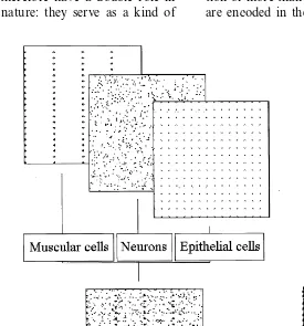
The body of the animat consists of three cell-types, epithelial cells, muscle cells and nerve cells. All of them take part of the formation of behavior.
5.2.1. Epithelial cells
The representation of this type of cell in our model is motivated by the fact that in animals
with primitive nervous systems, mainly Cnidari
and to conduct electric potential in a passive way and even to operate muscle cells (Anderson, 1980; Spencer, 1991; Ranganathan, 1994). In Porifera (which have no nervous system) these cells also play an important role in the conduc-tion of stimuli (Lawn, 1982). Many researchers derive nerve cells from them (Mackie, 1970; Robson, 1975). These cells are found mainly in the layer of cells that covers the outside of the body. In our model these cells give the ‘frame-work’ of the body, defining its shape and boundary. The layer of epithelial cells serves as anchor points for the muscle cells as so the movements of the animat will result in the change of position of the affected epithelial cells. These cells therefore have a double role in the model, as in nature: they serve as a kind of
skin, and they take part in receiving and con-ducting stimuli.
5.2.2. Muscle cells
The organization of muscle cells into functional units is discussed above. These cells form a ho-mogenous population similarly to the epithelial cells, which means that all of them have the same properties. One of these is the stimulus threshold, another is the number of nerve cells that can innervate the same muscle cell and a third is the ability of receiving stimuli from epithelial cells. (It
is well known that in Cnidarians more than one
motoneuron can innervate the same muscle cell and a motoneuron can take part in the innerva-tion of more than one muscle cell.) These features are encoded in the model genome.
5.2.3. Ner6e cells
Nerve cells can be receptors, interneurons or motoneurons by their functioning. But at lower levels of the evolution these functions have not differentiated from each other yet, so inCnidarians
-mainly in Hydra, which have the most primitive
nervous system — the same neuron can carry out more than one function (Westfall and Kinnamon, 1978). Evolution leads from less differentiated primitive nerve cells with multiple functions to highly specialized neurons. The less differentiated state is characteristic not only of the function but also of the anatomy of primitive nerve cells. The processes of the cells cannot be divided into den-drites and axons (Mackie, 1990b). Accordingly different functions do not preclude each other in the same nerve cell and the processes are considered equivalent in our model.
Connections between cells are not determined in advance, therefore synapses of a particular cell are not encoded in the genome (indirect encoding). From a biological point of view this solution is closer to reality than encoding of the complete connection-matrix in the genome (direct encoding) (Balakrishnan and Honavar, 1995). The formation of the synapses of a particular nerve cell is deter-mined by the number and the length of the pro-cesses of that cell and the number of potentially synaptic partner cells that are in reach.
Unlike epithelial and muscle cells, nerve cells do not necessarily form a homogenous cell population, they can develop several cell populations having different properties. Characteristics of cells belong-ing to the same group are uniform, therefore only the properties of the cell populations are encoded in the model genome, instead of the particular data of individual cells.
Based on the theory that nerve cells are derived from epithelial cells, we select the characteristics of nerve cells so that they are similar to epithelial cells (e.g. the conductivity of the nerve cells can be passive conduction with decrement).
Characteristics of passive conductivity of cells —
time-constant (t) and space-constant (l) — are
also built into the model. The time-constant is the length of time during which the value of membrane potential decreases to 1/e. The space-constant of the membrane is the distance where the membrane
potential decreases to 1/e(Ganong, 1987). Both of them are derivated from the cable-equation that describes the passive electric properties of biologi-cal membranes.
The object of this work is modeling the earliest evolutionary state of the nervous system. We in-clude no inhibitory interneurons, because their
existence is not proved in Cnidarians.
5.3. Genetics
The basic information about the construction and functioning of our animats are encoded in a ‘genome’. The ‘life’ of an animat begins with a short ‘ontogeny’, during which they develop on the basis of information derived from the genome. This phase is similar to that studied in the evolutionary modeling of neural networks (Nolfi and Parisi, 1994; Balakrishnan and Honavar, 1995). In our model however genotypes do not go through an evolutionary process directed by genetic al-gorithms. The possibility is included in the model for future work, but in this phase we only use the ability to create the ‘phenotypes’ (animats equipped with a simple body and a nervous system) on the basis of encoded data. This automates creating animats, and supports our objective of studying how a workable nervous system comes into being, based only on some general information about its construction.
We have applied the principle of indirect encod-ing. This means that not all the specific data of every cell are encoded, but only the most important general rules and data, on the basis of which the nerve cells and their connections can be created. In this case different phenotypes belong to a certain genotype (Nolfi et al., 1994b; Nolfi and Parisi, 1995). This solution stands closer to reality, and it also means having to store less data.
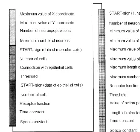
The genome consists of four main parts (Fig. 2.). General information about the size and construc-tion of the body is encoded in the first part, which determine the morphology of the animat. If the tube-like body is laid out, the position of the cells
can be described by x- and y-coordinates. Their
Fig. 2. ‘Gene-mapping’ of the animat. The basic information about the construction and functioning of the animats are encoded in a ‘genome’, so their ‘life’ begins with a short ‘ontogeny’, when they develop on the basis of this information. We have applied the principle of indirect encoding. It means that not all of the specific data of every cell are encoded, but only the most important general rules and data, on the basis of which the cells and their connections can be created. The genome consists of four main parts. General information about the size and construction of the body are encoded in the first part, which determine the morphology of the animal. In the second part the data of muscle cells are encoded, while in the third part the properties of epithelial cells. The largest part of the genome is the fourth, which stores data of nerve cells. These cells can form different populations, so this part consists of as many sections as there are populations. All of the data are encoded in a single ‘chromosome’, so every part of the genome begins with a ‘start’ sign, that shows the beginning of data belong to a cell type. In the recent model we do not use all of the data encoded in the genome, they are only possibilities for future works. Parameters in the first part of the genome: Maximum value of thex- andy-coordinate: if we ‘lay out’ the tube-like animat (see Fig. 1) thex-coordinate gives the horizontal
size and they-coordinate gives the vertical size of the body. All of the cells have to take place between these coordinates. Number of neuron-populations: This value gives the maximum number of types of nerve cells in an animal. A certain population
consists of nerve cells with the same properties (see the fourth part of the genome).
Maximum number of neurons: Gives the maximum number of nerve cells (all types) of a certain animal. The number of nerve cells of an animat can be less than this value, but cannot be more.
Parameters in the second part of the genome:
Number of cells: gives the number of muscular cells. (The cells form four regular longitudinal muscle-strips, so the size of the body determines their coordinates.)
Connection with epithelial cells: This parameter shows whether muscular cells are able to receive stimuli from epithelial cells or not.
Threshold: the strength of the stimulus that can cause the contraction of the muscle cell. Parameters in the third part of the genome:
Number of cells: gives the number of epithelial cells. (This type of cell gives the ‘framework’ of the body and they seat in a regular way, so the size of the body determines their coordinates.)
Receptor function: this parameter shows whether the epithelial cells are able to receive stimuli from the environment similarly to the most primitive nervous systems, or not.
Time-constant: the length of time during which the value of membrane potential decreases to 1/e.
The data of muscle cells are encoded in the second part. In the third part of the genome the properties of epithelial cells are encoded. Both of them make homogenous cell populations and play an important role in shaping the frame-work of the body. At the earliest evolutional state epithelial cells can take part in operating muscular cells, therefore data about this func-tion can be found in the genome.
The largest part of the genome is the fourth, which stores data of nerve cells. These cells can form different populations, so this part consists of as many sections as there are populations.
The value of the x- and y-coordinates of a
par-ticular cell is not encoded, just the range of co-ordinates where the cells of that population can be found. The coordinates are given random values between these limits when the phenotype is instantiated. The connection-matrix of the nerve cells is not encoded either, just the maxi-mum length and the maximaxi-mum number of the processes and the types of actual cells that can be potential synaptic partners for a particular nerve cell population. The actual connections are determined by the number of cells of appro-priate types within reach. All of the data are encoded in a single ‘chromosome’.
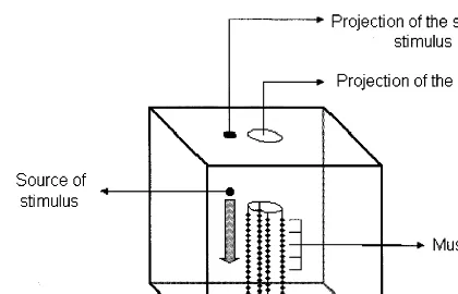
5.4. En6ironment
The environment and the task of the animat in our model is similar to those of ecological neural networks (Parisi et al., 1990; Nolfi et al.,
1994b). The environment is modeled as a box (Fig. 3.). As the animats are sessile, the interac-tion between the individuals would be minimal, so we perform the tests with only one individual in the box at a time.
The stimulus is the appearance of a piece of food, which slowly sinks to the bottom of the box. This is a chemical stimulus for the animat and its strength decreases exponentially with the distance. If the animat is able to catch at least one piece of food, the test is continued. The feeding is successful if the animat not only touches the piece of food, but it also ‘eats’ it, in
other words, it gets food particles into its coe
-lenteron through the mouth (Fig. 4.). The most successful individuals are those that can catch the most pieces of food.
6. Experiments
Investigation of the model consisted of several parts, during which the program was run with a number of different settings of the parameters. The purpose was to test and to ‘calibrate’ the abilities of the animat and to establish relations — if they exist — between the behavior and the values of these parameters. During the tests we studied the effect of the time-constant and space-constant of the epithelial cells and nerve cells, the number of nerve cells and the maximum length of neural processes on the adaptivity of the behavior of the animat.
Fig. 2. (Continued)
Parameters in the fourth part of the genome:
Number of neurons: gives the number of nerve cells belonging to a certain population.
Minimum and maximum value ofx- andy-coordinates: appoint the part of the body where the cells of a certain population can seat. (This can be the whole body, or a part of it, e.g. the hypostome or the basal disk.)
Maximum length of connections: gives the maximum length of neural processes. (InCnidarianswe cannot distinguish axons and dendrites, so we need only one parameter for the description.)
Maximum number of connections: gives the maximum number of the neural processes. (A certain nerve cell can have a smaller number of neurites than this value, but does not have more.)
Receptor function: this parameter shows whether nerve cells are able to receive stimuli from the environment.
Threshold; value of action potential; length of refractory period: these are the characteristics of action potential, but we did not use them in the model in question.
Time-constant; space-constant: see above.
Fig. 3. The animat and its environment The environment and the task of the animat in our model is similar to those of ecological neural networks. The environment is a box and the animats are put into it. As the animats are sessile, the interac-tion between the individuals would be minimal, so we perform the tests with only one individual in the box at a time. The environmental stimulus is the appearance of a piece of food that slowly sinks to the bottom of the box. It is a chemical stimulus for the animat and its strength is decreasing with the distance exponentially.
nervous system — can be sufficient to control the basic reactions of a simple living being to the environmental stimuli, which agrees with the re-sults of physiological experiments.
This fact probably had great importance during the early evolution of the nervous system, because it means that if a cell-type arises that is able to conduct electrical stimuli in addition to its origi-nal function, even if in a primitive way, it can benefit a simple organism by itself without having to organize into a specially constructed system. It can serve as a starting point to the evolution of a highly developed controlling system. Our model shows that the increase of the effectiveness of conducting stimuli alone is able to increase the adaptivity of the organism, which suggests a pos-sible direction for the evolution.
The relation between the fitness and the effec-tiveness of the conduction can be demonstrated by changing the time- and space-constant. In na-ture, however biophysical parameters can change only to a limited extent. Therefore evolution had to find another solution, discussed next.
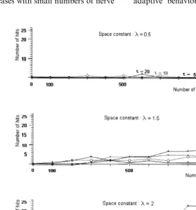
7.2. The effect of the number of ner6e cells on the beha6ior
In further experiments we studied the changes of the behavior of the animats when epithelial-like protoneural cells appear in the primitive nerve-free conducting system as created in the previous step. This corresponds to the evolutionary step during which the ancestors of nerve cells come into being from epithelial cells and begin to orga-nize to a simple network. In this phase nerve cells are rather similar to epithelial cells, because they are able to connect only with their close neigh-bors. The conduction is decremental, so the net-work is a ‘protoneuronal system’ that is similar to
the present Hexactinellida (Mackie et al., 1983).
The time- and space-constants of the nerve cells are similar to those of epithelial cells. They have such a low value that animats with nerve-free networks cannot feed successfully. Therefore the changes of the behavior must be explained by the appearance of nerve cells.
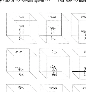
We found that the success of behavior is deter-mined by the space-constant and the number of 7. Results and discussion
7.1. The effect of the time- and space-constant of the epithelial cells on the beha6ior
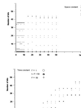
First we studied the behavior of animats that have only epithelial and muscle cells, but no nerve cells. This group forms our control group. The time-constant of the epithelial cells was studied in the range between 1 to 50 ms (the physiological value is about 1 – 20 ms) and the space-constant between 1 to 10 mm (the physiological value is about 1 – 5 mm).
Our results show that the excitability and the success of feeding are determined basically by the measure of the space-constant, and the time-con-stant modifies it only slightly (Fig. 5.). The success of feeding grows with the space-constant signifi-cantly if its measure is close to the physiological value. The epithelial cells are able to provide a minimally adaptive level of the behavior for the
animat, similar to the behavior of nerve-free Hy
-dra(Campbell et al., 1976).
On the basis of these results we can say that a
homogenous, diffuse conducting network —
nerve cells together (Fig. 6.). The number of hits (pieces of food that are caught and eaten) in-creases with the number of nerve cells and this tendency is more pronounced if we increase the value of the space-constant. Comparing this with the results of the nerve-free conducting system, we found that among animats with the same parameter value, those with a nerve-free con-ducting system are not able to respond to
envi-ronmental stimuli, but the appearance and
multiplication of nerve cells increases the success of feeding behavior. The nerve-free conducting system can have a similarly successful perfor-mance only if the space and time-constant have an extremely high value.
These results suggest that at the most primi-tive evolutionary state of the nervous system the
ability to respond to environmental stimuli can increase by increasing the number of elements of this system alone. It may have a great evolution-ary importance because at the beginning of the nervous organization increasing the number of cells may be the simplest way to increase the effectiveness. At this level of the evolution there is no need for developing complex centers in the nervous system to increase its effectiveness, so there is no need to store organizational informa-tion in the genome. Fitness can be increased merely by increasing the number of nerve cells. This could play an important role during the early evolution of the nervous system. Our tenet seems to be justified by the fact that it is not animals with the smallest number of nerve cells that have the most ancient nervous system.
Fig. 5. The number of hits depending on the time- and space-constant of the epithelial cells, in the case of the nerve-free network. The success of feeding is indicated by the number of hits (pieces of food that are caught and eaten). It is determined basically by the measure of the space-constant, and the time-constant can modify it only to a slight degree. The success of feeding grows with the space-constant significantly. If its measure is close to the physiological value (about 1 – 5 mm), the epithelial cells are able to ensure a minimally adaptive level of the behavior for the animal, similarly to the behavior of nerve-freeHydra.
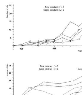
7.3. The effect of the connections of ner6e cells on the beha6ior
Next we studied how the length of the processes of nerve cells, which determines the maximum distance of connections, influence the success of behavior. The length of the processes is specified as a proportion of the cell size. During the simula-tion this length ranged between the 10- and 50-fold size of the nerve cells. In natural animals with
the simplest nervous systems we cannot find nerve cells with longer processes than this.
the main factor is not really the length of pro-cesses, but the ability to develop processes at all. If the cells are able to create them, the behavior is influenced favorably even with short processes. If the number of cells is between 200 – 500, the growth of processes has no additional effect. If the number of cells is more than 500, the adaptiv-ity increases with maximum process length. Therefore successful behavior depends first of all on the number of nerve cells, and secondarily on process length.
The effects due to the process size (these effects also depend on the numbers of nerve cells) can be explained by observing that nerve cells have to reach a critical density to organize a workable network. In the cases with small numbers of nerve
cells their density is not enough to get close to each other and they cannot form connections.
Though the number of connections increases with the size of the processes, they are insufficient to effect behavior. The effect of increasing the density of nerve cells at the first stage this growth is not due to the development of a well-function-ing neural net, but to the increaswell-function-ing number of nerve cells that get close to the muscle cells. These will function as motorneurons, innervating muscle
cells directly. It is common amongCnidariansthat
nerve cells have more than one function. They can serve as receptors and innervate a muscle cell (motoneuron function) at the same time (Westfall and Kinnamon, 1978). Therefore a certain level of adaptive behavior can be achieved even if the
Fig. 7. The number of hits depending on the number of nerve cells and the maximum length of connections. The success of feeding behavior strongly depends on the number of nerve cells, but increases only a little with the length of the maximum distance of connections (that is determined by the length of the processes of the nerve cells).
organization of the nerve cells into a network is at an early stage.
8. Summary
Summarizing the results we can say that in a network of homogenous epithelial-like cells, which is considered as the starting point of nervous organization, the changes that influence adaptivity are those that make the conductivity more effi-cient. We found that such changes can cause the increase of the effectiveness of the behavior by itself, without any differentiation in the network or any development of special cell-types. There are numerous ways to increase the conductivity in the network. One of them is the increase of the
space-constant of cells in case of passive decremental conduction. Biophysical parameters in living or-ganisms can change only to a limited degree, but we can experiment more freely during the simula-tion. Our results show that the effectiveness of the behavior increases significantly with conductivity. The model illustrates that evolution had found another solution to increase the effectiveness of forwarding electrical stimuli without increasing the space-constant in an extreme way, and it was action potential. Another way to increase the effectiveness of functioning is increasing the den-sity of nerve cells, which is perhaps the simplest way in biological systems to achieve this aim.
because at this stage the genome of the animals could not contain any information that encoded the construction and functioning of an effective nervous system, since one could not exist yet, only as a potentiality, because a new cell type had come into being that had the ability to do the job. Therefore the organization of this system pro-ceeded in an ad hoc way at the first stage, and this is the point where it has a great importance if this state of the system was able to increase the chance of surviving at all.
Our model supports the hypothesis that such a primitive system — without having detailed infor-mation about the organization — is able to influ-ence the behavior of a simple-constructed animal in an adaptive way.
The effectiveness of this simple system can in-crease significantly by changing merely
quantita-tive parameters, therefore the factors that
determine the adaptivity of the behavior can re-duce to quantitative attributes. All of these sug-gest that the way leading to the ancestral forms of the nervous system could be realized by simple steps. Perhaps this is not obvious if we only take an experimental look at the sophisticated ‘instru-ment’ that directs the behavior of even the sim-plest real-lifeHydra.
References
Anderson, P.A.V., 1980. Epithelial conduction: its properties and functions. Prog. Neurobiol. 15, 161 – 203.
Balakrishnan, K., Honavar, V., 1995. Evolutionary design of neural architectures — a preliminary taxonomy and guide to literature, Artificial Intelligence Research Group; Tech-nical Report CS TR c 95-01.
Beer, R., 1990. Intelligence as Adaptive Behavior. An Experi-ment in Computational Neuroethology. Academic Press, New York, p. 1990.
Bullock, T.H., Horridge, G.A., 1965. Structure and Function in the Nervous System of Invertebrates. Freeman, San Francisco, CA, pp. 450 – 506.
Campbell, R.D., Josephson, R.K., Schwab, W.E., Rushforth, N.B., 1976. Excitability of nerve-free hydra. Nature 262, 388 – 390.
Cecconi, F., Menczer, F., Belew, R.K., 1995. Maturation and the evolution of imitative learning in artificial organisms, Technical Report CSE 506; University of California, San Diego 1995.
Cliff, D., 1994. Computational Neuroethology, Technical Re-port CSRP 338; to appear in: Arbib M.A. (Ed.), Hand-book of Brain Theory and Neural Networks. MIT Press. Ganong, W.F., 1987. Review of Medical Physiology. Prentice
Hall, Appleton-Lange, pp. 87 – 111.
Guillot, A., Meyer, J.A., 1994. Computer simulation of adap-tive behavior in animats. In: Computer Animation. IEEE Computer Society Press.
Kodjabachian, J., Meyer, J.A., 1996. Evolution and develop-ment of control architectures in animats. Robot. Auton. Syst. 16, 2 – 4.
Lawn, I.D., 1982. Porifera. In: Shelton, G.A.B. (Ed.), Electri-cal Conduction and Behaviour in ‘Simple’ Invertebrates. Clarendon Press, Oxford, pp. 49 – 72.
Mackie, G.O., 1970. Neuroid conduction and the evolution of conducting tissues. Q. Rev. Biol. 45, 319 – 332.
Mackie, G.O., 1990a. The elementary nervous system revis-ited. Am. Zool. 30, 907 – 920.
Mackie, G.O., 1990b. Giant axons and control of jetting in the squidLoligoand the jellyfishAglantha. Can. J. Zool. 68, 799 – 805.
Mackie, G.O., Lawn, I.D., Pavans de Ceccatty, M., 1983. Studies on hexactinellid sponges II. Excitability, con-duction and coordination of responses inRhabdocalyptus dawsoni(Lambe 1873). Phil. Trans. R. Soc. B. 301, 401 – 418.
Meyer, J.A., Guillot, A., 1994. From SAB90 to SAB94: four years of animat research. In: Cliff, D., Husbands, P., Meyer, J.A., Wilson, S.W. (Eds.), From Animals to Ani-mats: Proceedings of the Third International Conference on Simulation of Adaptive Behavior, The MIT Press/ Brad-ford Books, Cambridge, Massachusetts.
Michel, O., Biondi, J., 1994. From the Chromosome to the Neural Network. Valbonne, p. 1994.
Nolfi, S., Elman, J.L., Parisi, D., 1994. Learning and evolution in neural networks, Technical Report 94 – 08; Departmen-tof Neural Systems and Artificial Life, Rome, 1994. Nolfi, S., Miglino, O., Parisi, D., 1994. Phenotypic plasticity in
evolving neural networks, Technical Report PCIA-94-05; To appear in: Proceeding of the First Conference From Perception to Action, 5 – 9 Sept. Lausanne.
Nolfi, S., Parisi, D., 1991. Growing neural networks, Technical Report PCIA-91-15; Department of Cognitive Processes and Artificial Intelligence, Rome 1991; to appear in Artifi-cial Life III.
Nolfi, S., Parisi, D., 1994. Phylogenetic recapitulation in the ontogeny of artificial neural networks, Technical Report 94; Institute of Psychology N.R.C. Rome.
Nolfi, S., Parisi, D., 1995. Genotypes’ for neural networks. In: Arbib, M.A. (Ed.), Handbook of Brain Theory and Neural Networks. MIT Press, p. 1995.
Parisi, D., Cecconi, F., Nolfi, S., 1990. Econets: neural net-works, that learn in an environment. Network 1, 149 – 168. Ranganathan, R., 1994. Evolutionary origins of ion channels.
Robson, E.A., 1975. The nervous system in coelenterates. In: Usherwood, P.N.R., Newth, D.R. (Eds.), Simple Nervous Systems. Edward Arnold, pp. 169 – 209.
Spencer, A.N., 1991. A protoneuronal system in sponges;
Cnidarian nervous systems. In: Ladd Prosser, C. (Ed.), Neural and Integrative Animal Physiology; Compara-tive Animal Physiology. Wiley-Liss Publication, pp. 554 – 567.
Westfall, J.A., Kinnamon, J.C., 1978. A second sensory-mo-tor-interneuron with neurosecretory granules inHydra. J. Neurocytol. 7, 365 – 379.
Wilson, S.W., 1991. The animat path to AI. In: Meyer, J.A., Wilson, S.W. (Eds.), From Animals to Animats: Proceed-ings of the First International Conference on Simulation of Adaptive Behavior, The MIT Press/Bradford Books, Cam-bridge, Massachusetts.
.