Journal of Chemical and Pharmaceutical Research, 2015, 7(1):124-130
Research Article
CODEN(USA) : JCPRC5
ISSN : 0975-7384
Lignocellulosic enzymes characterization and scanning electron microscope
analysis on rice bran surface structure changes
1,3
Mirni Lamid*,
1M. Anam Al-Arif,
2,3Ni Nyoman Tri Puspaningsih,
3Anita Kurniati,
3
One Asmarani and
1Sunaryo Hadi Warsito
1Animal Husbandry Department, Faculty of Veterinary Medicine, Airlangga University, Surabaya-Indonesia 2Chemistry Department, Faculty of Science and Technology, Airlangga University, Surabaya-Indonesia
3
Proteomics Laboratory, Institute of Tropical Disease, Airlangga University, Surabaya-Indonesia
_____________________________________________________________________________________________
ABSTRACT
Rice bran was one of livestock food ingredients with high crude fibers contents, including cellulose and hemicelluloses. Lignocellulosic enzymes such as xylanase and cellulase functioned in degrading cellulose and hemicelluloses. The objectives of this study were to qualitatively screen Actinobacillus sp. (G6) and Bacillus pumilus (G7) which produced xylanase and cellulase, determining molecular weight of xylanase and cellulase enzymes using SDS-PAGE and Zymogram, and analyzing structure changes on rice bran surface after Scanning Electron Microscope treatments using xylanase and cellulase enzymes. The findings of this study indicated xylanolitic indexes for G6 and G7 were 2 and 1.7 respectively while cellulosic index for G6 and G7 were 1.6 and 2. Enzymes produced by Actinobacillus sp. (G6) had molecular weight 25-35 kDa and above 100 kDa while enzymes produced by Bacillus pumilus (G7) had molecular weight 25 kDa up to above 100 kDa. The result of rice bran SEM analysis indicated profile change of the samples before and after enzymes treatment.
Keywords: rice bran, lignocellulosic enzymes, cellulose, hemicelluloses
_____________________________________________________________________________________________
INTRODUCTION
Rice bran is one of rice milling by-products. It is commonly used as an ingredient of livestock feed. The structure of primary cell wall of rice plant is complex, consisting of: 1) polysaccharide (β-1 polymer and 4-glucose polymer), hemicelluloses (xylose, galactose, and primary mannose), and pectin (consisting of polymethyl galacturonic acid and polygalacturonic acid; 2) Lignin (phenylpropane polymers) and: 3) Glycoproteins.[1].
the nutrition value of rice bran. Cellulosic enzyme consists of three components, namely C1 (β-1,4-glucan cellobiohydrolase or exo-β-1,4-glucanase), Cc (endo-β-1,4-glucanase), and cellobiase (β-glucocidase). Meanwhile, hemicellulosic enzymes consist of endo-β-1,4-xylanase, β-xylosidase, α-L-arabinofuranosidase, α-D-glucuronidase, and acetyl-xylan-esterase[3] There was only a little information regarding lignocelluloses as enzymatic process supplementation on agricultural waste (i.e. rice bran) used as poultry feed.
Cellulose biodegradation utilizing cellulosic enzyme produced by microbes was vital in processing agricultural waste (rice bran) onto poultry feed [4,5]. Supplementing cellulosic enzymes into poultry feed can improve feed efficiency and poultry performance by improving in-vitro[6,7], in–site[8,9], and in-vivo]10] fiber digestibility. This study was intended to determine qualitative screening index of Actinobacillus sp. and Bacillus pumilus bacteria as the producers of xylanase and cellulase, measuring molecular weight of xylanase and cellulase based on SDS-PAGE and Zymogram, and analyzing rice bran surface structure change after being treated using xylanase and cellulase by utilizing Scanning Electron Microscope (SEM).
EXPERIMENTAL SECTION
This research applied lignocellulosic enzymes obtained from Actinobacillus sp. (G6) and Bacillus pumilus (G7).
Research Procedures
Qualitative Screening of Xylanase and Cellulase Producing Bacteria
Qualitative examination on cellulase and xylanase enzymes activity was conducted by growing Actinobacillus sp. (G6) and Bacillus pumilus (G7) on solid medium Luria Bertani (LB) containing 1% Carboxyl Methyl Cellulose (CMC) and Oat-spelt Xylan (OX) substrates at 40° C temperatures for 16-18 hours. Enzymes activities was identified by halo zones (clearer areas around the bacteria growing locations. The medium was colored using Congo Red 0.1% and being washed in NaCl 1M to highlight halo zones in red background[11]. Xylanolitic and cellulolitic indexes were measured by:
Cellulase and Xylanase Production
Singular colonies of Actinobacillus sp. (G6) and Bacillus pumilus (G7) rumen bacteria isolates were cultured on 5
mL LB liquid medium containing 1% CMC substrate and 1% OX substrate at 40° C and being shaken using shaker
incubator at 150 rpm for 16-18 hours. About 1% of liquid culture were inoculated in 100 mL LB liquid medium at
40° C and being shaken on shaker incubator at 150 rpm for 16-18 hours. The suspensions were centrifuged at 3500
rpm and 4° C for 15 minutes. The resulted supernatants were raw extracts of cellulase and xylanase enzymes.
Enzymes Assay
Cellulase and xylanase enzymes activities were examined based on 3,5-dinitrosalicylic acid (DNS) method namely by mixing 100 µL enzymes and 100 µL substrate (i.e. CMC 1% and OX 1% substrates) in 100 mM pH 7 Phosphate-Citrate (PC) buffer solution incubated in water bath at 50° C for 60 minutes. The results of incubation were added into 600 µL DNS reagent and stored into boiled water bath for 15 minutes and being cooled down inside ice water bath for 20 minutes. Reduction sugar produced in these processes was measured using spectro-photometer
UV-Vis at 550 nm. One unit of enzyme activity is defined by amount of enzyme required to produce 1µmol product
per time unit and per mL enzyme [12].
SDS-PAGE and Zymogram Analysis
SDS polyacrylamide gel electrophoresis (SDS-PAGE) analyses were performed using 12% (w/v) polyacrylamide gel with a 5% stacking gel and the Mini-Protean II system (BioRad) according to the method described by Laemmli [13]
.After electrophoresis, the gel was stained for 30 min with Coomassie Blue R250 dye in a methanol-acetic
acid-water solution (4:1:5, by volume) and then destained in the same solution without the dye.
Zymogram was conducted by soaking the gels into 2.5% Triton X-100 solution for an hour at room temperature while constantly shaken on shaker machine. Cellulase and xylanase enzymes in gel measurement was conducted by
soaking the gels inside 100 mM PC buffer pH 7 and being incubated at 50° C temperatures for 4-5 hours. Active
cellulase and xylanase ribbons was identified by soaking the gels inside 0.1% Congo Red solution [14] for 30 minutes at room temperature followed by washing the gels several times in NaCl 1M solution. Colorless bands in red background indicated active protein ribbons.
Rice Bran Preparation
Rice bran as sample on this research was obtained from Surabaya, East Java-Indonesia. The sample was washed, powdered, and dried inside an oven at 40° C until reaching its constant weight.
Rice Bran Hydrolysis
40 mL enzymes were added into 4 grams rice bran. The mixture was incubated at 50° C for 24 hours. This process
applied three treatment variations, namely 4 grams rice bran was hydrolyzed using 40 mL xylanase; 4 grams rice bran was hydrolyzed using 40 mL cellulase; and 4 grams rice bran was hydrolyzed using 20 mL xylanase and 20 mL cellulase.
SEM Analysis
Changes on rice bran surface structure which had been hydrolyzed using enzymes at optimum condition were analyzed using SEM (phenon). Samples were layered by gold before being analyzed In this process, samples which had not treated using enzymes and samples which had been incubated using aquadest were also examined (used as control groups) to examine changes on rice bran surface structure.
RESULTS AND DISCUSSION
Qualitative Screening of Xylanase and Cellulase Producing Bacteria
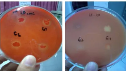
The activity of cellulase and xylanase enzymes produced by Actinobacillus sp.(G6) and Bacillus pumilus (G7) bacteria could be identified from the existing halo zones (clear/colorless area) around the location where bacteria grown (See Figure 1 below). The activity of cellulase enzyme could be seen on LB medium containing CMC 1% substrate while the activity of xylanase enzyme could be seen on LB medium containing OX 1% substrate. Xylanolitic index of G6 and G7 and cellulolitic index of G6 and G7 were presented on Figure 2 :
Figure 1: G6 and G7 Isolates Qualitative Screening Result
Figure 2: Cellulolitic and Xylanolitic Indexes of G6 and G7
utilized Congo Red dye for its strong interaction with polysaccharide containing D-glucopyranocyl and β-(1,4), β -(1,3)-D-glucan bonds and several hemicellulosic bonds, including galactoglucomannant. Based on this characteristics, Congo Red dye could be used to detect the existence of cellulose and hemicelluloses on agar plate [14]
. Halo zones formed through Congo Red coloration indicated substrates had been hydrolyzed into simple sugar products [15].. NaCl flushing was intended to preserve color stability and inhibit enzyme activities [16]. Qualitative data could be obtained by examining enzymes activities on CMC and OX substrates.
Enzymes Assay
The result of xylanase and cellulase activity was presented on Figure 3:
Figure 3: Graphic describing the activity of Xylanase and Cellulase produced by G6 and G7 bacteria
The activity of xylanase on G6 and G7 were higher compared to the activity of cellulase. Cellulase was examined by mixing CMC substrate with DNS reactor while xylanase activity was examined by mixing OX substrate with DNS reactor. The amount of reducing sugar was measured by applying spectrophotometric DNS method at wave-length (λ) 550 nm [12]. One activity unit of cellulose was defined as the amount of glucose (in µmol) produced by one mL enzyme per minute. Meanwhile, one activity unit of xylanase was defined as the amount of xylose (in µmol) produced by 1 mL enzyme per minute. Different results of cellulase and xylanase activity on G6 and G7 were related to enzymes different capability in hydrolyzing substrates.
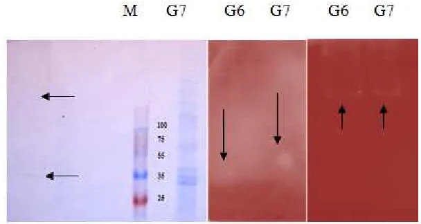
SDS-PAGE and Zymogram Analysis
The results of cellulase and xylanase enzymes analysis based on SDS-PAGE and zymogram were presented on Figure 4:
Figure 4: 1 (SDS-PAGE): M: Protein Marker (5 bands 100, 70, 55, 35, 25 kDa); G6 (1 band between 25 kDa and 35 kDa, 1 band above 100 kDa), G7 (2 bands between 25 kDa and 35 kDa, 1 band between 35 kDa and 55 kDa, 1 band between 55 and 70 kDa, 4 bands above 100 kDa). 2 (Xylanase zymogram): G6 (halo zone between 25 kDa band and 35 kDa), G7 (halo zone between 35 kDa and 55 kDa). 3
(Cellulase zymogram): G6 and G7 halo zone above 100 kDa
very clear. It may be caused by enzymes low protein contents. This issue can be solved by purifying the enzymes and increasing its concentration which made protein bands clearer during SDS-PAGE and zymogram process.
In this research, [17] reported that relative molecular weights of cellulase enzymes were varied, for example endoglucanase weighed 12.5 - 90 kDa, exoglucanase 41.8 – 65 kDa, and cellobiase 47 kDa. Cepeljnik et al (2001) [18]
also reported that relative molecular weight of β-1,4-endoxylanase was 58 kDa.
SEM Analysis
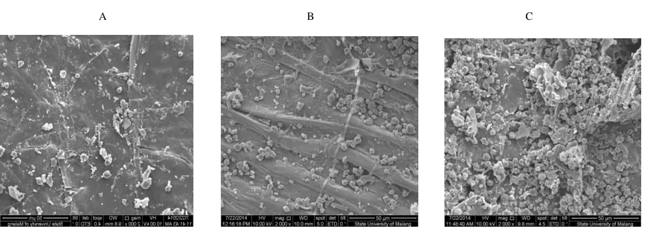
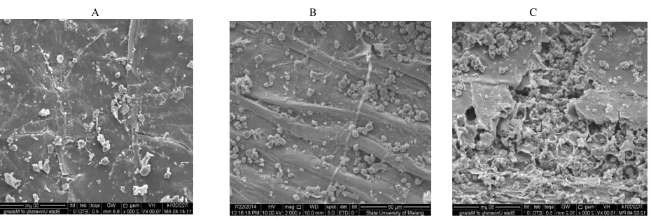
The results of SEM analysis indicated changes ob rice bran surface structure after enzyme treatment. These changes were presented on Figure 5, 6, 7, and 8 below:
A B C
A B C
A B C
Figure 5: Rice bran surface structures (2000x magnification). A: before treatment; B: treated using water; C treated using xylanase produced by G6
A B C
Scanning Electron Microscope (SEM)
SEM was equipment used to describe surface profile of an object by shooting electrons onto its surface. In order to obtain good description from an object, the object should be metallic. Eichhornia crassipes, rice stalks, and rice bran were organic object without metal contents. Therefore, in order to be able to analyze these objects under SEM, they should be metalized by layering them with metals, such as palladium gold. Samples which were analyzed consisted of rice bran samples which had been incubated for 24 hours. Rice bran sample which had not been treated using enzyme and rice bran sample which had been treated using aquadest were also analyzed as control groups.
The results of SEM analysis showed changes on surface profiles among the samples before and after being treated using enzymes (as described on Figure 5, 6, 7, and 8 above). There were changes on rice bran surface structure. Before being treated using enzymes, the surface structure of rice bran was compact and well-organized (See Figure 5A, 6A, 7A, 8A). After enzyme treatment, rice bran surface structures became more hollow and cracked (as seen on Figure 5C, 6C, 7C, and 8C above). There was no significant difference between rice bran which had not been treated (Figure 5A, 6A, 7A, and 8A) and rice bran which had been treated using aquadest (Figure 5B, 6B, 7B, 8B). this finding implied that temperature used during incubation process did not affect the cracks (damages) on rice bran surface structure. SEM analysis results on rice bran which had been treated using xylanase and cellulase (Figure 7C) indicated more cracks than rice bran which had been treated using xylanase (Figure 6C).
Cracks on rice bran surface structure was the effect of enzymes (cellulase and xylanase) activity in damaging rice bran cell wall which mainly composed by cellulose and hemicelluloses (particularly xylan). Xylan was main component of hemicelluloses which was located between lignin and lower cellulose fibers compounds [19]. Xylanase functioned to hydrolyze xylan into its monomers, such as xylose, xylooligosaccharide, and arabinose. Meanwhile, cellulase functioned to hydrolyze cellulose into glucose [20]. Previous studies found that enzymes (xylanase, cellulase, and combination of xylanase and cellulase) hydrolysis on rice stalks caused cracks on rice bran surface structure [21].
CONCLUSION
From the results of this research, it can be concluded that G6 (Actinobacillus sp.) and G7 (Bacillus pumilus) bacteria were able to produce xylanase and cellulase. The molecular weight of enzymes (xylanase and cellulase) produced by G6 (Actinobacillus sp.) bacteria was 25-35 kDa and above 100 kDa while the molecular weight of enzymes (xylanase and cellulase) was 25 kDa and above 100 kDa. The results of SEM analysis indicated changes on samples (rice bran) surface profile before and after enzymes treatment.
Acknowledg ement
This research was funded by DIPA-BOPTN financial year 2014 as stated on Airlangga University Rector Decree Number 1349/UN3/KR/2014 regarding Decentralized Research and Second-phase Seed Research of Airlangga University on May 9th, 2014.
REFERENCES
[1] T Juhasz; Z Szengyel; K Reczey; M Siika-Aho; L Viikari. Process Biochemistry., 2005, 40, 3519-3525. Figure 8: Rice bran surfade structures. A: before treatment; B: after being treated using water; C: after being treated using xylanase
[2] S de Vriest, Pustjens AM, Schols HA, Hendriks WH, Geritts WJJ, Animal Nutrition and Feed Technology,
2012, 178 (3-4): 123-138.
[3] GM Mathew; R Sukumaran; RR Singhania; A Pandey. Journal of Scientific and Industrial Research., 2005, Vol
67, 898-908.
[4] KS Azad; S Rahimi; KMA Torshizi. Iranian Journal of Veterinary Research., 2009, Vol. 10 (2),27.
[5] Murad HH, Hanfy MA, Kholif AM, Gawad MHA and Murad HA. J. Biol. Chem. Environ. Sci., 2009, 4:
791-809.
[6] Gado H.M., Salem AZM, Camacho LM, Elghandour MMY, Salazar MC, Animal Nutrition and Feed
Technology , 2013, 13: 569-574.
[7] Rodrigues MAM, Pinto P, Bezerra RMF, Dias AA and Guedes CVM. Anim. Feed Sci. Technol., 2008, 141: 326-338.
[8] Tricarico JM, Johnston JD, Dawson KA, Hanson KC, McLeod KR and Harmon DL. Anim. Sci., 2005, 81:
365-374.
[9] Krueger NA, Adesogan AT, Staples CR, Krueger WK, Kim SC, Littell RC and Sollenberger LE. J. Anim. Sci.,
2008,. 86:882–889.
[10] Gado HM, Salem AZM, Odongo NE, Borhami BE, Animal Feed Science and Technology 2011, 165 (1),
131-136.
[11] CAM Cordeiro; MLL Martins; AB Luciano; RF da Silva. Brazilian Archives of Biology and Biotechnology.,
2002, 45(4), 413-418
[12]LG Miller. Anal chem., 1959, 31, 426-428.
[13] Laemmli UK, Molbert E, Showe M, Kellenberger, E, J. Mol. Biol, 1970, 49, 99.. [14] R Teather; PJ Wood. Appl Environ Microbiol., 1982, 43, 777-780.
[15] J Pongjanta; A Utaipatanacheep; O Naivikul; K Piyachomkwan. Kasetsart J., 2008, 42, 198-205.
[16] NRGilkes; ML Langsford; DG Kilburn; RC Miller; RAJ Warren. The Journal of Biological Chemistry., 1984,
259(16), 10455-10459.
[17] Bijender K Bajaj, Himani Pangotra, Masood A Wani, Priyanka Sharma & Ajay Sharma, Indian Journal of Chemical Technology, 2009, Vol. 16 : 382-387.
[18] Cepeljnik T., Masa Zorec., Franc Viktor Nekrep., Romana Marinsek Logar. Actachim Slov., 2007, 48 :401-408. [19] S Subramaniyan; P Prema. Critical Rev Biotechnol., 2002, 22, 33-64.
[20] HJ Gilbert; GP Hazlewoo. Journal of General Microbiology., 1993, 139, 187-194.
[21] RG Medeiros; FG da Silva Jr; SN Báo; R Hanada; EXF Filho. Brazilian Archives of Biology and Technology.,