Molecular Characterization and Expression Analysis
of
Pathogenesis related
Protein 6 from
Panax ginseng
1D. Myagmarjav
a, 2, J. Sukweenadhi
a, 2, Y. J. Kim
b,*, M. G. Jang
b, S. Rahimi
a, J. Silva
a,
J. Y. Choi
b, P. Mohanan
a, W. S. Kwon
b, C. G. Kim
b, and D.-C. Yang
a,b,**
aGraduate School of Biotechnology and Ginseng Bank, College of Life Science, Kyung Hee University, Yongin, 446-701 Korea bDepartment of Oriental Medicinal Biotechnology, College of Life Science, Kyung Hee University, Yongin, 446-701 Korea
*e-mail: [email protected] **e-mail: [email protected]
Received December 26, 2016
Abstract—Panax ginseng Meyer is one of the important medicinal plants in the world, particularly in Asian countries. Ginseng encounters many stress exposure during its long cultivation period. However, the molec-ular mechanism of stress resistance is still poorly understood in spite of its importance. In this study, patho-genesis-related protein 6 (PR6), also called proteinase inhibitor (PI), was isolated from ginseng embryogenic callus, named PgPR6. The small size of PR6, containing an open reading frame of 219 bp encoding 72 amino acids, the typical characteristic of PR6 protein, shares the highest sequence similarity to PR6 of Theobroma cacao (69% identity). Sequence and structural analysis indicated that PgPR6 belongs to class Kunitz-type PI family. This is the first report pertaining to the identification of PR6 gene from the P. ginseng genome. The high-level expression of PgPR6 was observed in root as revealed by quantitative real-time PCR. The temporal expression analysis demonstrated that PgPR6 expression was highly up-regulated by signaling molecules, heavy metals, mechanical wounding, chilling, salt, sucrose, and mannitol stress, indicating that PgPR6 may play an important role in the molecular defense response of ginseng to a various range of environmental stresses.
Keywords: Panax ginseng, abiotic stress, biotic stress, pathogenesis-related protein 6, proteinase inhibitor pro-tein, gene expression
DOI: 10.1134/S1022795417110060
INTRODUCTION
Plants are continuously threatened by biotic or abi-otic stresses during its growth and development. Therefore, they developed unique defense mecha-nisms by rapid changes in gene expression, resulting in the synthesis of specific proteins to protect them from various stresses such as pathogen attack, insect, herbi-vores and also other harsh growing conditions. Most inducible plant proteins under these circumstances are pathogenesis-related (PR) proteins, which are induced and accumulated in host plants as a result of biotic and abiotic stresses [1]. From the first isolation of PR protein in leaves of Nicotiana tabacum after tobacco mosaic virus infection, many proteins with similar properties have been isolated from various plants [2]. PR proteins can be divided into 17 groups (PR-1 to PR-17) on the basis of their functions, sequence characteristics, immunological relation-ships, enzymatic activities or other biological
prop-erties and numbered in the order in which they belong [3].
Among the diverse PR families, the PR6 family is considered as the proteinase inhibitors (PIs) [4]. Plant PIs are generally small proteins with a molecular mass of 8–20 kDa, and inhibit proteolytic enzymes of ani-mal and fungal origin, which are widespread in both monocots and dicots [4, 5]. Among four major sub-classes including serine, cysteine, aspartate and metallo-PIs based on their amino acid in the active site of the proteinases [6], the serine PIs were well characterized in various dicot species, especially in potato and tomato [5]. Serine PIs are the most abun-dant PR6 which is sub-divided further into Kunitz Type PI (KTI) and Bowmin-Birk PI (BBI), according to their molecular mass, location of disulfide bridge and cysteine content [6, 7].
The exact physiological role of PR6 according to their classes has not yet been well understood. PR6 implicates in defense against the herbivores, insects, and nematode or mechanical wounding, via interac-tion with the proteinases in all plant organs [6–8].
1
The article is published in the original.
2
These authors contributed equally to this work.
Even though most of PR6 studies have been focused on its role in plant-insect interactions, it has been sug-gested that it plays many roles in the plant response to abiotic stresses such as salt, heavy metal, water-deficit and mechanical wounding [9–12]. The identification and characterization of PR6 have been demonstrated particularly in Gramineae [5], Leguminosae [13], Solanaceae families [14] and they were primarily found as a result of nutritional interest in inhibitors of animal digestive enzymes in important agricultural crops. However, most of the studies have been carried out on crop plants (cereals, legumes and solanaceous species) due to their economic importance, thus, non-crop species being comparatively neglected [15]. Korean ginseng (Panax ginseng Meyer) has been used for thousands of years as one of the most important sources of medicinal herb in Asia and is cultivated for its highly valued root used for medicinal purposes.
P. ginseng requires 4 to 6 years cultivation period to provide the high-quality roots [16] and the root dis-eases are the major threat to the quality of its roots. The consecutive planting of ginseng in the same field may cause serious root diseases [17]. Therefore, to effectively manage ginseng diseases and other disor-ders, it is important to study and identify functional genes related to ginseng defense mechanism. Previ-ously, we identified and characterized PR4 [16], PR10 [18] family in P. ginseng. In the present study, we report the cloning of PR6 gene from P. ginseng and provide the analysis on the expression profile of this gene in the defense against various environmental stresses.
MATERIALS AND METHODS
Isolation of PgPR6 and Sequence Analysis
PgPR6 ESTs were searched against the GenBank database using BLASTX algorithm. Total RNA iso-lated from the embryogenic callus of ginseng was pre-viously constructed into the cDNA library [19]. Size-selected cDNA was ligated into a λ TriplEx2 vector and pTriplEx phagemids were excised from the λ pTri-plEx2 and used as templates for sequence analysis. The 5' ends of the cDNA inserts were sequenced by an automatic DNA sequencer (ABI Prism 3700,
Perkin-Elmer, USA). Homologous sequences of PR6
expressed sequence tag (EST) were searched against the GenBank databases using a BLASTX algorithm. A
pTriplEx phagemid for PR6 cDNA was excised from
the λpTriplEx2 and used as a template for sequence analysis. Nucleotide and amino acid sequence analy-ses were performed using DNASIS program (Hita-chi). Secondary structure analysis for PR6 homolo-gous from plants was conducted by SOPMA [20].
These deduced amino acid sequences were utilized to search for homologous proteins via BLAST network services at the NCBI (http://blast.ncbi.nlm.nih.gov/ Blast.cgi). ClustalX [21] with default gap penalties were used to perform multiple alignments of PR6 gene
sequence isolated in ginseng with other previously reported sequences from different species. A phyloge-netic tree was constructed by the neighbor-joining method, and the reliability of each node was estab-lished by bootstrap methods using MEGA4 [22] soft-ware. The protein properties were estimated using ProtParam [23] and the hydropathy value, identifica-tion of conserved motifs within PR6 was accomplished with MEME [24] and a three-dimensional (3-D) model was prepared using SWISS-MODEL [25] Workspace and UCSF Chimera package, as described previously [16].
Plant Materials and Application of Environmental Stresses
P. ginseng cv. Hwang-Sook seeds (provided by Gin-seng Bank) were used and cultured three-week-old plantlets were used for the treatments and RNA was extracted, as described previously [16]. For chemical stress or plant hormone treatments, the plantlets were placed for various time periods in Murashige and Skoog (MS) medium containing the indicated con-centrations of chemicals: 100 mM NaCl, 10 mM H2O2, 0.2 mM jasmonic acid (JA), 0.1 mM abscisic acid (ABA) [16], 1 mM salicylic acid (SA) [18], 500 μM CuSO4, 500 μM CdSO4, 11% mannitol, 20% sucrose
[39]. Chilling stress was applied by exposing the plant-lets to 4°C [16]. For the UV treatment, the plantlets were irradiated under UVC lamps at 1.35 μE m–2 s–1
(below 280 nm). To monitor the effect of light stress, plants were kept in the white light with an intensity of about 500 μE m–2 s–1 [18]. Anoxic conditions were
generated by submerging whole potted plants for the duration of the experiment. For mechanical wounding stress, healthy leaves and stems of plantlets were wounded with a sterile scalpel [16]. In all cases, stress treatments were carried out in the MS media and ten plantlets were treated with each stress for 1, 4, 8, 24, 48, or 72 hours. Control plants were held in a growth room at 25°C under a 16 h photoperiod.
Real-Time Quantitative RT-PCR
Total RNA was extracted from seedlings of P. gin-seng using the RNeasy mini kit (Qiagen, Valencia, CA, USA). For RT-PCR, 200 ng of total RNA was used as a template for reverse transcription using oligo(dT)15
primer (0.2 mM) and AMV reverse transcriptase
(10 U/µL) (INTRON Biotechnology, Inc., South
Korea) according to the manufacturer’s instructions. Real-time quantitative PCR was performed using 100 ng
of cDNA in a 10-µL reaction volume using SYBR®
10 min at 95°C, followed 40 cycles of 95°C for 10 s, 60°C for 10 s, and 72°C 20 s. Amplification, detection, and data analysis were carried out with a Rotor-Gene 6000 real-time rotary analyzer (Corbett Life Science, Sydney, Australia). The threshold cycle (Ct) rep-resents the number of cycles at which the f luorescence intensity was significantly higher than the background f luorescence at the initial exponential phase of PCR amplification. To determine the relative fold differ-ences in template abundance for each sample, the Ct value for PgPR6 was normalized to the Ct value for
PgActin and calculated relative to a calibrator using the formula 2–ΔΔCt. The actin gene was selected as a
refer-ence gene [40] and its primers were designed accord-ing to the conserved region of PgActin mRNA sequences (GenBank accession no. KF699319). Three independent experiments were performed.
RESULTS
Isolation and Amino Acid Sequence Analysis of PgPR6
From the EST analysis of a cDNA library [19], we
identified a cDNA clone encoding a PR6 gene. We
named this gene as PgPR6 (P. ginseng pathogenesis-related protein 6); the sequence data of PgPR6 have been deposited in GenBank under accession number
KP893146. The full-length cDNA of PgPR6 was 725
nucleotides long and had the putative open reading frame of 219 bp. This ORF encodes PR6 protein of 72 amino acids, beginning at the start codon ATG tion 70) and terminating at the stop codon TAA (posi-tion 288) of the cDNA.The predicted molecular weight is 17.33 kDa and its isoelectric point is 5.38, which is the typical characteristic of KTI [8, 13]. The instability index (II) is calculated 24.94 and the ali-phatic index, defined as a positive factor for increased thermostability was computed as 25.75 using Prot-Param [23].
Homology Analysis
A GenBank BlastX search revealed that the amino acid sequence of PgPR6 has a high degree of sequence homology with PR6 from other plants. PgPR6 shows the highest sequence identity of KTI-type PR6 from
Theobroma cacao (TcPR6, XP_007050380.1, 69%
identity), followed by Ricinus communis (RcPR6,
EEF41422.1, 65% identity), and Cucurbita maxima
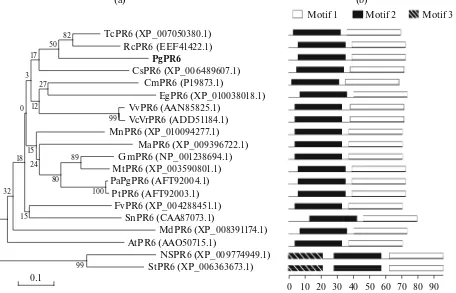
Fig. 1. Sequence homology analysis of PgPR6 with other PR6s. (a) A phylogenetic tree of PgPR6 with PR6 isozymes from various
organisms including plants shown in Fig. 1. The neighbor-joining method was used, and the branch lengths are proportional to the divergence, with the scale of 0.1 representing 10% changes. (b) Organization of putative motifs in PR6 identified by MEME. Numbered color boxes represent different putative motifs. Motifs 1, 2, and 3 are indicated by the white (first), black (middle), and diagonal lines (last) boxes, respectively. Motif sizes are indicated at the bottom of the figure.
99 99
100 89 82
80 50
27
32 12 17
24 15 3
15 0
18
0.1
(a)
Motif 1 Motif 2 Motif 3
(b)
0 10 20 30 40 50 60 70 80 90 TcPR6 (XP_007050380.1)
RcPR6 (EEF41422.1) PgPR6
CmPR6 (P19873.1)
EgPR6 (XP_010038018.1) VvPR6 (AAN85825.1)
VcVrPR6 (ADD51184.1) MnPR6 (XP_010094277.1)
MaPR6 (XP_009396722.1) GmPR6 (NP_001238694.1) MtPR6 (XP_003590801.1) PaPgPR6 (AFT92004.1)
PtPR6 (AFT92003.1) FvPR6 (XP_004288451.1)
SnPR6 (CAA87073.1)
MdPR6 (XP_008391174.1) AtPR6 (AAO50715.1)
(CmPR6, P19873.1, 60% identity). The protein sequence of PgPR6 and the similar proteins from other plants were used for doing multiple sequence alignment (Sup. Fig. S1) and phylogenetic tree con-struction (Fig. 1a). Multiple sequence alignment revealed that all amino sequences are well conserved among the known PR6 (Sup. Fig. S1). However, the N-terminal region of PR6 protein sequences showed variance between species, except conserved Cys resi-due, which probably forms disulfide bridge with Cys in C-terminal. Consistently, two motifs were conserved
well in all PR6s except N-terminal-located motif 3, which was shown specific to just two PR6s (Fig. 1b).
The hydrophilic profile (Sup. Fig. S2a) and sec-ondary structure (Sup. Fig. S2b) of the estimated PR6 proteins support their similar profiles, but also shows the species-specific difference in N-terminal regions. The secondary structure analysis showed that PgPR6 consists of 14 α-helices, 11 β-turns linked by 18 extended strands and 29 random coils. The composi-tion is highly comparable to the secondary structure of EgPR6 (Eucalyptus grandis, XP_010038018.1) that
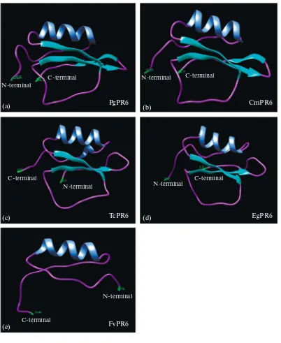
Fig. 2. The comparison of a predicted three-dimensional (3d) model of PR6. (a) P. ginseng PR6 (PgPR6), (b) C. maxima (CmPR6), (c) T. cacao (TcPR6), (d) E. grandis (EgPR6).
TcPR6
(c) (d) EgPR6
FvPR6 (e)
PgPR6
N-terminal
N-terminal N-terminal
N-terminal
N-terminal
C-terminal C-terminal
C-terminal C-terminal
C-terminal
contains 19 α-helices, 10 β-turns jointed by 16 extended strands, and 28 random coils and FvPR6 (Fragaria vesca, XP_004284511.1) which contains 15
α-helices, 12 β-turns linked by 18 extended strands, and 25 random coils. However, the structure profile appears more similar to the secondary structure of CmPR6 that contains 8 α-helices, 12 β-turns linked by 21 extended strands, and 27 random coils (Sup. Fig. S2b) and its tertiary structure also showed most similar construction with PgPR6, having one helix chain and three turns (Figs. 4a, 4b), rather than TcPR6, EgPR6, and FvPR6 (Figs. 2c–2e).
Expression Analysis of PgPR6 Gene in Diverse Organs
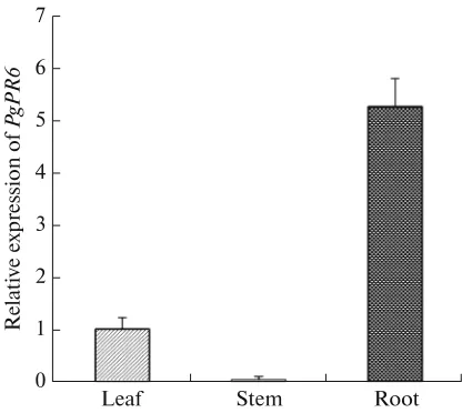
To examine the expression profile of PgPR6, the quantitative RT-PCR was carried out using the cDNA templates from three organs such as leaf, stem, and root. As shown in Fig. 3, PgPR6 was highly expressed in ginseng root and very weekly expressed in the stem.
Temporal Expression of PgPR6 Gene in Response to Abiotic Stresses
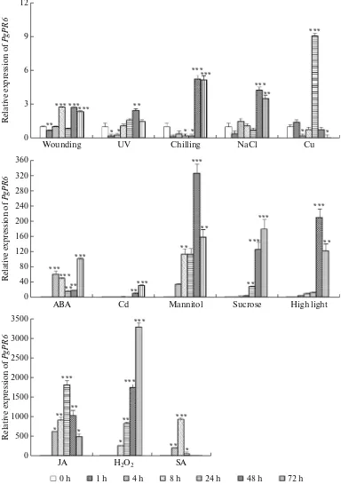
The expression pattern of PgPR6 against different stresses at different time points after treatments were analyzed by real-time PCR (Fig. 4). The PgPR6
response against abiotic stress and hormone treatment demonstrated the up-regulations by all of the treat-ments compared to the controls (0 h). High light and UV exposure increased PgPR6 mRNA level up to 200-fold and 2.5-200-fold, respectively, at 48 h post-treatment. Under chilling stress, PgPR6 expression decreased rapidly but accumulated at 48 h (5-fold) and main-tained. Similarly, salt stress also increased PgPR6
expression at 48 h (4.2-fold). Osmotic stresses such as sucrose and mannitol highly up-regulated PgPR6 after 48 h post treatment (>100-fold and >200-fold, respec-tively). Heavy metal stresses including copper and cadmium treatments increased the expression of
PgPR6 at 24 and 72 h, respectively. ABA rapidly accu-mulated PgPR6 mRNA at 4 h and maintained its up-regulation until 72 h of treatment. Moreover, hormone treatment more strongly accumulated PgPR6 mRNA, suggesting its high response to the signal molecules. JA caused the rapid and strong up-regulation of PgPR6 in all the harvesting times and peaked at 24 h (1700-fold), in spite of the weak response against wounding treat-ment. PgPR6 was also strongly up-regulated but decreased to the control level after 8 h of SA treatment. The expression of PgPR6 against H2O2 was
up-regu-lated from 8 h and gradually increased until 72 h post-treatment (3500-fold).
DISCUSSION
In this study, we firstly report the isolation and characterization of the gene encoding PR6 from
P. ginseng and its expression in response to signaling molecules, heavy metals, mechanical wounding, chill-ing, salt, sucrose and mannitol stresses. Sequence analysis of PgPR6 showed that the full-length cDNA encoding 72 amino acids, showing typical small size of KTI-type PR6. Structural pattern of most plant KTIs are single polypeptide chain of approximately 20 kDa with low cysteine residues (usually four), two disulfide bridges and single reactive site. The protein sequence analysis showing that PgPR6 has a single polypeptide chain of 17.35 kDa predicted molecular mass with 2 Cys residues which can make single disulfide bridge, led us to classify PgPR6 into KTI-type PR6. Plant KTIs are not so structurally follow the Cys-Cys pattern. For example, KTI isolated from Cajanus cajan contains five Cys residues and four of them are involved in two disulfide bridges [26], but KTI isolated from Inga lau-rina contains only two cysteine residues [27] which are similar to our result.
The putative amino acid sequence of PgPR6 exhib-ited high similarity to other PR6, particularly KTI-type from other plant species, T. cacao, R. communis, and C. maxima containing two conserved motifs (Sup. Fig. S1). In spite of its distance on phylogenetic tree (Fig. 1a), PgPR6 and CmPR6 shares very similar ter-tiary structure. However, other relatives of PR6 shows different construct (Fig. 2), even though its hydropathy and secondary structures are similar (Sup. Fig. S2). This result suggests its rapid adaptive evolution of structure according to the species-dependent environment or development, which could contribute to its diversity and function of KTI [28].
The exact physiological role of PR6 has not been well reported in other species. In addition to the clas-sical studies of PR6s related to the seed development and storage protein [4, 6], several reports have shown
Fig. 3. Expression of PgPR6 gene in leaves, stems, and roots of Panax ginseng. Vertical bars indicate the mean value ±SE from three independent experiments.
Fig. 4. Relative quantities of PgPR6 mRNA at various time points (h) post-treatment with various abiotic stresses: wounding, UV light, chilling, 100 mM NaCl, 0.1 mM abscisic acid, heavy metal treatment with 500 μM CuSO4, and 500 μM CdSO4, 11% man-nitol, 20% sucrose, high light, 0.2 mM jasmonic acid, 10 mM H2O2, and 1 mM salicylic acid. The error bars represent the stan-dard error of the means of three independent replicates. Statistical significance using Student’s t-test was assigned at * for P < 0.05, ** for P < 0.01, and *** for P < 0.001, respectively, compared to non-treated plants.
12
9
6
3
0
Wounding UV Chilling NaCl Cu
R
ABA Cd Mannitol Sucrose High light
their expression in all plant organs [6, 8] which may link with the general role for defense. Similar to the
Populus deltoides KTI [12], the expression of PgPR6
was observed strongly in roots and weakly in leaves (Fig. 3), suggesting PgPR6 expression possibly relate to the preferred defense role in the root, as a storage protein.
The wound-induced response of PR6 has been extensively studied related with insect-resistance [29] and the up-regulation of PgPR6 to wounding would be strongly mediated by hormones, such as SA, JA, and ABA [30–32]. The response to wound signaling path-way from release to transduction occurs within few minutes [33] and the high expression peak at 8 h and falling off after 24 h of PgPR6 is similar with the pat-tern of soybean PR6 [32]. ABA acts as a primary signal in the systemic wound signaling which in turn acti-vates the biosynthesis of JA signaling pathway, that trigger the expression of PR6 [34].
The defense response of PgPR6 against heavy met-als was supported by the recent reports that P. deltoids
KTI could interact with the metal ions and up-regu-lated by copper stress [12], and serine PIs from Coptis japonica and Capsicum annuum contributes to the toler-ance to various toxic compounds including Cd [11, 35]. The late and strong response of PgPR6 under high light exposure is consistent with BBI from Oryza sativa
[5], and KTI from Amaranthus hypochondriacus [10]. Tomato PR6 was also shown to accumulate in response to UVC radiation [36] as well as sucrose [37]. Furthermore, PgPR6 expression could be positively
responsive to abiotic stresses such as H2O2, chilling,
sodium chloride, sucrose, and mannitol. The involve-ment of PR6 in the response of plants to abiotic stresses is connected with the enhanced proteolysis that involves during senescence of plant tissues [6].
Taken together, our results indicate that PgPR6
could play a critical role in signaling molecules, heavy metals, mechanical wounding, chilling, salt, sucrose and mannitol stress implying its involvement in the defense mechanism against biotic and abiotic stresses in ginseng. Ginseng cultivation suffers difficulties with abiotic and biotic stresses and its cultivation requires special consideration during long duration [38]. By contrast to the lack of information about ginseng genome, more PR genes involved in stress have been detected and some of them identified and character-ized in ginseng. Consequently, the puzzle is starting to fill although still far from complete; much more research will be needed to link them together.
ACKNOWLEDGMENTS
This research was supported by a grant from the Basic Science Research Program through the National Research Foundation (NRF), Ministry of Education, Republic of Korea to Y.J. Kim (2016R1A6A3A11931858) and Korea Institute of Planning and Evaluation for Technology in Food, Agriculture, Forestry and Fisheries IPET to D.C. Yang (112142-05-4-SB010).
Sup. Fig. S1. Comparison of the putative amino acid sequences of PgPR6 with those of PR6s from other plants: Theobroma cacao (XP_007050380.1), Ricinus communis (EEF41422), Sambucus nigra (CAA87073.1), Vitis vinifera (AAN85825.1), Vitis cineria (ADD51184.1), Glycine max (NP_001238694.1), Medicago truncatula (XP_003590801.1), Populus alba (AFT92016.1), Populus tamentasa (AFT92015.1), Morus notabilus (XP_010094276.1), Cucurbita maxima (P19873.1,) Arabidopsis thaliana (AAO50715). A hyphen was inserted within the amino acid sequence to denote a gap. Shadowbox indicates well-conserved residues, * rep-resents a conserved amino acid, and: reprep-resents a very similar amino acid. Arrows show the two conserved cysteines in positions 28, 83. The conserved α helix domain indicated above the alignment. Three boxes show that conserved motifs obtained by MEME analysis.
Motif 3 Motif 2 Motif 1
10 20 30 40 50 60 70 80 90 100
PgPR6 72
TcPR6 69
RcPR6 72
CsPR6 71
FvPR6 70
SnPR6 79
CmPR6 68
EgPR6 73
VvPR6 71
VcPR6 71
GmPR6 70
MtPR6 72
PaPR6 72
PtPR6 72
MaPR6 70
MnPR6 70
AtPR6 70
MdPR6 73
NsPR6 95
StPR6 95
: : * * . * * . * : : : : : . * * * * : : : . *
REFERENCES
1. Sarowar, S., Kim, Y.J., Kim, E.N., Kim, K.D., et al., Overexpression of a pepper basic pathogenesis-related protein 1 gene in tobacco plants enhances resistance to heavy metal and pathogen stresses, Plant Cell Rep., 2005, vol. 24, no. 4, pp. 216—224.
2. Niderman, T., Genetet, I., Bruyere, T., Gees, R., et al., Pathogenesis-related PR-1 proteins are antifungal (iso-lation and characterization of three 14-kilodalton pro-teins of tomato and of basic PR-1 of tobacco with inhib-itory activity against Phytophtora infestans), Plant Physiol.,1995, vol. 108, no. 1, pp. 17—27.
Sup. Fig. S2. Superimposed hydrophobicity profile of PgPR6 and secondary structure predictions for PgPR6 and homologous plant PR6s. (a) Hydrophobic domains are indicated by positive numbers; hydrophilic domains are above the line and hydrophilic domains are below the line. (b) Comparison of PR6 secondary structures by SOPMA. The helix, sheet, turn, and coil are indi-cated in the order from the longest to the shortest.
(a)
TcPR6 RcPR6 PgPR6
EgPR6 FvPR6
CmPR6 2.0
1.0 1.5
0.5
0
–0.5
–1.0
–1.5
–2.0
0 2 4 6 8 10 12 14 16 18 20 22 24 26 28 30 32 34 36 38 40 42 44 46 48 50 52 54 56 58 60
TcPR6
RcPR6 PgPR6 (b)
EgPR6
FvPR6 CmPR6
10 20 30 40 50 60
10 20 30 40 50
10 20 30 40 50 60
10 20 30 40 50 60
10 20 30 40 50
10 20 30 40 50
62 64 66 68 70 72 74 76 78 80 82 84 86 88 90 92 94 96 98 100102 Position
M
e
a
n
h
y
d
ro
p
h
o
bi
c
ity
3. Sinha, M., Singh, R.P., Kushwaha, G.S., Iqbal, N., et al., Current overview of allergens of plant pathogen-esis related protein families, Sci. World J., 2014, vol. 2014, no. 2014, pp. 1—19.
4. Sels, J., Mathys, J., Coninck B.M.A.De., Cammue, B.P.A., et al., Plant pathogenesis-related (PR) proteins: a focus on PR peptides, Plant Physiol. Biochem., 2008, vol. 46, no. 11, pp. 941—950.
5. Rakwal, R., Agrawal, G.K., and Jwa, N.S., Character-ization of a rice (Oryza sativa L.) Bowman—Birk pro-teinase inhibitor: tightly light regulated induction in response to cut, jasmonic acid, ethylene and protein phosphatase 2A inhibitors, Gene., 2001, vol. 263, nos. 1—2, pp. 189—198.
6. Kidric, M., Kos, J., and Sabotic, J., Proteases and their endogenous inhibitors in the plant response to abiotic stress, Bot. Serbica, 2014, vol. 31, pp. 139—158. 7. Oliva, M.L.V., Silva M.C.C., Sallai, R.C., Brito, V.M.,
et al., A novel subclassification for Kunitz proteinase inhibitor from leguminous seeds, Biochimie, 2010, vol. 92, no. 11, pp. 1667—1673.
8. Kuhar, K., Kansal, R., Mishra, A., Koundal, K.R., et al., Cloning, characterization and expression analy-sis of a novel gene encoding Kunitz-type protease inhibitor from Dolichos biflorus,Biotech., 2012, vol. 2, no. 3, pp. 199—209.
9. Dombrowski, J.E., Salt stress activation of wound-related genes in tomato plants, Plant Physiol., 2003, vol. 132, no. 4, pp. 2098—2107.
10. Sanchez-Hernandez, C., Martinez-Gallardo, N., Guerrero-Rangel, A., Valdes-Rodriguez, S., et al., Trypsin and α-amylase inhibitors are differentially induced in leaves of amaranth (Amaranthus hypochon-driacus) in response to biotic and abiotic stress, Physiol. Plant., 2004, vol. 122, no. 2, pp. 254—264.
11. Joshi, R.S., Tanpure, R.S, Singh, R.K., Gupta, V.S., et al., Resistance through inhibition: ectopic expression of serine protease inhibitor offers stress tolerance via delayed senescence in yeast cell, Biochem. Biophys. Res. Commun., 2014, vol. 452, no. 3, pp. 361—368.
12. Guerra, F. P., Reyes, L., Vergara-Jaque, A., Campos-Hernández, C., et al., Populus deltoides Kunitz trypsin inhibitor 3 confers metal tolerance and binds copper, revealing a new defensive role against heavy metal stress, Environ. Exp. Bot., 2015, vol. 115, pp. 28—37. 13. Jamal, F., Pandey, P.K., Singh, D., and Ahmed, W., A
Kunitz-type serine protease inhibitor from Butea mono-sperma seed and its inf luence on developmental physi-ology of Helicoverpa armigera, Process Biochem., 2014, vol. 50, no. 2015, pp. 311—316.
14. Zhang, H.Y., Xie, X.Z., Xu, Y.Z., and Wu, N.H., Iso-lation and functional assesment of tomato proteinase inhibitor II gene, Plant Physiol. Biochem., 2004, vol. 42, no. 5, pp. 437—444.
15. Konarev, A.V., Griffin, J., Konechnaya, G.Yu., and Shewrey, P.R., The distribution of serine proteinase inhibitors in seeds of the Asteridae, Phytochem., 2004, vol. 65, no. 22, pp. 3003—3020.
16. Kim, Y.J., Lee, H.J., Jang, M.G., Kwon, W.S., et al., Cloning and characterization of pathogenesis-related protein 4 gene from Panax ginseng, Russ. J. Plant Phyiol., 2014, vol. 61, no. 5, pp. 664—671.
17. Song, M., Yun, H.Y., Kim, Y.H., Antagonistic Bacillus
species as a biological control of ginseng root rot caused by Fusarium cf. incarnate,J. Ginseng Res., 2014, vol. 38, no. 2, pp. 136—145.
18. Kim, Y.J., Jang, M.G., Lee, H.J., Jang, G.H., et al., Functional characterization of the pathogenesis-related protein family 10 gene, PgPR10-4, from Panax ginseng in response to environmental stresses, Plant Cell Tiss. Organ Cult., 2014, vol. 118, no. 3, pp. 531—543. 19. Sathiyamoorthy, S., In, J.G., Lee, B.S., Kwon, W.S.,
et al., In silico analysis for expressed sequence tags from embryogenic callus and f lower buds of Panax ginseng
C.A. Meyer, J. Ginseng Res., 2011, vol. 35, no. 1, pp. 21—30.
20. Geourjon, C. and Deléage, G., SOPMA: significant improvements in protein secondary structure predic-tion by consensus predicpredic-tion from multiple alignments,
Comput. Appl. Biosci., 1995, vol. 11, no. 6, pp. 681—684. 21. Larkin, M.A., Blackshields, G., Brown, N.P., Chenna, R., et al., Clustal W and Clustal X version 2.0. Bioinf. Appl. Note, 2007, vol. 23, no. 21, pp. 2947–2948.
22. Tamura, K., Dudley, J., Nei, M., and Kumar, S., Mega4: molecular ecolutionary genetics analysis (MEGA) software version 4.0, Mol. Biol. Evol., 2007, vol. 24, no. 8, pp. 1596—1599.
23. Walker, J.M., The Proteomics Protocols Handbook,
Totowa, NJ: Humana, 2005.
24. Atman, R., Brutlag, D., Karp, P., Lathrop, R., et al.,
Proceedings of the Second International Conference on Intelligent Systems for Molecular Biology, Stanford, California: AAAI, 1994, p. 28.
25. Arnold, K., Bordoli, L., Kopp, J., and Schwede, T., The SWISS-MODEL workspace: a web-based envi-ronment for protein structure homology modelling,
Bioinformatics, 2006, vol. 22, no. 2, pp. 195—201. 26. Haq, S.K. and Khan, R.H., Characterization of a
pro-teinase inhibitor from Cajanus cajan (L.), J. Protein Chem., 2003, vol. 22, no. 6, pp. 543—553.
27. Macedo, M.L.R., Carcia, V.A., Freire, M.G.M., and Richardson, M., Characterization of a Kunitz trypsin inhibitor with a single disulfide bridge from seeds of
Inga laurina (SW.) Willd, Phytochem., 2007, vol. 68, no. 8, pp. 1104—1111.
28. Major, I.T. and Constabel, C.P., Functional analysis of the Kunitz trypsin inhibitor family in poplar reveals biochemical diversity and multiplicity in defense against herbivores, Plant Physiol., 2008, vol. 146, no. 3, pp. 888—903.
29. Oliva, M.L.V., Silva, M.C.C., Sallai, R.C., Brito, V.M., et al., A novel subclassification for Kunitz proteinase inhibitor from leguminous seeds, Biochimie, 2010, vol. 92, no. 11, pp. 1667—1673.
30. Doares, S.H., Narvaez-Vasquez, J., Conconi, A. and Ryan, CA., Salicylic acid inhibits synthesis of protein-ase inhibitors in tomato leaves induced by systemin and jasmonic acid, Plant Physiol., 1995, vol. 108, no. 4, pp. 1741—1746.
phosphatase 2A inhibitors, Gene, 2001, vol. 263, nos. 1‒2, pp. 189—198.
32. Graham, M.Y., Weidner, J., Wheeler, K., Pelow, M.J., et al., Induced expression of pathogenesis-related pro-tein genes in soybean by wounding and the Phytoph-thora sojae cell wall glucan elicitor, Physiol. Mol. Plant Pathol., 2003, vol. 63, no. 3, pp. 141—149.
33. Leon, J., Rojo, E., and Sanchez-Serrano, J.J., Wound signaling in plants, J. Exp. Bot., 2001, vol. 52, no. 354, pp. 1—9.
34. Dammann, C., Rojo, E., Jose, J., and Serrano, S., Abscisic acid and jasmonic acid activate wound-induc-ible genes in potato through separate, organ-specific signal transduction pathways, Plant J., 1997, vol. 11, no. 4, pp. 773—782.
35. Shitan, N., Horiuchi, KI., Sato, F., and Yazaki, K., Bowmin—Birk proteinase inhibitor confers heavy metal and multiple drug tolerance in yeast, Plant Cell Physiol., 2007, vol. 48, no. 1, pp. 193—197.
36. Yruela, I., Pueyo., J.J., Alonsos, P.J., and Picorel, R., Photoinhibitiona of photosystem II from higher plants
effect of copper inhibition, J. Biol. Chem.,1996, vol. 271, no. 44, pp. 27408—27415.
37. Johnson, R. and Ryan, C.A., Wound-inducible potato inhibitor II genes: enhancement of expression by sucrose, Plant. Mol. Biol., 1990, vol. 14, no. 4, pp. 527— 536.
38. Kim, Y.J., Jeon, J.N., Jang, M.G., Oh, J.Y., et al., Gin-senoside profiles and related gene expression during foliation in Panax ginseng Meyer, J. Ginseng Res., 2014, vol. 38, no. 1, pp. 66—72.
39. Purev, M., Kim, Y.J., Kim, M.K., Pulla, R.K. and Yang, D.C., Isolation of a novel catalase (Cat1) gene from Panax ginseng and analysis of the response of this gene to various stresses, Plant Physiol. Biochem., 2010, vol. 48, no. 6, pp. 451—460.