Corresponding author: [email protected]
Mechanism of cytotoxic activity of
chalcone derivatives against K562
leukemia cell lines
Arina Novilla,1,2*, Indwiani Astuti3, Jumina4, Hery Suwito5, Mustofa3
1Doctoral Program of Faculty of Medicine, Universitas Gadjah Mada, Yogyakarta, 2Department of Medical Laboratory Technology, School of Health Sciences Jenderal
Achmad Yani, Cimahi, 3Department of Pharmacology and Therapy, Faculty of Medicine,
Universitas Gadjah Mada, Yogyakarta, 4Department of Chemistry, Faculty of Mathematics
and Natural Sciences, Universitas Gadjah Mada, Yogyakarta, 5Department of Biology,
Faculty of Science and Technology, Airlangga University, Surabaya, Indonesia
DOI: http://dx.doi.org/10.19106/JMedSci004904201701
ABSTRACT
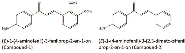
Two chalcone derivatives i.e. (E)-1-(4-aminophenyl)-3-(2,3dimethoxyphenyl)-prop-2-en-1-one (Compound-1), and (E)-1-(4-aminophenyl)-3-phenylprop-2-en-1-(E)-1-(4-aminophenyl)-3-(2,3dimethoxyphenyl)-prop-2-en-1-one) (Compound-2), has been proven to have potential cytotoxic activity. The aim of this study was to evaluate the effect of these compounds on PI3K/Akt signalling pathway in K562 cell lines. After incubation with the tested compounds, AKT, caspase-3, STAT3 and cyclin D1 concentrations were measured using ELISA. Furthermore, cell cycle was analysed using lowcytometry. Imatinib and isotretinoin were used as positive control, whereas cell culture without treatment was used as negative control. The AKT concentration after treatment with Compound-1 and -2 was signiicantly lower than that control, imatinib and isotretinoin (p<0.05). The apoptotic indices after treatment with Compound-1 and -2 were signiicantly higher than control, however they were lower than imatinib and isotretinoin (p<0.05). The caspase-3 concentration after treatment with Compound-1 at 5 and 10 µg/mL and Compound-2 at 10 µg/mL was signiicantly higher than that control and imatinib, however it was lower than isotretinoin (p<0.05). The STAT3 concentration after treatment with Compound-1 and -2 was signiicantly lower than that control and isotretinoin at 50 µg/mL (p<0.05) and similar with imatinib (p>0.05). The cyclin D1 concentration after treatment with Compound-1 and -2 was signiicantly lower than that control, imatinib and isotretinoin (p<0.05). In addition, Compound-1 and -2 arrested G0/ G1 and G2/M phase in K562 cell lines, with comparable results to imatinib and isotretinoin. In conclusion, the mechanism of cytotoxic activity of Compound-1 and -2 are through the PI3K/Akt signalling pathway inhibition, apoptosis induction by upregulation of apoptotic markers, and inhibition of cell cycle progression by regulating cell cycle-related factors.
ABSTRAK
diinkubasikan dengan senyawa uji, kadar AKT, caspase-3, STAT3 dan siklin D1 diukur dengan ELISA. Selanjutnya siklus sel dianalisis dengan lowcyometry. Imatinib dan isotretinoin digunakan sebagai kontrol positif dan kultur sel tanpa perlakuan sebagai kontrol negatif. Kadar AKT setelah perlakuan dengn Senyawa-1 dan -2 lebah rendah secara nyata dibaningkan dengan kontrol, imatiib dan isotretinoin (p<0.05). Indeks apoptosis setelah perlakuan dengan Senyawa-1 dan -2 lebih tinggi secara nyata dibandingkan kontrol, tetapi lebih rendah dibandingkan imatinib dan isotretinoin (p<0.05). Kadar caspase-3 setelah perlakuan Senyawa-1 dengan kadar 5 dan 10 µg/mL dan Senyawa-2 dengan kadar 10 µg/mL lebih tinggi secara nyata dibandingkan kontrol dan imatinib, tetapi lebih rendah dibandingkan dengan isotretinoin (p<0.05). Kadar STAT3 setelah perlakuan dengan Senyawa-1 dan -2 lebih rendah secara nyata dibandingkan control dan isotretinoin kadar 50 µg/mL (p<0.05) dan setara dengan imatinib (p>0.05). Kadar siklin D1 setelah perlakuan dengan Senyawa-1 dan -2 lebih rendah secara nyata dibandingkan dengan kontrol, imatinib dan isotretinoin (p<0.05). Senyawa-1 dan -2 terbukti menghambat siklus sel K562 pada fase G0/G1 dan G2/M yang sebanding dengan imatinib dan isotretinoin. Dapat disimpulkan, mekannisne aktivitas sitotoksik senyawa-1 dan -2 melalui penghambatan jalur sinyal PI3K/ Akt, induksi apoptosis melalui upregulasi penanda apoptosis dan penghambatan siklus sel melalui pengaruhnya terhadap factor-faktor yang berhubungan dengan pengaturan siklus sel.
Keywords : anticancer - chalcone derivatives – leukemia – K562 cell line - PI3K/Akt signaling
INTRODUCTION
Leukemia is a cancer of the white blood cells characterized by the widespread, rapid, and disorderly proliferation of leukocytes.1,2
Leukemia is a rare disease; however, it exceeds the number of deaths caused by acute communicable diseases due to its fatal character.2 In 2012, leukemia was suffered
by approximately 352,000 people around the world and caused 265,000 deaths.3
The PI3K/Akt signaling pathway plays an important role in both normal and malignant hematopoiesis.4,5 The activity of AKT is
regulated by P13K, which brings AKT into the cell membrane upon PIP3 binding, which is activated by PDK1.6 Activated AKT is
an essential survival factor which inhibits apoptosis through phosphorylation of several targets, including Bad, FoxO transcription factors, Raf-1 and caspase-9.7 AKT also
activates the transcription activator (STAT3),
and proliferation.8 Activation of STAT3 leads
to tumor-promoting gene products, including cleavage caspase-3, cleavage poly ADP-ribose polymerase (PARP), cyclin D1, and survivin. Thus, these pathways are commonly targeted in cancer treatments.
Until now, chemotherapy is still one of the effective strategies for the treatment of cancer.9 However, the use of anticancer
treatments as chemotherapy is limited. All of the current anticancer drugs have severe side effects in normal cells due to their
non-speciicity. In addition, the resistance of cancer cells to anticancer drugs remains a signiicant
challenge to successful chemotherapy.10
Therefore, the discovery and development of
new anticancer treatments with speciic targets
and novel mechanisms are urgently needed.
Chalcones are precursors of lavonoids,
and their anticancer activity in cancer animal models have been reported.11,12 Furthermore,
the molecular mechanisms of the anticancer action of chalcones have also been investigated. The chalcones exhibited anticancer activity through multiple mechanisms including cell cycle disruption, angiogenesis inhibition, tubulin polymerization inhibition, apoptosis induction and blockade of the nuclear factor-kappa B (NF-κB) signaling pathway.12,13
We previously evaluated the cytotoxic activity of several chalcone derivatives on the K562 leukemia cell line, resulting in two chalcones showing the highest activity i.e. (E)-1-(4-aminophenyl)-3-(2,3 dimethoxyphenyl)-prop-2-en-1-one (Compound-1), and
(E)-1-(4-aminophenyl)-3-phenylprop-2-en-1-one)
(Compound-2). In this study we reported
the mechanism of cytotoxic activity of these compounds through their effect on the level of AKT, caspase-3, STAT3, cyclin D1, and cell cycle on K562 leukemia cell lines. Furthermore, imatinib and isotretinoin were used as positive control in this study.
MATERIALS AND METHODS Tested compounds
The methoxy amino chalcone derivatives
were synthesized by Suwito et al.14 The
structure of these compounds is presented in FIGURE 1.
FIGURE 1. Chemical structure chalcone derivatives
Cell culture
The K562 cell lines were obtained from Stem Cells and Cancer Institute (SCI) Jakarta, Indonesia. The cell lines were maintained in
iscove’s modiied dulbeco’s medium (Biowest
L0190-500) supplemented by 10% fetal bovine serum (FBS) (Biowest S181H2), and 2% penicilin-streptomycin (Biowest L0022-100) and incubated at 37oC in a 5% CO
2
atmosphere with 95% humidity. After 24 hours of incubation, the number of viable cells was counted using a haemocytometer with tryphan blue staining.
Measurement of AKT, caspase-3, STAT3 and cyclin D1 concentrations in K562 cell lines culture
The concentration of AKT was determined using an ELISA kit (ab176657). After pre-incubation of K562 cell lines treated with imatinib (10 and 50 µg/mL), isotretinoin (25 and 50 µg/mL), Compound-1 and -2 (5 and
10 µg/mL), 50 μL of sample was added to wells, and then 50 μL of the antibody was
added into each well, and incubated for 1 hour at room temperature. Each well was washed
with 350 μL of wash buffer. One hundred μL
of TMB substrate was added into each well, and incubated for 15 minutes in the dark. The
reaction was stopped by adding 100 μL of
Spectrophotometer at 450 nm. Untreated K562 cell lines served as control.
The similar methods were applied to determine the caspase-3 and STAT3 concentrations using different antibody i.e. ELISA kit ab181418 for caspase-3, ELISA kit ab176666 for STAT3 and biotinylated detection antibody for cyclin D1.
Cell cycle analysis
Cells (2 × 10⁶ cells/well) were cultured
in 24-well plates, and then treated with 0.1% DMSO, and 100 µL tested compounds at an
appropriate inal concentration (10 and 20
µg/mL for imatinib; 25 and 50 µg/mL for isotretinoin; 5 and 10 µg/mL for Compound-1 and -2, before being incubated for 24 hr. Cells
were harvested, washed using PBS, ixed in
70% ethanol overnight and stained with 50 μg/ mL PI and 20 µg/mL RNAse. Data acquisition and analysis were performed on a MACSQuant
Analyzer 10 lowcytometer (Miltenyil Biotec),
and data from 500,000 cells were collected
for each data ile. Cell cycle analysis was
performed with MACSQuantify™ Software (Miltenyil Biotec).
Statistical analysis
Data were presented as means ± standard deviation (SD). SPSS (Version 16, SPSS Inc., Chicago, IL, USA) was used to perform
one-way analysis of variance (ANOVA) to analysis the different among group means. Duncan’s multiple range test was used to validate
signiicant differences for all treatments. A p value <0.05 was considered signiicantly
different.
RESULTS
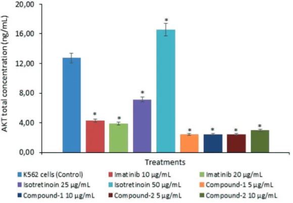
AKT concentration in K562 cell lines
AKT concentration on K562 cell lines of all treatments is presented in FIGURE 2. The AKT concentration of all treatments was
signiicantly lower than control (p<0.05)
except on isotretinoin treatment at 50 µg/mL
which it was signiicantly higher (p<0.05).
FIGURE 2. AKT total concentration on K562 cell lines after treatment with imatinib, isotretinoin, Compound-1 and -2. (*) indicates a
signiicant difference between treatments compared to control
(p<0.05).
Apoptotic index and caspase-3 in K562 cell lines
Effects of chalcone derivatives on apoptosis in K562 cell lines as indicated in the caspase-3 concentration and apoptotic index are shown in FIGURE 3 and 4. The apoptotic indices in K562 cell lines of all treatments
were signiicantly higher than control
(p<0.05). The highest apoptotic indices were observed after treatment with imatinib 20 µg/mL (6.89%) and isotretinoin 50 µg/mL (6.84%). The apoptotic indices after treatment with Compound-1 at 10 µg/mL, Compound-2 at 10 µg/mL and control were 3.89, 4.03 and 2.33%, respectively.
FIGURE 3. Apoptotic index on K562 cell lines after treatment with imatinib, isotretinoin,
Compound-1 and -2. (*) indicates a signiicant difference between treatments
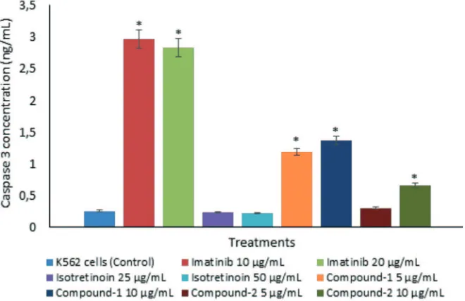
The caspase-3 concentrations on K562 cell lines after treatment with Compound-1 at 5 µg/mL (1.19 ng/mL), Compound-1 at 10 µg/mL (1.37 ng/mL) and Compound-2 at 10
µg/mL (0.66 ng/mL) were signiicantly higher
than control (0.26 ng/mL) (p<0.05). The similar results were observed after treatment
with imatinib at 10 and 20 µg/mL in which the caspase-3 concentrations were 2.97 and 2.83
ng/mL, respectively (p<0.05). No signiicantly
different in caspase-3 concentrations compare to control were observed treatment with isotretinoin at 25 µg/mL (0.24 ng/mL) and at 50 µg/mL (0.22 ng/mL) (p>0.05).
FIGURE 4. Caspase-3 concentration on K562 cell lines after treatment with imatinib,
isotretinoin, Compound-1 and -2. (*) indicates a signiicant difference
between treatments compared to control (p<0.05).
STAT3 concentration in K562 cell lines
STAT3 concentration on K562 cell lines of all treatments is presented in FIGURE 5. The STAT3 concentration of all treatments
was signiicantly lower than control (p<0.05)
except on isotretinoin treatment at 25 µg/
mL which it was not signiicantly different
(p>0.05). The STAT3 concentrations after treatment with Compound-1 at 5 µg/mL (9.00 ng/mL), Compound-1 at 10 µg/mL (5.48 ng/ mL), Compound-2 at 5 µg/mL (8.62 ng/mL)
and Compound-2 at 10 µg/mL (14.77 ng/
mL) were signiicantly lower than control
(37.56 ng/mL) (p<0.05). The similar results were observed after treatment with imatinib at 10 and 20 µg/mL and isotretinoin at 50 µg/ mL in which the STAT3 concentrations were 10.11, 7.83 and 21.51 ng/mL, respectively
(p<0.05). No signiicantly different in STAT3
FIGURE 5. STAT3 concentration on K562 cell lines after treatment with imatinib,
isotretinoin, Compound-1 and -2. (*) indicates a signiicant difference
between treatments compared to control (p<0.05).
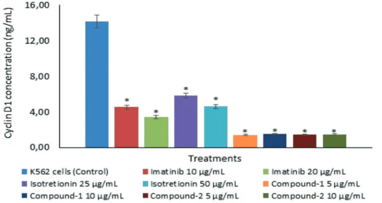
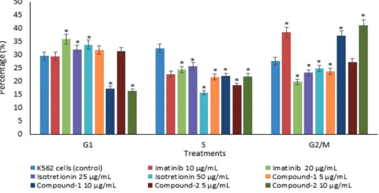
Cyclin D1 concentration in K562 cell lines and cell cycle
Cyclin D1 concentration on K562 cell lines and its cell cycle of all treatments are presented in FIGURE 6 and 7. The cyclin D1 concentration of all treatments was
signiicantly lower than control (p<0.05).
The cyclin D1 concentrations after treatment
with Compound-1 at 5 µg/mL (1.41 ng/mL), Compound-1 at 10 µg/mL (1.51 ng/mL), Compound-2 at 5 µg/mL (1.45 ng/mL) and Compound-2 at 10 µg/mL (1.47 ng/mL) were
also signiicantly lower than imatinib 10 µg/
mL (4.53 ng/mL), imatinib 20 µg/mL (3.43 ng/ mL), isotretinoin 25 µg/mL (5.85 ng/mL) and isotretinoin 50 µg/mL (4.62 ng/mL) (p<0.05).
FIGURE 6. Cyclin D1 concentration on K562 cell lines after treatment with imatinib,
isotretinoin, Compound-1 and -2. (*) indicates a signiicant difference between
As shown in FIGURE 7, Compound-2 at 5 µg/mL was the only compound that
signiicantly arrested G0/G1 phase (32.42%),
and it was comparable to imatinib of 20 µg/ mL (35.99%) and isotretinoin of 25 and 50 µg/mL (32.15 and 33.87%, respectively). Both Compound-1 at 5 and 10 µg/mL (21.73
and 22.01%, respectively) and Compound-2 at 5 and 10 µg/mL (18.50 and 21.94%,
respectively) signiicantly decreased S phase
compared to control (32.52%). Compound-1
and -2 at 10 µg/mL arrested G2/M phase
(37.21 and 41.07%, respectively), which were comparable to imatinib at 10 µg/mL (38.45%).
FIGURE 7. K562 cell cycle after treatment with imatinib, isotretinoin, Compound-1 and -2. (*)
indicates a signiicant difference between treatments compared to control (p<0.05).
DISCUSSION
Chemotherapy is one of the most potent and effective strategies to treat cancer.9
However, all of the anticancer therapies that are currently available have severe side effects on normal cells due to their
non-speciicity, leading to the development of new anticancer therapies with speciic targets.
Molecular mechanisms of cancer have been studied in drug discovery. The PI3K/Akt signaling pathway plays an important role in both normal and malignant hematopoiesis.4,5
Activated AKT is critical for leukemia cell survival and proliferation,8 and is known to
function as an essential survival factor by
inhibiting apoptosis.7 These pathways are
commonly targeted in cancer treatments. Almost all treatments in the present study
signiicantly decreased AKT levels on K562
cells, except isotretinoin at 25 µg/mL, which increased the AKT level. Both Compound-1
and -2 showed the most signiicant decrease among treatments. These indings are in
accordance with previous studies that reported on chalcone derivatives as anticancer agents. Xu et al.,15 showed chalcone derivative,
L2H17, inactivated NF-κB and AKT signaling
proteins and processes; phosphorylation of Bad,16,17 that promotes release of Bad
from heterodimeric of Bcl-2 and Bcl-XL; phosphorylation of MDM218,19 stabilizing
it and promoting its translocation to the nucleus, where it triggers p53 degradation; and phosphorylation of XIAP, an inhibitor of caspase cascade, and thus inhibiting its degradation.20
In this study, Compound-1 and -2 also
signiicantly increased caspase-3 on K562, as conirmed by the higher apoptotic index
compared to the control. Chalcones have also been reported to induce apoptosis in cancer cells. Referring to a previous study, chalcones (1.3-diphenyl-2-propenone) in a human diet rich in fruits and vegetables inhibit the proliferation of T24 and HT-1376 cells by inducing apoptosis. Chalcone increased the expression of Bax and Bak, but decreased the levels of Bcl-2 and Bcl-X(L) and subsequently triggered the mitochondrial apoptotic pathway (release of cytochrome c and activation of caspase-9 and caspase-3).21
Apoptosis is by far the best-characterized type
of cell death and is deined by morphologic modiications (chromatin condensation,
loss of mitochondrial membrane potential, plasma membrane asymmetry, overall cell shrinkage, blebbing of the plasma membrane, and detachment from the cellular matrix), all occurring before the loss of plasma membrane
integrity. Those modiications occur due to
executioner caspase activation.22
The results of the present study showed that all treatments decreased STAT3 on
K562. These indings are validated by a
previous study which showed that a chalcone
derivative, 4,3′,4′,5′-tetramethoxychalcone,
inhibits the phosphorylation of STAT3 and its upstream protein tyrosine kinase c-Src.23
STAT3, an oncogenic transcription factor, is often constitutively active in human cancer
cells.24 Activated STAT3 may up-regulate
the expression of genes such as apoptosis inhibitors (Bcl-xl, Bcl-2), cell cycle regulators (cyclin D1) and oncogenic transcription factors (c-myc) in tumorigenesis.25,26
In this study, all treatments signiicantly
decreased cyclin D1 levels on K562 cells. Compound-1 and -2 showed the highest decrease among treatments. The cyclin D1/CDK4 complex is responsible for cell cycle progression in early G1 phase, and is frequently overexpressed in various human carcinomas.27-29 Compound-1 and -2 also
arrested G
0/G1 and G2/M phase in K562 cells,
with the results being comparable to imatinib and accutane. These indings are in agreement
with a previous study that shows chalcone inhibits the proliferation of T24 and HT-1376 cells by blocking cell cycle progression in the G
2/M phase.
21Another study investigated
the effect of a synthetic chalcone derivative,
4,3′,4′,5′- tetramethoxychalcone, that resulted
in G0/G1 cell cycle arrest through the down-regulation of cyclin D1 and CDK4, and the up-regulation of p16, p21 and p27 proteins in A2780 cells.23 The p16 protein is a
speciic inhibitor of CDK-cyclin D complex,
preventing the phosphorylation of Rb and cell cycle reentry at G0/G1 phase.27Eukaryotic
cell cycle progression involves sequential activation of cyclin-dependent kinases (CDK), whose activation is dependent on their association with cyclins.30 A complex
formed by the association of Cdc2 (also known as Cdk1 or p34Cdc2) and cyclin D1 plays a major role at entry into mitosis.30 The
phosphorylation of Tyr15 of Cdc2 suppresses the activity of Cdk1/cyclin B1 kinase complex. Dephosphorylation of Tyr15 of Cdc2 is catalysed by Cdc25 phosphatases, and this reaction is believed to be the ratelimiting step for entry into mitosis.31 Cell cycle progression
between the cellular concentration of CDK inhibitors, such as members of the CDK-interacting protein/CDK-inhibitory protein (CIP/KIP) and inhibitor of CDK families, and that of cyclin-CDK complexes. The CIP/ KIP family, including CIP/p21, and KIP/p27, bind to cyclin-CDK complexes and prevent kinase activation and subsequently blocking the progression of the cell cycle at the G0/G1 or G2/M phases.30,32
CONCLUSION
It can be concluded that (E)-1-(4-aminophenyl)-3-(2,3 dimethoxyphenyl)-prop-2-en-1-one (Compound-1), and (E)-1-(4-aminophenyl)-3-phenylprop-2-en-1-one)
(Compound-2) may be potential compounds
to be developed as anticancer against leukemia. The molecular mechanisms of cytotoxic activity of these compounds in the K562 cell lines involved i) inhibition of the PI3K/Akt signaling pathway; ii) induction of apoptosis through the up-regulation of apoptotic markers; and iii) inhibition of cell cycle progression by regulating cell cycle-related factors.
ACKNOWLEDGMENTS
This research was supported by Biomolecular and Biomedical Research Center, Aretha Medika Utama, Bandung, Indonesia for laboratory facilities, and research methodology. We would like to thank staff of Biomolecular and Biomedical Research Center, Aretha Medika Utama, Bandung for their valuable assistance. We also would like to thank the Stem Cell and Cancer Institute, Kalbe Farma, Jakarta for the K562 cell lines.
REFERENCES
1. Campana D. Childhood leukemia. In: Abeloff MD, Armitage JO, Niederhuber JE, Kastan MB, McKena WG, editors. Abeloff’s Clinical Oncology 4th ed. Philadelphia, Pa: Elsevier Churchill Livingstone, 2008; chap 101. http://dx.doi.org/10.1016/b978-0-443-06694-8.50105-6
2. Modak H, Kulkarni SS, Kadakol GS, Hiremath SV, Patil BR, Hallikeri U, et al.
Prevalence and risk of leukemia in the multi-ethnic population of North Karnataka. Asian Pac J Cancer Prev 2011; 12(3):671-5.
3. Stewart BW & Wild CP editors. World Cancer Report 2014. Lyon, France: International Agency for Research on Cancer, 2014.
4. Martelli AM, Evangelisti C, Chiarini F, McCubrey JA. The phosphatidylinositol 3-kinase/Akt/mTOR signaling network as a therapeutic target in acute myelogenous leukemia patients. Oncotarget 2010; 1(2):89-103. http://dx.doi.org/10.18632/ oncotarget.114
5. Polak R & Buitenhuis M. The PI3K/ PKB signaling module as key regulator of hematopoiesis: implications for therapeutic strategies in leukemia. Blood 2012; 119(4):911-23. http://dx.doi.org/10.1182/ blood-2011-07-366203
6. Kandel ES & Hay N. The regulation and activities of the multifunctional serine/ threonine kinase Akt/PKB. Exp Cell Res 1999; 253(1):210-29. http://dx.doi.org/10.1006/ excr.1999.4690
7. Hanada M, Feng J, Hemmings BA. Structure, regulation and function of PKB/AKT--a major therapeutic target. Biochim Biophys Acta 2004; 1697(1-2):3-16. http://dx.doi. org/10.1016/j.bbapap.2003.11.009
mTOR inhibitor, with clofarabine as a new therapeutic option for patients with acute myeloid leukemia. Oncotarget 2012; 3(12):1615-28. http://dx.doi.org/10.18632/ oncotarget.762
9. Cook AM, Lesterhuis WJ, Nowak AK, Lake RA. Chemotherapy and immunotherapy: mapping the road ahead. Curr Opin Immunol 2016; 39:23-9. http://dx.doi.org/10.1016/j. coi.2015.12.003
10. Liang XJ, Chen C, Zhao Y, Wang PC. Circumventing tumor resistance to chemotherapy by nanotechnology. Methods Mol Biol 2010; 596:467-88. http://dx.doi. org/10.1007/978-1-60761-416-6_21
11. Saydam G, Aydin HH, Sahin F, Kucukoglu O, Erciyas E, Terzioglu E, et al. Cytotoxic
and inhibitory effects of 4,4′-dihydroxy
chalcone (RVC-588) on proliferation of human leukemic HL-60 cells. Leuk Res 2003; 27(1):57-64. http://dx.doi.org/10.1016/ S0145-2126(02)00058-9
12. Karthikeyan C, Moorthy NS, Ramasamy S, Vanam U, Manivannan E, Karunagaran D,
et al. Advances in chalcones with anticancer activities. Recent Pat Anticancer Drug Discov 2015; 10(1):97-115. http://dx.doi.org/10.217 4/1574892809666140819153902
13. Mandge S, Singh HP, Gupta SD, Moorthy NSHN. Synthesis and characterization of some chalcone derivatives. Trends Appl Sci Res 2007; 2(1):52-6. http://dx.doi. org/10.3923/tasr.2007.52.56
14. Suwito H, Jumina, Mustofa, Ni’matuzahroh, Puspaningsih, NNT. Anticancer and antimicrobial activity of methoxy amino chalcone derivatives. Der Pharma Chemica, 2015, 7(3):89-94
15. Xu S, Chen M, Chen W, Hui J, Ji J, Hu S,
et al. Chemopreventive effect of chalcone derivative, L2H17, in colon cancer development. BMC Cancer 2015; 15:870. http://dx.doi.org/10.1186/s12885-015-1901-x
16. Datta SR, Dudek H, Tao X, Masters S, Fu HA, Gotoh Y, et al. Akt phosphorylation of BAD couples survival signals to the cell-intrinsic death machinery. Cell 1997; 91(2):231-41. http://dx.doi.org/10.1016/ S0092-8674(00)80405-5
17. del Peso L, Gonzalez-Garcia M, Page C, Herrera R, Nunez G. Interleukin-3-induced phosphorylation of BAD through the protein kinase Akt. Science 1997; 278(5338):687-9. http://dx.doi.org/10.1126/ science.278.5338.687
18. Ogawara Y, Kishishita S, Obata T, Isazawa Y, Suzuki T, Tanaka K, et al. Akt enhances Mdm2-mediated ubiquitination and degradation of p53. J Biol Chem 2002; 277(24):21843-50. http://dx.doi.org/10.1074/jbc.M109745200 19. Feng J, Tamaskovic R, Yang Z, Brazil
DP, Merlo A, Hess D, et al. Stabilization of Mdm2 via decreased ubiquitination is mediated by protein kinase B/Akt-dependent phosphorylation. J Biol Chem 2004; 279(34):35510-7. http://dx.doi.org/10.1074/ jbc.M404936200
20. Dan HC, Sun M, Kaneko S, Feldman
RI, Nicosia SV, Wang HG, et al. Akt
phosphorylation and stabilization of X-linked inhibitor of apoptosis protein (XIAP). J Biol Chem 2004; 279(7):5405-12. http://dx.doi. org/10.1074/jbc.M312044200
21. Shen KH, Chang JK, Hsu YL, Kuo PL. Chalcone arrests cell cycle progression and induces apoptosis through induction of mitochondrial pathway and inhibition of nuclear factor kappa B signalling in human bladder cancer cells. Basic Clin Pharmacol Toxicol 2007; 101(4):254-61. http://dx.doi. org/10.1111/j.1742-7843.2007.00120.x
22. Green DR. Apoptotic pathways: the roads to ruin. Cell 1998; 94(6):695-8. http://dx.doi. org/10.1016/S0092-8674(00)81728-6
suppresses proliferation, blocks cell cycle progression, and induces apoptosis of human ovarian cancer cells. PloS one 2014; 9(9):e106206. http://dx.doi.org/10.1371/ journal.pone.0106206
24. Bromberg JF, Wrzeszczynska MH, Devgan G, Zhao Y, Pestell RG, et al. Stat3 as an oncogene. Cell 1999; 98(3):295-303. http:// dx.doi.org/10.1016/S0092-8674(00)81959-5 25. Chan KS, Sano S, Kiguchi K, Anders J,
Komazawa N, Takeda J, et al. Disruption of Stat3 reveals a critical role in both the initiation and the promotion stages of epithelial carcinogenesis. J Clin Invest 2004; 114(5):720-8. http://dx.doi.org/10.1172/ JCI21032
26. Yu H, Kortylewski M, Pardoll D. Crosstalk between cancer and immune cells: role of STAT3 in the tumour microenvironment. Nat Rev Immunol 2007; 7(1):41-51. http://dx.doi. org/10.1038/nri1995
27. Kusume T, Tsuda H, Kawabata M, Inoue T, Umesaki N, Suzuki T, et al. The p16-cyclin D1/CDK4-pRb pathway and clinical outcome in epithelial ovarian cancer. Clin Cancer Res 1999; 5(12):4152-7.
28. Wolter F, Akoglu B, Clausnitzer A, Stein J. Downregulation of the cyclin D1/Cdk4 complex occurs during resveratrol-induced
cell cycle arrest in colon cancer cell lines. J Nutr 2001; 131(8):2197-203.
29. Bali A, O′Brien PM, Edwards LS, Sutherland
RL, Hacker NF, Henshall SM. Cyclin D1, p53, and p21Waf1/Cip1 expression is predictive of poor clinical outcome in serous epithelial ovarian cancer. Clin Cancer Res 2004; 10(15):5168-77. http://dx.doi. org/10.1158/1078-0432.CCR-03-0751
30. Sancar A, Lindsey-Boltz LA,
Unsal-Kacmaz K, Linn S. Molecular mechanisms of mammalian DNA repair and the DNA damage checkpoints. Annu Rev Biochem 2004; 73:39-85. http://dx.doi.org/10.1146/ annurev.biochem.73.011303.073723
31. De Souza CP, Ellem KA, Gabrielli BG. Centrosomal and cytoplasmic Cdc2/cyclin B1 activation precedes nuclear mitotic events. Exp Cell Res 2000; 257(1):11-21. http:// dx.doi.org/10.1006/excr.2000.4872