Effects of atorvastatin and vitamin C on endothelial function of
hypercholesterolemic patients
Francesco Perticone *, Roberto Ceravolo, Raffaele Maio, Cosima Cloro,
Mafalda Candigliota, Angela Scozzafava, Annalisa Mongiardo,
Pasquale Mastroroberto
a, Massimo Chello
a, Pier L. Mattioli
1Cardio6ascular Diseases Unit,Department of Medicina Sperimentale e Clinica‘G Sal6atore’,Policlinico Mater Domini,Via Tommaso Campanella,
Uni6ersity of Catanzaro,88100Catanzaro,Italy
aCardiac Surgery Unit,Department of Medicina Sperimentale e Clinica‘G Sal6atore’,Policlinico Mater Domini,Via Tommaso Campanella, Uni6ersity of Catanzaro,88100Catanzaro,Italy
Received 2 August 1999; received in revised form 12 November 1999; accepted 16 December 1999
Abstract
We tested the effects of vitamin C and atorvastatin treatment on endothelium-dependent and endothelium-independent vasodilation in 18 hypercholesterolemic patients (ten men and eight women, aged 20 – 46 years) in comparison with 12 normal volunteers (seven men and five women, aged 20 – 45 years). The responses of the forearm blood flow (FBF) to acetylcholine (ACh) (7.5, 15 and 30mg/min), sodium nitroprusside (SNP) (0.8, 1.6, 3.2mg/min) and L-NMMA (2, 4, 8mmol/min) were evaluated at baseline and after 1 month of atorvastatin (10 mg/day) treatment. Drugs were infused into the brachial artery and FBF was measured by strain-gauge plethysmography. At baseline, the response to ACh was significantly attenuated in hypercholes-terolemics versus controls: at the highest dose (30 mg/min), FBF was 27.093.4 versus 11.591.9 ml·100 ml tissue−1·min−1
respectively (PB0.0001). No significant differences were found between groups during SNP infusion. The atorvastatin treatment significantly improved ACh-stimulated FBF: at highest dose the FBF increased to 14.991.5 ml·100 ml tissue−1·min−1
(PB0.0001). Similarly, the L-NMMA endothelial effects were significantly enhanced by lipid-lowering treatment, supporting the improvement of basal nitric oxide. Vitamin C increased ACh-vasodilation in the same way before and after atorvastatin treatment. In conclusion, the endothelial dysfunction in hypercholesterolemics is due to an oxidative stress and atorvastatin rapidly improves both basal and stimulated endothelium-dependent vasodilation. © 2000 Elsevier Science Ireland Ltd. All rights reserved.
Keywords:Hypercholesterolemia; Endothelium; Atorvastatin; Atherosclerosis; Vitamin C
www.elsevier.com/locate/atherosclerosis
1. Introduction
The normal endothelium plays a key role in the local regulation of the vascular tone by producing and releas-ing contractreleas-ing and relaxreleas-ing agents [1]. One of these factors is the nitric oxide (NO) [2 – 4] that is released after stimulation of endothelial cells by shear stress [5], as well as some agonists such as acetylcholine (ACh), bradykinin, substance P and serotonin [4]. On the other
hand, sodium nitroprusside (SNP) is an endothelium-independent vasodilator compound that produces va-sodilation by direct activation of guanylate cyclase in vascular smooth-muscle cells by providing inorganic source of NO [6].
Endothelium-dependent vasodilation has been shown to occur in most mammalian species [2] and in humans, by in vitro studies using arterial preparations [7,8]. Human studies demonstrated that this regulatory ac-tion of the endothelium is exerted on resistance vessels too [9]. Moreover, this endothelial function is impaired in the presence of different risk factors such as hyper-tension [10 – 12], cigarette smoking [13], diabetes [14] and hypercholesterolemia [12,15]. A dysfunctioning en-dothelium reduces its ability to exert a protective effect 1Professor Mattioli suddenly died on June 23, but he actively
participated in the present study.
* Corresponding author. Tel.: +39-0961-712264; fax: + 39-0961-775564.
E-mail address:[email protected] (F. Perticone).
on the vascular system by keeping vessels in a dilatory state and playing, thus, a key pathophysiological role in the development of atherosclerosis. Recently, improve-ment of endothelial dysfunction was demonstrated after various treatments such as antioxidants [16,17], estro-gen replacement [18], lipid lowering drugs [17,19,20], angiotensin-converting enzyme inhibitors [21] and cal-cium-channel blockers [22,23].
Inhibitors of HMG-CoA reductase are currently ex-tensively used to lower serum cholesterol levels and to improve long-term morbidity and mortality related to coronary artery disease (CAD) [24,25]. Atorvastatin, a new HMG-CoA reductase inhibitor, demonstrated great effectiveness in reducing total and LDL-choles-terol and triglyceride levels and in raising HDL-choles-terol [26]. Recently, Simons demonstrated, by reactive hyperemia, that atorvastatin improves forearm blood flow (FBF) in hypercholesterolemics [27]. However, at present no data are available about the atorvastatin effects on ACh-stimulated vasodilation by strain-gauge plethysmography.
Based on these observations, the aims of this study were to test in hypercholesterolemics without overt vascular disease: (1) the effect of vitamin C by intra-ar-terial infusion; (2) the effects on both basal endothe-lium-dependent vasodilation and ACh-induced vasodilation in the forearm vasculature after atorvas-tatin treatment; and (3) the combined effect of vitamin C and atorvastatin administration.
2. Methods
2.1. Study population
2.1.1. Hypercholesterolemic patients
We enrolled 18 hypercholesterolemic outpatients, ten men and eight women, aged 20 – 46 years (mean9S.D., 4096 years). Patients were eligible if they had total serum cholesterol levels\6.2 mmol/l and were not receiving cholesterol-lowering medications. Patients were excluded if any of the following were present: diabetes mellitus, primary or secondary hypertension, CAD, heart failure, peripheral vascular disease, cigarette smoking, vasoactive drugs, antioxidant com-pounds, post-menopausal status or hormone replace-ment therapy.
2.1.2. Control subjects
Twelve normal volunteers, seven men and five women, aged 20 – 45 years (mean9S.D., 3995 years), were selected as a control group. Each patient under-went clinical history, physical examination, standard electrocardiogram, routine chemical analyses and chest X-rays. None had evidence of present or past hyperten-sion, cardiovascular disease, hyperlipidemia, or any
other systemic condition and none was taking drugs at the time of the study.
2.2. Study protocol
The protocol was designed as a randomized, double blind, placebo-controlled crossover study. At first, FBF dose – response curves were evaluated in both groups (normal subjects and hypercholesterolemic patients) for ACh and SNP infusions before and during co-adminis-tration of vitamin C.
After the initial evaluation of vascular function, the hypercholesterolemic patients were randomized in equal numbers (n=9) to receive for 4 weeks the same diet plus atorvastatin (10 mg daily) or placebo. At the end of this period, the vascular function was reevaluated in each patient. Then, after crossover, they were again studied, 4 weeks later. Biochemical and hematological parameters were obtained with the patients fasting at randomization and at the end of each treatment period. To assess the reproducibility of endothelium-depen-dent and endothelium-indepenendothelium-depen-dent vasodilation, vascu-lar function in the control group was reevaluated 2 months later.
The local ethical committee approved the study,and all participants gave written informed consent for all procedures.
2.3. Vascular function
All studies were performed at 9:00 a.m. after overnight fasting, with the subjects lying supine in a quiet air-conditioned room (22 – 24°C), using the same protocol previously described by Panza [10] and succes-sively employed by ourselves [23,28].
All hypercholesterolemics and controls underwent measurement of FBF and blood pressure (BP) during intra-arterial infusion of saline and increasing doses of ACh and SNP. All participants rested at least 30 min after artery cannulation to obtain a stable baseline before data collection; FBF and vascular resistance (VR) were repeated every 5 min until stable.
Endothelium-dependent vasodilation was evaluated by dose – response curve to intra-arterial ACh infusions (7.5, 15 and 30 mg/min, each for 5 min). Endothelium-independent vasodilation was assessed by dose – re-sponse-curve to intra-arterial SNP infusions (0.8, 1.6, 3.2 mg/min, each for 5 min).
Finally, vitamin C administration was followed, in hypercholesterolemic patients, by L-NMMA infusions (2, 4 and 8 mmol/min, each for 5 min) to assess the basal NO-mediated endothelium-dependent vasodila-tion before and after atorvastatin treatment.
The FBF and VR of the opposite arm were also evaluated during the study.
Infusion rate for each drug was 1 ml/min.
2.4. Drugs
ACh (Sigma, Milan, Italy), vitamin C (Bracco, Mi-lan, Italy) and L-NMMA (Clinalfa, San Diego, CA) were obtained from commercially available sources and diluted in saline immediately before each infusion. SNP (Malesci, Florence, Italy) was diluted in 5% glucose solution and protected from light with aluminum foil.
2.5. Statistical analysis
The goal of the present study was the increase in FBF from the baseline values. Based on a single mea-surement in each step, the sample sizes required were previously calculated in order to detect a given im-provement in FBF. A sample of eight patients is suffi-cient to detect a 20% improvement in FBF, a sample of 12 patients is sufficient to detect a 15% improvement in FBF with a power of 90% and a=0.05. Differences between means were compared by paired and unpaired Student’st-test, as appropriate. The responses to SNP, ACh, L-NMMA and vitamin C were compared by ANOVA for repeated measurements and, when analy-sis was significant, Tukey’s test was applied. Reproduci-bility of FBF and VR measurements were tested by using Pearson’s coefficient. All calculated P values are
two tailed. Significant differences were assumed to be present at PB0.05. All group data are reported as mean9S.D.
3. Results
Baseline demographic, hemodynamic and humoral characteristics are summarized in Table 1. According to inclusion criteria, serum cholesterol values were signifi-cantly lower in controls in comparison with hyperc-holesterolemics (4.590.8 mmol/l vs 7.190.9 mmol/l; PB0.0001).
All participants were never smokers and no women were in postmenopausal status or taking estrogen replacement.
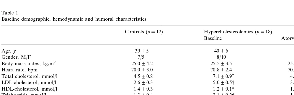
3.1.Reproducibility of endothelium-dependent 6asodilation
The FBF and VR values, reevaluated in control subjects 2 months later and correlation coefficient are reported in Table 2. Similarly, the BP and heart rate (HR) did not change either.
3.2.Control subjects 6ersus hypercholesterolemic patients
The basal FBF and forearm VR were not different in normal subjects and hypercholesterolemic patients; FBF mean values were: 3.690.4 and 3.590.6 ml·100 ml tissue−1·min−1 (P=NS), respectively; VR mean
values were 26.1+4.5 and 27.2+5.6 U (P=NS), respectively.
Table 1
Baseline demographic, hemodynamic and humoral characteristics
Controls (n=12) Hypercholesterolemics (n=18)
Baseline Atorvastatin
Age,y 3995 4096
7/5
Gender, M/F 8/10
25.593.5 25.494.0
25.094.2 Body mass index, kg/m2
70.093.0 70.192.5
Heart rate, bpm 70.892.4
4.590.8
Total cholesterol, mmol/l 7.190.9† 4.890.7†
LDL-cholesterol, mmol/l 2.690.3 5.090.5† 3.390.3†
HDL-cholesterol, mmol/l 1.490.3 1.290.1* 1.490.2†
1.390.4
Triglyceride, mmol/l 2.190.2† 1.690.3†
Glucose, mmol/l 5.090.2 5.190.4 5.090.5
Blood pressure, mm Hg 125/7696/5 125/7497/6 126/7595/6 3.690.8 3.590.6
FBFa, ml·100 ml tissue−1·min−1 3.690.4
27.295.6 27.194.3
Vascular resistance, U 26.194.5
aforearm blood flow.
*PB0.05 hypercholesterolemics versus controls.
the highest dose of ACh (30mg/min), FBF increased to 11.591.9 ml·100 ml tissue−1·min−1 in
hypercholes-terolemics compared with 27.093.4 ml·100 ml tissue−1·min−1 in normal subjects (PB0.0001).
Forearm VR during ACh infusions significantly de-creased in hypercholesterolemics (decrements: 9.692.3, 15.293.7, 19.094.7 U) and in controls (decrements: 9.293.4, 16.297.4, 22.997.3 U). At the highest dose of ACh (30 mg/min), VR was 3.290.5 U in controls and 8.292.0 U (PB0.0001) in hypercholesterolemics. FBF and VR in the contralateral arm did not change during incremental ACh infusions in either group of subjects. Similarly, ACh infusions did not affect BP or HR.
3.2.2. Endothelium-independent 6asodilation
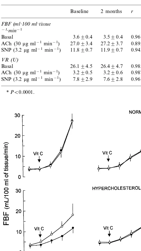
Significant increases in FBF as well as decreases in forearm VR were observed in both normal subjects and hypercholesterolemics during SNP infusions, though no significant differences were found between groups (Fig. 1). At the highest dose (3.2 mg/min), FBF increased to 11.890.7 ml·100 ml tissue−1·min−1 in the control
group and to 11.690.8 ml·100 ml tissue−1·min−1 in
hypercholesterolemics, respectively (P=NS). At this dose, the forearm VR decreased to 7.892.9 U in control group compared with 7.992.1 U in hyperc-holesterolemics (P=NS).
SNP infusions did not affect FBF and VR in the contralateral arm and did not change BP or HR.
3.3. Vitamin C administration
Intra-arterial infusion of vitamin C significantly in-creased basal FBF in hypercholesterolemics from 3.59 0.9 to 4.790.9 ml·100 ml tissue−1·min−1 (P
B0.0001); no significant differences were observed in control group, 3.690.6 – 3.790.9 ml·100 ml tissue−1·min−1 (P=NS). Basal VR in
hypercholes-terolemics decreased from 27.796.2 to 20.494.2 U (PB0.0001), but was unchanged in normal subjects, 25.594.1 to 24.994.7 U (P=NS).
3.3.1. Endothelium-dependent 6asodilation
The vasodilator response to ACh in hypercholes-terolemics significantly increased during co-administra-tion of vitamin C (PB0.0001 by ANOVA) (Fig. 1). At the highest dose of ACh (30mg/min) plus vitamin C (24 mg/min), the FBF increased to 18.095.6 ml·100 ml tissue−1·min−1(PB0.0001); forearm VR decreased to
5.791.8 U (PB0.0001).
On the contrary, in control group the ACh-stimu-lated vasodilation was not significantly affected by co-administration of vitamin C (Fig. 1). At the highest dose of ACh (30mg/min), before and during co-infusion of vitamin C (24 mg/min), the FBF was 27.093.4 ml·100 ml tissue−1·min−1 and 27.194.0 ml·100 ml
Table 2
Forearm blood flow (FBF) and vascular resistance (VR) in 12 normal volunteers at baseline and 2 months later
Baseline 2 months r
FBF(ml·100ml·tissue
−1·min−1
Basal 3.690.4 3.590.4 0.961* ACh (30mg ml−1min−1) 27.093.4 27.293.7 0.895* SNP (3.2mg ml−1min−1) 11.890.7 11.990.7 0.943*
VR(U)
Basal 26.194.5 26.494.7 0.985* 0.987* 3.290.6
3.290.5 ACh (30mg ml−1min−1)
7.892.9 7.692.8
SNP (3.2mg ml−1min−1) 0.961*
*PB0.0001.
Fig. 1. Forearm blood flow (FBF) dose – response curves to acetyl-choline (ACh) and sodium nitroprusside (SNP) in normal control subjects and hypercholesterolemic patients during saline ( ) and vitamin C () infusion are reported. The ACh-stimulated FBF is significantly attenuated in hypercholesterolemics (PB0.0001 by ANOVA), but significantly improved (PB0.0001 by ANOVA) by vitamin C coinfusion. Data are shown as mean9S.D. and expressed as absolute values. Non significant differences were found during SNP infusions.
3.2.1. Endothelium-dependent Vasodilation
The forearm ACh-stimulated vasodilation was sig-nificantly (PB0.0001 by ANOVA) attenuated in hyper-cholesterolemics compared with controls (Fig. 1). The increments from basal in hypercholesterolemics and control subjects were 1.991.2, 4.292.1, 8.091.2 ml·100 ml tissue−1·min−1 and 2.191.5, 9.395.1,
tissue−1·min−1(
P=NS), respectively. At this dose, VR was unaffected by co-infusion of vitamin C, 26.192.5 to 25.993.2 U (P=NS).
3.3.2. Endothelium-independent 6asodilation
In both normal subjects and in hypercholesterolemics, the vasodilator response to SNP was not significantly modified during co-administration of vitamin C (Fig. 1). In hypercholesterolemics, at the highest dose of SNP (3.2 mg/min), the FBF was 11.690.8 ml·100 ml tissue−1
·min−1
before and 11.790.9 ml·100 ml tissue−1
·min– 1
during co-infusion of vitamin C (P= NS). At this dose, the forearm VR before and during
co-administration of vitamin C was 7.891.9 and 7.79 2.1 U (P=NS), respectively.
3.4. Ator6astatin treatment
3.4.1. Effects on lipid profile and hemodynamic parameters
Atorvastatin treatment significantly lowered both serum cholesterol and triglyceride values. After 4 weeks, in comparison with placebo, total cholesterol was 32% lower (PB0.0001), LDL-cholesterol was 34% lower (PB0.0001), triglyceride were 24% lower (PB0.0001) and HDL-cholesterol was 17% higher (PB0.0001).
The basal FBF and VR were not different between atorvastatin and placebo treatment periods: 3.690.8 versus 3.590.9 ml·100 ml tissue−1·min−1(
P=NS) and 27.194.3 versus 27.393.4 U (P=NS), respectively.
3.4.2. Effects on ACh-6asodilation
The ACh-mediated vasodilation significantly in-creased between placebo and 4 weeks (PB0.0001 by ANOVA) of atorvastatin treatment. At the highest dose (30mg/min), FBF significantly (PB0.0001 by ANOVA) increased to 14.991.5 ml·100 ml tissue−1·min−1 (+
313.8%) (Fig. 2). At this dose, forearm VR significantly decreased to 7.291.6 U (PB0.0001). However, it is necessary to remark that even if atorvastatin treatment markedly improved the FBF response to ACh, it still remained depressed in hypercholesterolemics when com-pared with control subjects.
The ACh-stimulated FBF and VR mean values after four weeks of placebo treatment were not significantly different when compared to basal study. At the highest dose of ACh, the FBF and forearm VR were:11.792.1 ml·100 ml tissue−1·min−1 and 8.192.2 U (P=NS).
Thus, hypercholesterolemic patients who had previously received given to atorvastatin treatment, did not show a residual effect on endothelial function 4 weeks into placebo.
The ACh-stimulated vasodilation significantly in-creased during co-infusion of vitamin C (PB0.0001 by ANOVA). At the maximal dose of ACh (30mg/min) plus vitamin C (24 mg/min), FBF raised from 4.890.8 to 18.794.2 ml·100 ml tissue−1·min−1 (
+289.5%) (Fig. 2). At this dose, forearm VR decreased to 5.191.2 U. Thus, the peak percent increase in FBF after atorvastatin treatment and during vitamin C co-infusion were not significantly different.
3.4.3. Effects on SNP-6asodilation
Significant increase on FBF as well as decrease on VR were observed during SNP infusions in hypercholes-terolemic patients after lipid lowering treatment. How-ever, no significant differences with placebo group were found at the different time points. Similarly, responses to endothelium-independent vasodilator SNP were not Fig. 2. Forearm blood flow (FBF) dose – response curves to
acetyl-choline (ACh) and sodium nitroprusside (SNP) in hypercholes-terolemic patients in baseline (2), after 4 weeks of atorvastatin treatment ( ) and plus vitamin C co-infusion (). Data are shown as mean9S.D. and expressed as absolute values.
significantly modified by co-infusion of vitamin C (Fig. 2).
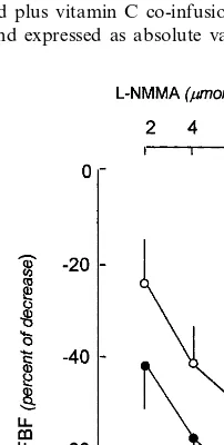
3.4.4. Effects on L-NMMA infusion
After 4 weeks of atorvastatin treatment the decrease in FBF induced by L-NMMA infusions was significantly greater than at baseline conditions, as reported in Fig. 3.
4. Discussion
Results obtained in this study confirm in hypercholes-terolemics that: (1) endothelium-dependent vasodilation is blunted compared with normal subjects; (2) intra-arte-rial infusion of vitamin C, an antioxidant compound, improves the impaired ACh-mediated vasodilation; (3) atorvastatin is very effective in lowering rapidly serum cholesterol and triglyceride.
Moreover, after 4 weeks of atorvastatin treatment, our results provide the first evidence that ACh-stimulated vasodilation was significantly increased, as was L-NMMA-mediated vasoconstriction, indicating that both stimulated and basal endothelium-dependent vasodila-tion were improved. These data are consistent with those of Simons et al. [27] who in a previous study demon-strated that atorvastatin treatment in patients with severe primary hypercholesterolemia improved flow-mediated endothelium-dependent dilation in response to reactive hyperaemia. However, it is necessary to remark that the vascular response to hypereamia may be influenced by different stimuli and, therefore, did not test the ACh-only stimulated vasodilation, as performed in our study. In agreement with data on simvastatin and vascular func-tion reported by O’Driscoll [20], our study also demon-strates, for the first time, that L-NMMA-mediated vasoconstriction is enhanced after atorvastatin treat-ment, indicative of an increased basal effect of NO. The disagreement with Stroes [19] who reported unchanged L-NMMA responses after combined simvastatin and cholestyramine therapy, may be explained by differences in patients’ characteristics, study design and analysis as, reported by O’Driscoll also [20].
4.1. Nitric oxide and atherosclerosis
NO is a major factor involved in the antiatheroscle-rotic properties of the normal endothelium. In fact, NO interferes with key events in the appearance and progres-sion of atherosclerosis, such as monocytes and leukocytes adhesion as well as platelet-vessel wall interaction [32,33]. NO also reduces vascular tone and decreases endothelial permeability [34]; finally, it inhibits vascular smooth muscle cells migration and proliferation in vitro as well as in vivo [35]. Major risk factors for atherosclerotic vascular disease, such as hypertension, smoking, diabetes
and hypercholesterolemia have been associated with impaired NO activity [10 – 15].
4.2. Hypercholesterolemia and 6ascular function
The vascular response to muscarinic agonists depends on the integrity of the endothelial cells and the L-arginine-NO pathway. A depressed ACh-stimulated va-sodilation was reported in animals with experimental hypercholesterolemia [36] and in the coronary and fore-arm vasculature of hypercholesterolemic humans [15,17]. This endothelial dysfunction can be attributed to de-creased bioavailability of NO due to either dede-creased synthesis by endothelial cells or increased degradation by vascular oxidative stress. Recent experimental data sup-port that the reduced NO bioavailability is more likely to be secondary to increased degradation by oxygen-derived free radicals rather than decreased production by endothelial cells.
In agreement with previous reports [12,15,17,19,20], the ACh-stimulated response, but not that for SNP, was depressed in our hypercholesterolemics compared with control group, confirming the presence of endothelial dysfunction in hypercholesterolemia. However, the de-pressed vasodilation was significantly improved by intra-arterial infusion of vitamin C; this effect of oxygen-free radical scavenger is probably specific because it was obtained neither in normal subjects nor on endothelium-independent vasodilation. Thus, these findings suggest that superoxide anion production in hypercholes-terolemia accounts for endothelial dysfunction and indi-cate that vitamin C improves endothelial function, probably by direct scavenging oxygen-free radicals.
Vitamin C may also improve ACh-stimulated vasodi-lation by sparing from oxidative degradation intracellu-lar glutathione that represents an important source of intracellular reduced thiols. An intracellular depletion of reduced thiols affects NO synthase activity in cultured endothelial cells [37].
4.3. Ator6astatin and endothelial function
[39] and with data recently published by Aviram [40]. In fact, in our study peak percent increase from base-line in ACh-stimulated vasodilation after atorvastatin treatment and during co-infusion of vitamin C was similar (313.8 vs 289.5%). Thus, because the potentia-tion of atorvastatin in ACh-induced vasodilapotentia-tion is similar to that exerted by vitamin C co-infusion and no additive effect was detected, oxidative stress may account, at least partly, for endothelial dysfunction in human hypercholesterolemia.
Another interesting evidence provided by our study is that lipid-lowering therapy improves, but does not completely normalize the endothelial response to ACh, suggesting that the restoration of normal en-dothelium-mediated vasodilation may probably re-quire even more extended therapy, as has been reported in animal [41] and human studies [20].
4.4. Clinical implications
Controlled clinical studies of the statins demon-strate the significant benefit of these drugs in reducing CAD risk in hypercholesterolemics. Plaque rupture is a mechanical event within the vessel and it is integral to many pathophysiologic processes in vascular dis-ease, including acute myocardial infarction, stroke and other acute vascular syndromes. Although the ex-act mechanism of ex-action of plaque stabilization in response to cholesterol-lowering treatment is still un-clear, improvement of endothelial function represents a critical component. The reduction of vascular tone and structural stabilization of atherosclerotic plaques, undetectable on angiogram, may effect a reduction in vascular events rate. Therefore, because elevated cholesterol levels induce deleterious effects on the lipid core composition of the plaque, an increase in mechanical load and a decrease in endothelial func-tion, it is to be hoped that clinicians should institute lipid-lowering therapy in all patients with clinical or preclinical manifestation of atherosclerotic vascular disease.
5. Conclusions
This study provides the first evidence that atorvas-tatin rapidly improves but does not normalize the ACh-stimulated vasodilation in hypercholesterolemics. The study also demonstrates that superoxide anion production accounts for this endothelial dysfunction and indicates that vitamin C induces a significant im-provement on endothelium-dependent vasodilation, probably by direct scavenging oxygen-free radicals. Our data are consistent with the proposed antioxidant action of statins because percent peak increase in ACh-stimulated FBF after atorvastatin treatment and
during co-infusion of vitamin C are similar. Thus, the beneficial effects exerted by atorvastatin treatment on hypercholesterolemics may be explained by lipid-low-ering action and also by the recently proposed pleiotropic effects, including the improvement of en-dothelial dysfunction that represents the earlier atherosclerotic lesion.
Acknowledgements
We’ll be forever grateful to Prof. Mattioli, our un-forgettable magister, for his lessons and affection.
References
[1] Vane JR, Anggard EE, Botting RM. Regulatory functions of the vascular endothelium. N Engl J Med 1990;323:27 – 36. [2] Furchgott RF, Zawadzki JV. The obligatory role of the
endothe-lial cells in the relaxation of arterial smooth muscle by acetyl-choline. Nature 1980;288:373 – 6.
[3] Palmer RMJ, Ferridge AG, Moncada S. Nitric oxide release accounts for the biological activity of endothelium-derived relax-ing factor. Nature 1987;327:524 – 6.
[4] Lu¨sher TF, Vanhoutte PM. The Endothelium: Modulator of Cardiovascular Function. Boca Raton, FL: CRC Press, 1990. [5] Vanhoutte PM, Rubanyi GM, Miller VM, Houston DS.
Modu-lation of vascular smooth muscle contraction by the endothe-lium. Ann Rev Physiol 1986;48:307 – 20.
[6] Smith RP, Kruszyna H. Nitroprusside produces cyanide poison-ing via reaction with hemoglobin. J Pharmacol Exp Ther 1974;191:557 – 63.
[7] Lu¨sher TF, Cooke JP, Houston DS, Neves RJ, Vanhoutte PM. Endothelium-dependent relaxations in human arteries. Mayo Clin Proc 1987;62:601 – 6.
[8] Thom S, Hughes A, Martin G, Sever PS. Endothelium-depen-dent relaxation in isolated human arteries and veins. Clin Sci 1987;73:547 – 52.
[9] Vallance P, Collier J, Moncada S. Effects of endothelium-derived nitric oxide on peripheral arteriolar tone in man. Lancet 1989;2:997 – 1000.
[10] Panza JA, Quyyumi AA, Brush JE, Epstein SE. Abnormal endothelium-dependent vascular relaxation in patients with es-sential hypertension. N Engl J Med 1990;323:22 – 7.
[11] Linder L, Kiowski W, Bu¨hler FR, Lu¨sher TF. Indirect evidence for release of endothelium-derived relaxing factor in human forearm circulation in vivo: blunted response in essential hyper-tension. Circulation 1990;81:1762 – 7.
[12] Zeither AM, Drexler H, Saubier B, Just H. Endothelium-medi-ated coronary blood flow modulation in humans: effects of age, atherosclerosis, hypercholesterolaemia and hypertension. J Clin Invest 1993;92:652 – 62.
[13] Zeiher AM, Schachinger V, Minners J. Long-term cigarette smoking impairs endothelium-dependent coronary arterial va-sodilation function. Circulation 1995;92:1094 – 100.
[14] Johnstone MT, Creager SJ, Scales KM, et al. Impaired endothe-lium-dependent vasodilation in patients with insulin-dependent diabetes mellitus. Circulation 1993;88:2510 – 6.
[16] Ting HH, Timini FK, Haley EA, et al. Vitamin C improves endothelium-dependent vasodilation in forearm resistance vessels of humans with hypercholesterolemia. Circulation 1997;95:2617 – 22.
[17] Anderson TJ, Meredith IT, Yeung AC. The effect of cholesterol-lowering and anti-oxidant therapy on endothelium-dependent coronary vasomotion. N Engl J Med 1995;332:488 – 93. [18] McCrohon JA, Adams MR, McCredie RJ, et al. Hormone
replacement therapy is associated with improved arterial physiol-ogy in healthy postmenopausal women. Clin Endocrinol 1996;45:435 – 41.
[19] Stroes ESG, Koomans HA, de Bruin TWA, Rabelink TJ. Vascular function in the forearm of hypercholesterolemic patients off and on lipid-lowering medication. Lancet 1995;346:467 – 71. [20] O’Driscoll G, Green D, Taylor RR. Simvastatin, an
HMG-Coen-zyme A reductase inhibitor, improve endothelial function in 1 month. Circulation 1997;95:1126 – 31.
[21] Mancini GBJ, Hanry GC, Macaya C, et al. Angiotensin-convert-ing enzyme inhibition with quinapril improves endothelial vaso-motor dysfunction in patients with coronary artery disease. Circulation 1996;94:258 – 65.
[22] Taddei S, Virdis A, Ghiadoni L, et al. Lacidipine restores endothelium-dependent vasodilation in essential hypertensive pa-tients. Hypertension 1997;30:1606 – 12.
[23] Perticone F, Ceravolo R, Mario R, et al. Calcium antagonist isradipine improves abnormal endothelium-dependent vasodila-tion in never treated hypertensive patients. Cardiovasc Res 1999;41:299 – 306.
[24] Scandinavian Simvastatin Survival Study: Investigators Ran-domised trial of cholesterol lowering in 4444 patients with coronary heart disease: the Scandinavian Simvastatin Survival Study (4S). Lancet 1994;344:1383 – 1389
[25] Shepherd J, Cobbe SM, Ford I, et al. Prevention of coronary heart disease with pravastatin in men with hypercholesterolemia: West of Scotland Coronary Prevention Study. N Engl J Med 1995;333:1301 – 7.
[26] Alanpovic P, Heinonen T, Shurzinske L, Black DM. Effect of a new HMG-CoA reductase inhibitor, atorvastatin, on lipids, apolipoproteins and lipoprotein particles in patients with elevated serum cholesterol and triglyceride levels. Atherosclerosis 1997;133:123 – 33.
[27] Simons LA, Sullivan D, Simons J, Celermajer DS. Effects of atorvastatin monotherapy and simvastatin plus cholestyramine on arterial endothelial function in patients with severe primary hypercholesterolemia. Atherosclerosis 1998;137:197 – 203. [28] Perticone F, Ceravolo R, Maio R, et al. Angiotensin-converting
enzyme gene polymorphism is associated with
endothelium-depen-dent vasodilation in never treated hypertensive patients. Hyperten-sion 1998;31:900 – 5.
[29] Heitzer T, Just H, Munzel T. Antioxidant vitamin C improves endothelial dysfunction in chronic smokers. Circulation 1996;94:6 – 9.
[30] Taddei S, Virdis A, Ghiadoni L, Magagna A, Salvetti A. Vitamin C improves endothelium-dependent vasodilation by restoring nitric oxide activity in essential hypertension. Circulation 1998;97:2222 – 9.
[31] Timini FK, Ting HH, Haley EA, et al. Vitamin C improves endothelium-dependent vasodilation in patients with insulin-de-pendent diabetes mellitus. J Am Coll Cardiol 1998;31:552 – 7. [32] Gauthier TW, Scalia R, Murohara T, Guo JP, Lefer AM. Nitric
oxide protects against leukocyte-endothelium interactions in the early stages of hypercholesterolemia. Arterioscl Thromb Vasc Biol 1995;15:1652 – 9.
[33] de Graaf JC, Banga JD, Moncada S, et al. Nitric oxide inhibitor of platelet adhesion under flow conditions. Circulation 1992;85:2284 – 90.
[34] Draijier R, Atsma DE, van der Laarse A, van Hinsbergh VW. cGMP and nitric oxide modulate thrombin-induced endothelial permeability: regulation via different pathways in human aortic and umbilical vein endothelial cells. Circ Res 1995;76:199 – 208. [35] Sarkar R, Webb RC, Stanley JC. Nitric oxide inhibition of endothelial mitogenesis and proliferation. Surgery 1995;118:274 – 9.
[36] Andrews HE, Bruckdorfer KR, Dunn RC, Jacobs M. Low density lipoproteins inhibit endothelium-dependent relaxation in rabbit aorta. Nature 1987;327:237 – 9.
[37] Murphy ME, Piper HM, Watanabe H, Sies H. Nitric oxide production by cultured aortic endothelial cells in response to thiol depletion and replenishment. J Biol Chem 1991;266:19378 – 83. [38] Gellmann J, Ezekowitz MD, Sarembock IJ, et al. Effect of
lovastatin on intima hyperplasia after balloon angioplasty. A study in an atherosclerotic hypercholesterolemic rabbit. J Am Coll Cardiol 1991;17:251 – 9.
[39] Giroux LM, Davignon J, Naruszewicz M. Simvastatin inhibits the oxidation of low-density lipoproteins by activated human mocyte-derived macrophages. Biochim Biophys Acta 1993;1165:335 – 8. [40] Aviram M, Rosenblat M, Bisgaier CL, Newton RS. Atorvastatin
and gemfibrozil metabolites, but not the parent drugs, are potent antioxidants against lipoprotein oxidation. Atherosclerosis 1998;138:271 – 80.
[41] Harrison DG, Armstrong ML, Freiman PC, Heistad DD. Restoration of endothelium-dependent relaxation by dietary treat-ment of atherosclerosis. J Clin Invest 1987;80:1808 – 11.