Virulence 5:1, 170–178; January 1, 2014; © 2014 Landes Bioscience REVIEW
170 Virulence Volume 5 Issue 1
REVIEW
Neonatal Sepsis
Neonatal sepsis remains one of the leading causes of morbidity and mortality both among term and preterm infants.1 Although
advances in neonatal care have improved survival and reduced complications in preterm infants, sepsis still contributes signifi-cantly to mortality and morbidity among very-low-birth-weight (VLBW, <1500 g) infants in Neonatal Intensive Care Units (NICUs).2,3
The signs and symptoms of neonatal sepsis are nonspecific.4
These include fever or hypothermia, respiratory distress including cyanosis and apnea, feeding difficulties, lethargy or irritability, hypotonia, seizures, bulging fontanel, poor perfusion, bleeding problems, abdominal distention, hepatomegaly, gauiac-positive
stools, unexplained jaundice, or more importantly, “just not looking right”.5,6 Infants with hypoxia–acidosis may gasp in utero
and lead to pneumonia and meconium aspiration.7
The incidence of neonatal sepsis or bacteremia in asymp-tomatic infants is low, but not negligible.4 Voora et al. reported
a 1% prevalence of fever in term newborns with 10% of the febrile (≥37.8 °C rectal or core body temperature) infants hav-ing culture-proven sepsis.8 While term newborns were described
as being more likely to react to a bacterial infection with fever, preterm newborns were more likely to react with hypothermia, because of transitional difficulty with temperature control espe-cially in the first 2 d.9,10 In contrast, the lack of clinical relevance
of body temperature in diagnosing sepsis later in preterm infants might be attributable to the use of incubators.11 However,
neo-nates with core body temperature elevation sustained for more than 1 h, not due to environmental causes and greater than 39 °C are more likely to have bacteremia, meningitis, pneumonia, and also associated with viral disease, particularly herpes simplex encephalitis and therefore evaluation should include lumber puncture.12 Respiratory distress including tachypnea, grunting,
nasal flaring, and retraction of respiratory muscles can be the sole manifestation of sepsis with or without pneumonia and can be confused with transient tachypnea of newborn initially. Rapid clinical deterioration ensues unless prompt antibiotic manage-ment is started in neonates with sepsis. Neonatal sepsis can be complicated by metastatic foci of infection, disseminated intra-vascular coagulation, congestive heart failure and shock.13
Necrotizing enterocolitis (NEC) is an acute inflammatory necrosis of the bowel and may be the underlying cause of neo-natal sepsis. The probability of the latter is high when a neonate presents with gram-negative sepsis and has nonspecific intesti-nal and radiological signs.14-16 Chaaban et al. reported 12 of 51
neonates with nonspecific abdominal findings had positive blood cultures.17 Rates are especially higher in premature sick infants.
Thirty-four percent of infants with <1000 g birth weight and 51% of infants with <29 week gestational age had concurrent bloodstream infections in a study of NEC.15 In fact, depending
on severity, 40–60% of NEC cases have concurrent bloodstream infections.18-20 Gram-negative bacteremia and sepsis are the most
common.14,15
*Correspondence to: James K Padbury; Email: [email protected] Submitted: 07/03/2013; Revised: 10/21/2013; Accepted: 10/22/2013 http://dx.doi.org/10.4161/viru.26906
Neonatal sepsis
An old problem with new insights
Birju A Shah1 and James F Padbury2,*
1Instructor of Pediatrics; Neonatal-Perinatal Medicine; Warren Alpert Medical School of Brown University; Women & Infants Hospital of Rhode Island; Providence, RI USA; 2Pediatrician-in-Chief, Professor of Pediatrics; Warren Alpert Medical School of Brown University; Women & Infants Hospital of Rhode Island; Providence, RI USA
Keywords: neonatal sepsis, epidemiology, microbiology, biomarkers, algorithms, newer tests, screening, group B streptococcus,
antibiotic prophylaxis
Abbreviations: CDC, Centers for Disease Control and Prevention; GBS, group B streptococcus; EOS, early-onset sepsis; IAIP,
inter alpha inhibitor protein(s); LOS, late-onset sepsis; NEC, necrotizing enterocolitis; NICHD, Eunice Kennedy Shriver National Institute of Child Health and Human Development; NICU, neonatal intensive care unit; VLBW, very low birth weight
Neonatal sepsis continues to be a common and significant health care burden, especially in very-low-birth-weight infants (VLBW <1500 g). Though intrapartum antibiotic prophylaxis has decreased the incidence of early-onset group B strepto-coccal infection dramatically, it still remains a major cause of neonatal sepsis. Moreover, some studies among VLBW preterm infants have shown an increase in early-onset sepsis caused by
Based on the timing of the infection neonatal sepsis has been classified into early-onset sepsis (EOS) and late-onset sepsis (LOS).2 This classification helps to guide antibiotic therapy as
it implies differences in the presumed mode of transmission and predominant organisms. EOS is defined as onset of sepsis in the first 3 d and is mostly the result of vertical transmission of bac-teria from mothers to infants during the intrapartum period.21
LOS is defined as infection occurring after 1 week of life is attrib-uted to the horizontal transmission of pathogens acquired post-natally and is often more insidious in onset.2 One investigative
group classified neonatal sepsis into early-onset (≤4 d), late-onset (5–30 d), and late, late-onset (>30 d) according to the infant’s age when positive blood culture obtained.2 VLBW preterm infants
are at particularly high risk for LOS in part because of imma-turity of the immune system, prolonged mechanical ventilation, prolonged hospitalization, use of indwelling catheters, endotra-cheal tubes, and other invasive procedures.22
Microbiology of Neonatal Sepsis
Longitudinal trends in the demographics, pathogens, and outcome were observed in a single-center database on neonatal sepsis at Yale–New Haven Hospital from 1928.2 Streptococcus
pneumoniae and group A streptococci were the major causes of neonatal sepsis from 1933 to 1943. From the late 1940s to the mid-1960s, Gram-negative organisms, especially Escherichia coli (E. coli), were the most common causes of neonatal sepsis.23
Thereafter, group B streptococci infections emerged as the fore-most cause of EOS in the1970s.1
Organisms associated with early-onset sepsis (EOS)
Group B streptococcus (GBS, Streptococcus agalactiae) is a gram-positive encapsulated bacterium and remains the leading cause of neonatal sepsis and meningitis in the United States. Stoll et al. has recently described Escherichia coli (E. coli) to have emerged as the major pathogen of neonatal sepsis in preterm infants and the second most common cause in term infants.24
E. coli is frequently associated with severe infections and men-ingitis and is the leading cause of sepsis related mortality among VLBW infants (24.5%).25 GBS and E. coli together account for
about 70% of cases of EOS in the neonatal period.2,26 Although
less common, Listeria monocytogenes is associated with invasive disease in the newborn, spontaneous abortions or stillbirth if acquired during pregnancy.
Organisms associated with late-onset sepsis (LOS)
With improved survival of preterm infants, LOS has become an important cause of morbidity and mortality among low birth weight infants.27 LOS is mainly associated with the organisms
acquired from the environment after birth. In a study on 6215 infants admitted to National Institute of Child Health and Human Development (NICHD) Neonatal Research Network (NRN) centers, 70% of first episode late-onset infections were caused by gram-positive organisms, with coagulase-negative staphylococci accounting for 48% of the infections.22 Death rates
were highest for infants infected with Pseudomonas aeruginosa,
Candida albicans, Serratia marcescens, and E. coli.28 The incidence
of late-onset GBS disease has remained unchanged despite intra-partum antibiotic prophylaxis. Meningitis remains a common presentation of late-onset GBS disease, with serious neurologic sequelae and permanent impairment among many survivors.29,30
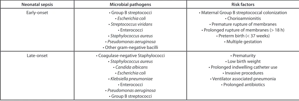
The microbial pathogens and risk factors associated with neona-tal sepsis are shown in Table 1.
Prevention of Early-Onset Group B Streptococcal (GBS) Disease
Early onset GBS infection has a case fatality of 5–20%; a mul-tistate active surveillance system demonstrated that 6% of early-onset GBS infections resulted in death.31 Neonatal infection can
occur when GBS ascends from the vagina to the amniotic fluid after the onset of labor or rupture of membranes.32,33 It is more
commonly the result of vertical transmission from mother to infant in women with recto-vaginal colonization. Colonization with GBS occurs in roughly 10–30% of pregnant woman in their vagina or rectum.34
The only intervention proven to decrease the incidence of early-onset neonatal GBS sepsis is maternal treatment with intra-partum intravenous antibiotics. Adequate prophylaxis is expo-sure to penicillin (preferred agent), ampicillin, or cefazolin given
Table 1. Microbial pathogens and risk factors associated with neonatal sepsis
Neonatal sepsis Microbial pathogens Risk factors
Early-onset • Group B streptococci
• Escherichia coli
• Streptococcus viridans
• Enterococci • Staphylococcus aureus
• Pseudomonas aeruginosa
• Other gram-negative bacilli
• Maternal Group B streptococcal colonization • Chorioamnionitis
• Premature rupture of membranes • Prolonged rupture of membranes (> 18 h)
• Preterm birth (< 37 weeks) • Multiple gestation
Late-onset • Coagulase-negative Staphylococci
• Staphylococcus aureus
• Candida albicans
• Escherichia coli
• Klebsiella pneumoniae
• Enterococci • Pseudomonas aeruginosa
• Group B streptococci
• Prematurity • Low birth weight • Prolonged indwelling catheter use
• Invasive procedures • Ventilator associated pneumonia
• Prolonged antibiotics
172 Virulence Volume 5 Issue 1
for >4 h before delivery.31,35 Erythromycin is no longer
recom-mended for prophylaxis because of high resistance rates. In those women with a non-serious penicillin allergy, cefazolin is the drug of choice. For a mother with a history of life-threatening peni-cillin allergy (anaphylaxis, rash, angioedema, respiratory symp-toms), clindamycin is the substitute for penicillin, but should only be used if the rectovaginal GBS isolate is tested and found to be susceptible. If the clindamycin sensitivity is unknown or the GBS isolate is resistant to clindamycin, vancomycin is the substitute for prophylaxis. Intrapartum antibiotics are indicated in the following situations:35
1) Positive antenatal cultures or molecular testing at admis-sion for GBS (except for women who have a cesarean delivery without labor or membrane rupture),
2) Unknown maternal colonization status with gesta-tion <37 weeks, rupture of membranes >18 h, or temperature >100.4 °F (>38 °C),
3) GBS bacteriuria during current pregnancy, 4) Previous infant with invasive GBS disease.
Risk-Based Approach vs. Universal Screening Approach
The epidemiology and management of neonatal GBS dis-ease has evolved significantly over the last two decades. Initially, screening approaches based on risk factors for EOS were tested. Later, the effectiveness of universal screening was compared with risk-based approaches in preventing early-onset GBS disease in a multistate retrospective cohort study. The risk of early-onset GBS sepsis was notably lower in the infants of women who underwent universal screening than among those in the risk-based group.36
With universal screening, it is possible to identify GBS colonized woman even without obstetric risk factors and reach more of the population at risk than with the risk based approach. After con-trolling for risk factors associated with early-onset GBS disease (preterm delivery, prolonged ROM, young maternal age, black race), the protective effect of the universal screening approach has been shown to be robust in subsequent prospective studies.37
Intrapartum antibiotic prophylaxis is approximately 90% effec-tive in preventing early-onset GBS disease.37
Changes in Microbial Pathogens in the Post Chemoprophylaxis Era
Decreased incidence of neonatal group B streptococcal (GBS) disease
After the nationwide implementation of intrapartum antibi-otic prophylaxis, a striking 80% decline in the incidence of inva-sive early-onset GBS disease was observed.35 Before prophylaxis,
the incidence of GBS in the United States was 2–3 cases per 1000 live births.38 After the 1996 GBS prevention guidelines were
issued, incidence of early-onset GBS sepsis declined significantly in the following 2 y and then reached a plateau of approximately 0.5 cases per 1000 live births during the period from 1999– 2001.35 Upon issuance of the 2002 guidelines published in the
landmark Morbidity and Mortality Weekly Report (MMWR) by
CDC, the incidence further declined to 0.3–0.4 cases per 1000 live births.35 This additional decline is consistent with the
transi-tion from the 1996 preventransi-tion strategy to the universal screening approach recommended in 2002.
Increased proportion of non-group B streptococcal pathogens
A potential increase in false-negative neonatal blood cul-tures as well as sepsis caused by pathogens other than GBS is a potential concern with the extensive use of maternal intrapartum antibiotics. Such a change would be important as several studies have demonstrated increased severity of disease and risk of death in the neonates with gram-negative infections.24 In a study
con-ducted by the NICHD between 1998 and 2000 on 5447 VLBW infants (those weighing between 401 and 1500 g), there was a significant reduction in the incidence of EOS caused by GBS. However, there was also a significant increase in the proportion of E. coli infections among VLBW infants.23 Though there has
been a dramatic decline in the incidence of EOS due to GBS, the increasing incidence of ampicillin-resistant neonatal sepsis among VLBW infants is concerning.39 Nonetheless, the benefits
from the use of antepartum antibiotic chemoprophylaxis still off-set the risks of resistant bacterial infections.36
Neonatal Early-Onset Sepsis Risk Algorithms
Pediatricians currently use the CDC 2010 and American Academy of Pediatrics Committee on the Fetus and Newborn (COFN) 2012 algorithms for evaluation and management of infants at risk for EOS born at or near term gestation.35,40 While
both sources acknowledge maternal chorioamnionitis as a signifi-cant risk, they do not offer a standard definition of this clinical diagnosis.24 Additionally, there are differences in their
recom-mendations for the evaluation of infants who received inadequate intrapartum GBS prophylaxis and with the rupture of mem-branes at least equal to 18 h. Puopolo et al. proposed a multivari-ate predictive model of EOS risk developed for infants born at or above 34 weeks’ gestation based on objective clinical data avail-able at the time of birth.41,42 This could decrease the number of
infants evaluated and empirically treated for EOS but studies are needed for validation of this computational model.
Biomarkers of Neonatal Sepsis
Since sepsis is a systemic inflammatory response to infection, isolation of bacteria from blood is considered the gold standard for the diagnosis of sepsis.43 However, it takes 24–48 h for
cul-ture results. Inoculation of only 0.5–1.0 ml of blood decreases its sensitivity, as approximately 60–70% of infants have a low level of bacteremia.44 Theoretically, for optimal results, 6 ml of blood
would be required which is not feasible.45 Sepsis cannot always
diagnostic tests based on evaluation of the immune system are being evaluated to help resolve ambiguities in these situations.46
Complete blood count (CBC)
In order to improve the outcome associated with neonatal sep-sis, it is necessary for a diagnostic test to be rapid and sensitive to decrease delay in treatment. At the same time in order to avoid unnecessary exposure to antibiotics and invasive procedures, a test with higher specificity is needed. A large number of stud-ies have been performed to evaluate the use of complete blood count (CBC), differential count, and immature to total leuko-cyte ratio (I:T) for the diagnosis of neonatal sepsis. Although the CBC has a poor predictive value, serial normal values can be used to enhance the prediction that bacterial sepsis is not present.47,48
Low WBC and absolute neutrophil counts, as well as high immature-to-total neutrophil ratio (I:T) are associated with an increased risk of infection (odds ratios 5.38, 6.84, and 7.90 respectively).44 However, the sensitivity for detection of sepsis is
low. Two serial normal CBCs, performed 8 to 12 h apart, and a negative blood culture at 24 h improve the predictive power to rule out EOS in the first 24 h after birth.49 This strategy has been
associated with a negative predictive value as high as 100%, but the specificity and positive predictive values may be too low to guide therapy decisions.49 Components of the white cell count,
including absolute neutrophil count (ANC) and immature to total neutrophil ratio (I:T) have also been shown to be more use-ful for excluding infants without infection rather than identifying newborns who are infected.40 The maximal (I:T) ratio in
unin-fected newborns is 0.16 in the first 24 h, which by 120 h decreases to 0.12.50 I:T ratio of >0.2 is suggestive of sepsis. However, the I:T
ratio can be affected by various noninfectious processes like labor, prolonged induction with oxytocin, and even prolonged crying.49
A total leukocyte count of <5000 to 7500/mm3 can be used to
infer the diagnosis of neonatal sepsis.6 Many infected newborns
may have higher counts. However, the sensitivity of a low leuko-cyte count is 29%, though the specificity is as high as 91%.6 There
are important gestational age effects on the leukocyte count in the newborn period. In newborns >36 weeks gestation, the lower limit of normal for ANC at birth is 3500/mm3. The lower limit
of normal in infants born between 28–36 weeks is 1000/mm3
and 500/mm3 in infants <28 weeks gestation.51 Total neutrophil
counts rise after birth and reach their peak levels at 6 to 8 h of life. The lower limits of normal at that time are 7500/mm3, 3500/mm3,
and 1500/mm3 for infants born at >36 weeks, 28–36 weeks, and
<28 weeks respectively.51 Thus it is more effective to obtain total
leukocyte counts at 6–12 h after birth, as they are more likely to be reliable. Factors such as maternal hypertension or perinatal asphyxia may cause neutropenia or an elevated I/T ratio.50 Also,
leukocyte counts may be normal in the early course of neonatal sepsis. In summary, the WBC, ANC, and I/T ratio have signifi-cant limitations in the diagnosis of neonatal sepsis.
C reactive protein (CRP)
CRP is one of the most extensively studied, most available, and most frequently used laboratory tests for the diagnosis of neonatal sepsis.52 CRP is an acute phase reactant synthesized by the liver.
It has a half- life of 24–48 h. It takes 10–12 h for CRP to change significantly after onset of infection. Serial determination of CRP
24–48 h after the onset of symptoms increases its sensitivity.52,53
Serial CRP measurements may also be helpful in monitoring the response to treatment in infected neonates and thus may help cli-nicians guide the duration of antibiotic therapy.53-55 The
specific-ity and positive predictive value of CRP ranges from 93–100%.56
Thus, CRP can be considered as a “specific” but “late” marker of neonatal infection. If the CRP levels remain persistently normal, it correlates strongly with the absence of infection thereby guiding safe discontinuation of antibiotic therapy.57
Preterm infants have lower CRP baseline values and a lower rise in response to infection. A variety of non-infectious condi-tions like meconium aspiration syndrome, traumatic or ischemic tissue injuries, hemolysis, or histologic chorioamnionitis may cause an elevation in the CRP levels.52 Because it takes 10–12 h to
change significantly after the onset of infection; the sensitivity of CRP is low during the early phase of sepsis. Due to noninfectious CRP elevations, the influence of gestational age and birth weight on kinetics of CRP, and the lack of reliable age specific reference values, the use of CRP requires further research to cover these pitfalls and falls short as an ideal marker.
Procalcitonin (PCT)
PCT is an acute phase reactant produced both by hepatocytes and macrophages that has been studied since the mid-1990s. Serum concentrations of PCT begin to rise 4 h after exposure to bacterial endotoxin, peak at 6 to 8 h, and remain elevated for at least 24 h.58 The half-life is about 25–30 h, and the serum
con-centration is not affected by gestational age. Nonetheless, in non-infected newborns, serum PCT concentrations vary widely. It is low soon after birth, rises to a peak at 24 h and returns to baseline at 48 h.56 Serum PCT concentrations increase appreciably in the
presence of systemic bacterial infection and necrotizing enteroco-litis during early- and late-onset neonatal infection.59,60 The PCT
response is more rapid than the elevation of CRP, thus it is an attractive alternative for the detection of EOS. Because PCT levels remain high compared with TNF-α and IL-6, PCT is also useful in predicting severity of infection, response to treatment, and out-come.59,60 In contrast to CRP, infants with trauma, viral infections,
meconium aspiration, and hypoxemia have normal or minimal elevation in PCT.60 The sensitivity and specificity of PCT varies
between 83–100% and 70–100% respectively.61 The sensitivity
and specificity of PCT is greater than CRP or interleukin 6 if dif-ferent cutoff points at birth and at 24 h and 48 h of life are used.61,62
Nonetheless, PCT has its own limitations as it is increased in newborns requiring neonatal resuscitation and in infants born to mothers with chorioamnionitis in the absence of neonatal infection.61 In healthy neonates it has been shown that PCT
con-centrations are affected by maternal GBS colonization and pro-longed rupture of membranes ≥18 h.63 Therefore, PCT needs to
be studied further in larger groups of infants so as to improve its diagnostic accuracy.
Cytokines
174 Virulence Volume 5 Issue 1
to rise early in response to bacterial infection in neonates. The rise occurs before the newborn develops signs or symptoms of sepsis and even before previously described laboratory tests become positive.57 Cytokines do not cross placental barrier and elevations
have been found in umbilical cord blood suggesting the possibil-ity of predicting infants who are going to develop sepsis in first few hours of their life.64,65 In addition, cytokine analysis may be
useful in predicting late-onset infection.66 IL-6
In response to exposure to bacterial endotoxins, IL-6 concen-trations rise before that of CRP.67 Umbilical cord IL-6 is
con-sistently increased in newborns with EOS.65,68 The sensitivity
of cord blood IL-6 in predicting neonatal sepsis was found to be 87–100%, with the negative predictive value of 93–100% in some cohorts.65,68-70 However, the half-life of IL-6 is very short
and the levels fall to undetectable values quickly with treatment; thus sensitivity falls at 24 and 48 h (67% and 58% respectively).71
Therefore, IL-6 can be considered as an early and sensitive marker of neonatal infection.56 Diagnostic accuracy is further improved
by combining IL-6 (early and sensitive) and CRP (late and spe-cific) in first 48 h of presumed clinical sepsis.72
TNF-α
The concentrations of TNF-α were shown to be significantly higher in infected as compared with non-infected newborns in multiple studies.71,73,74 TNF-α has very similar kinetics to IL-6.67
Silveira et al. observed the diagnostic accuracy of TNF-α was equivalent to PCT.74 Sensitivity and specificity increases to 60%
and 100% respectively when TNF-α and IL-6 levels are com-bined for the diagnosis of neonatal sepsis.75
IL-8
IL-8 is a pro-inflammatory cytokine which aids in the acti-vation and chemotaxis of neutrophils.76 IL-8 not only serves as
a marker for sepsis but is also associated with severity of infec-tion. It is produced as a result of infection by monocytes, macro-phages, and endothelial cells with kinetics similar to IL-6.76 IL-8
has sensitivity and specificity ranging from 80 to 91% and 76 to 100% respectively.67,70 In a study performed by Boskabadi et al.
in 93 neonates greater than 72 h of age, serum concentrations of IL-8 in non-surviving neonates were 3.3 times higher than surviving neonates.77 The combination of IL-8 and CRP is more
reliable for early diagnosis of neonatal sepsis, with a sensitivity and specificity of 91% and 73% respectively.55,65,67 Thus,
combin-ing CRP and IL-8 may reduce excessive use of antibiotics.55
Though IL-6 and IL-8 increase very rapidly with bacte-rial invasion, their levels promptly normalize in serum (within the first 24 h), limiting their ability to be used as ideal mark-ers. Therefore, operational difficulties in detection of cytokines, elevation of cytokines in non-specific settings, and lack of avail-ability in many centers are limitations for their use in day-to-day practice.65 Studies with larger sample sizes are needed before
cytokines can be endorsed as valid diagnostic markers.
Cell Surface Markers
With the advances in flow cytometric technology, cell sur-face antigens can be detected in blood cells. Such tests are readily
performed requiring a very low volume (0.05 ml) of whole blood. Neutrophil cluster of differentiation (CD) CD11β and CD64 have been found to be reliable markers for detecting early- and late-onset neonatal sepsis respectively with a high sensitivity and specificity.56 Their expression increases within minutes following
exposure to bacterial products. Further, as the biological activi-ties of the cytokines may not be revealed by their circulating con-centrations, measuring the cellular response to cytokines may be a better way of recognizing an early immunological response to infection.
CD11β
CD11β is the α-subunit of the β2-integrin adhesion molecule involved in neutrophil adhesion, diapedesis, and phagocytosis. It is detectable within 5 min in response to bacterial infection.78,79
The sensitivity and specificity are as high as 96–100% and 100% in two studies.80,81 It has better diagnostic accuracy for early than
late-onset neonatal sepsis.56 The variable diagnostic accuracy of
CD11β in late-onset neonatal sepsis may be related to different infant population being evaluated, time interval between phle-botomy and sample processing and at which phase of infection blood is obtained.82,83
CD64
The high affinity antibody receptor CD64 is expressed at a very low level on the surface of neutrophils in the absence of an infection.84,85 The expression of CD64 on activated neutrophils
markedly increases after an episode of bacterial infection.86-88
CD64 has a sensitivity of 95–97% and a negative predictive value of 97–99%.56,73 The addition of IL-6 or C-reactive protein
to CD64 further enhances its sensitivity and negative predictive value to 100%, with the specificity and positive predictive value exceeding 88% and 80%, respectively.82 With the use of CD64
it may be possible for clinicians to discontinue antibiotics within 24 h in non-infected newborns.82
In a prospective study performed by Streimish et al. at Yale University, using a cut-point CD64 index value of 2.38 for EOS, the test had a sensitivity of 100%, a specificity of 68%, and an NPV of 100%; while using a cut-point value of 3.62 for LOS sepsis, the test had a sensitivity of 75%, a specificity of 77%, and an NPV of 96%. Due to the large sample size, this study was able to demonstrate the potential of CD64 to influence the initiation and duration of antibiotic therapy.89 However, the cost, the need
for sophisticated equipment and the processing time are barriers to the use of these markers in clinical practice.
Genomics, Proteomics, and Nucleic Acid-Based Molecular Techniques
Esparcia et al. employed a gene-based molecular technique using 16S rDNA for diagnostic accuracy of bacterial meningitis and early-onset neonatal sepsis.90 Ng et al. used a score based
on proapolipoprotein CII and a des-arginine variant of serum amyloid to withhold antibiotics in 45% of infants with suspected infection and to discontinue antibiotics in 16%.91,92 Kasper et al.
in premature infants.93 The limitations of these studies includes
failure to provide information about antibiotic resistance, inabil-ity to differentiate the false-positive results because of potential contamination during blood sampling or processing from true positive cases and high cost. Prospective evaluation is needed to determine accuracy and safety of these exciting new approaches. Therefore, these are currently adjunctive methods with the excep-tion of HSV PCR, which remains gold standard for the diagnosis of HSV encephalitis.1
None of the markers including hematologic indices, acute phase reactants, cytokines, and cell surface markers have shown sensitivity, specificity, positive and negative predictive value that are sufficiently powerful to guide the clinical management of neo-natal sepsis.4,94 Different biomarkers have been used to diagnose
neonatal sepsis, but with inconclusive results, because of small sample size, lack of clear reference values and lack of homogene-ity in the study group. Thus, there remains a need for a marker with high sensitivity, specificity, positive and negative predictive accuracy which is able to detect infection at an early stage.
Inter α inhibitor proteins (IAIP)
The inter alpha inhibitor family of proteins (IAIP) are serine protease inhibitors which provide protection from the increased protease activity associated with systemic immune system acti-vation that accompanies sepsis and inflammation. They are involved in extracellular matrix stabilization, inflammation, wound healing, and play an important anti-inflammatory and regulatory role in infection.95 IAIP is one of the important serine
protease inhibitors secreted by the liver. IAIP is a heterotrimeric, 250 kd protein complex composed of two heavy chains and one light chain held together by glycosaminoglycan bonds. The light chain, Bikunin, has a molecular weight of 30 kd and is the active, anti-protease component. In the presence of serine proteases, Bikunin is released and it provides protective effects.95,96 The
half-life of Bikunin is very short and it is rapidly excreted by the kidneys.
IAIP concentration is independent of gestational age, postna-tal age, and is similar to adult levels.97 However, IAIP levels are
significantly lower in septic neonates as compared with non-sep-tic age matched controls. Receiver operating curve analysis has shown IAIP measurement to have a sensitivity of 89.5%, a speci-ficity of 99%, a positive predictive value of 95% and a negative predictive value of 98% in a pilot study of 573 neonates.98 The
levels of IAIP not only decrease in neonatal sepsis but also rise in response to antibiotic treatment.97 Yang and colleagues showed
that low levels of IAIP are highly predictive of mortality in septic adult patients.99 Because the levels of IAIP decrease with severe
sepsis, measurement may also help to guide the prognosis as lower levels are associated with adverse outcome.100 Chaaban et al.
dem-onstrated that the levels of IAIP also decrease significantly in patients with necrotizing enterocolitis (NEC, stage II/III accord-ing to modified the Bell criteria) and thus can be useful to diag-nose patients with NEC at an early stage.17 Singh et al. showed
an immunomodulatory and protective role of administration of IAIP in a septic newborn mice.101 This underscores potential role
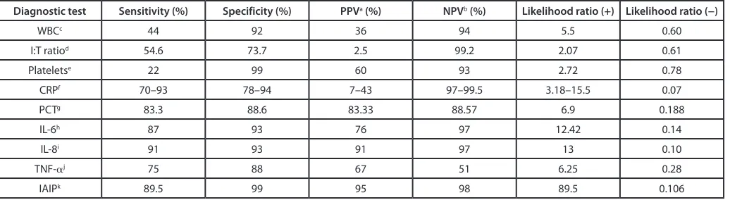
of IAIP as a theranostic marker in infants with sepsis. The diag-nostic performance of IAIP and other adjunctive tests of neonatal sepsis is shown in Table 2.
Conclusion
Systemic bacterial infection in the newborn creates a signifi-cant burden due to its impact on neonatal mortality and long-term morbidity. In spite of ongoing efforts in early diagnosis, treatment, and prevention, neonatal sepsis still remains an enig-matic area for neonatologists due to changes in epidemiology and the lack of ideal diagnostic markers. The need for a biomarker with high diagnostic accuracy and reliability is paramount as a guiding tool for physicians to assess the risk of infection and need for antibiotic therapy. Studies are currently ongoing in the search of a novel marker for neonatal sepsis. Inter α inhibitor proteins are among the candidates with significant promise.
Disclosure of Potential Conflicts of Interest
No potential conflicts of interest were disclosed.
Table 2. Diagnostic performance of adjunctive tests of neonatal sepsis
Diagnostic test Sensitivity (%) Specificity (%) PPVa (%) NPVb (%) Likelihood ratio (+) Likelihood ratio (−)
WBCc 44 92 36 94 5.5 0.60
I:T ratiod 54.6 73.7 2.5 99.2 2.07 0.61
Plateletse 22 99 60 93 2.72 0.78
CRPf 70–93 78–94 7–43 97–99.5 3.18–15.5 0.07
PCTg 83.3 88.6 83.33 88.57 6.9 0.188
IL-6h 87 93 76 97 12.42 0.14
IL-8i 91 93 91 97 13 0.10
TNF-αj 75 88 67 51 6.25 0.28
IAIPk 89.5 99 95 98 89.5 0.106
aPPV, positive predictive value; bNPV, negative predictive value; cwhite blood cell (WBC) counts ≤5000 or ≥25 000, 30 000, or 21 000 per mm3 at birth, 12–24 h
and day 2 or after, respectively57; dI:T ratio (ratio of immature to absolute neutrophil count) >0.244; eplatelets <150 000 cells/mm3,57; fC-reactive protein (CRP)
176 Virulence Volume 5 Issue 1
References
1. Camacho-Gonzalez A, Spearman PW, Stoll BJ. Neonatal infectious diseases: evaluation of neona-tal sepsis. Pediatr Clin North Am 2013; 60:367-89; PMID:23481106; http://dx.doi.org/10.1016/j. pcl.2012.12.003
2. Bizzarro MJ, Raskind C, Baltimore RS, Gallagher PG. Seventy-five years of neonatal sepsis at Yale: 1928-2003. Pediatrics 2005; 116:595-602; PMID:16140698; http://dx.doi.org/10.1542/ peds.2005-0552
3. Hornik CP, Fort P, Clark RH, Watt K, Benjamin DK Jr., Smith PB, Manzoni P, Jacqz-Aigrain E, Kaguelidou F, Cohen-Wolkowiez M. Early and late onset sepsis in very-low-birth-weight infants from a large group of neonatal intensive care units. Early Hum Dev 2012; 88(Suppl 2):S69-74; PMID:22633519; http://dx.doi.org/10.1016/ S0378-3782(12)70019-1
4. Gerdes JS. Diagnosis and management of bacte-rial infections in the neonate. [viii-ix.]. Pediatr Clin North Am 2004; 51:939-59, viii-ix; PMID:15275982; http://dx.doi.org/10.1016/j.pcl.2004.03.009 5. Bonadio WA, Hennes H, Smith D, Ruffing R,
Melzer-Lange M, Lye P, Isaacman D. Reliability of observation variables in distinguishing infectious outcome of febrile young infants. Pediatr Infect Dis J 1993; 12:111-4; PMID:8426766; http://dx.doi. org/10.1097/00006454-199302000-00001 6. Gerdes JS. Clinicopathologic approach to the
diagno-sis of neonatal sepdiagno-sis. Clin Perinatol 1991; 18:361-81; PMID:1879113
7. Gleason CA, Devaskar SU, Avery ME. Avery’s dis-eases of the newborn / [edited by] Christine A. Gleason, Sherin U. Devaskar. Philadelphia, PA: Elsevier/Saunders, 2012.
8. Voora S, Srinivasan G, Lilien LD, Yeh TF, Pildes RS. Fever in full-term newborns in the first four days of life. Pediatrics 1982; 69:40-4; PMID:7033912 9. Weisman LE, Stoll BJ, Cruess DF, Hall RT,
Merenstein GB, Hemming VG, Fischer GW. Early-onset group B streptococcal sepsis: a current assess-ment. J Pediatr 1992; 121:428-33; PMID:1517922; http://dx.doi.org/10.1016/S0022-3476(05)81801-3 10. Hofer N, Müller W, Resch B. Neonates
present-ing with temperature symptoms: role in the diagnosis of early onset sepsis. Pediatr Int 2012; 54:486-90; PMID:22299645; http://dx.doi. org/10.1111/j.1442-200X.2012.03570.x
11. Bekhof J, Reitsma JB, Kok JH, Van Straaten IH. Clinical signs to identify late-onset sepsis in pre-term infants. Eur J Pediatr 2013; 172:501-8; PMID:23271492; http://dx.doi.org/10.1007/ s00431-012-1910-6
12. Remington JS. Infectious diseases of the fetus and newborn infant. Philadelphia, PA: Saunders/Elsevier, 2011.
13. Martin RJ, Fanaroff AA, Walsh MC. Fanaroff and Martin’s neonatal-perinatal medicine: diseases of the fetus and infant. Philadelphia: Saunders/Elsevier, 2011.
14. Sharma R, Tepas JJ 3rd, Hudak ML, Wludyka PS,
Mollitt DL, Garrison RD, Bradshaw JA, Sharma M. Portal venous gas and surgical outcome of neonatal necrotizing enterocolitis. J Pediatr Surg 2005; 40:371-6; PMID:15750931; http://dx.doi. org/10.1016/j.jpedsurg.2004.10.022
15. Sharma R, Tepas JJ 3rd, Hudak ML, Pieper P, Teng
RJ, Raja S, Sharma M. Neonatal gut injury and infec-tion rate: impact of surgical debridement on outcome. Pediatr Surg Int 2005; 21:977-82; PMID:16211416; http://dx.doi.org/10.1007/s00383-005-1539-x
16. Cole CR, Hansen NI, Higgins RD, Bell EF, Shankaran S, Laptook AR, Walsh MC, Hale EC, Newman NS, Das A, et al.; Eunice Kennedy Shriver National Institute of Child Health and Human Development’s Neonatal Research Network. Bloodstream infections in very low birth weight infants with intestinal failure. J Pediatr 2012; 160:54, e2; PMID:21840538; http://dx.doi.org/10.1016/j. jpeds.2011.06.034
17. Chaaban H, Shin M, Sirya E, Lim YP, Caplan M, Padbury JF. Inter-alpha inhibitor protein level in neonates predicts necrotizing enterocolitis. J Pediatr 2010; 157:757-61; PMID:20955849; http://dx.doi. org/10.1016/j.jpeds.2010.04.075
18. Uauy RD, Fanaroff AA, Korones SB, Phillips EA, Phillips JB, Wright LL; National Institute of Child Health and Human Development Neonatal Research Network. Necrotizing enterocolitis in very low birth weight infants: biodemographic and clinical cor-relates. J Pediatr 1991; 119:630-8; PMID:1919897; http://dx.doi.org/10.1016/S0022-3476(05)82418-7 19. Sharma R, Hudak ML. A clinical perspective of
nec-rotizing enterocolitis: past, present, and future. Clin Perinatol 2013; 40:27-51; PMID:23415262; http:// dx.doi.org/10.1016/j.clp.2012.12.012
20. Kaufman D, Fairchild KD. Clinical microbiology of bacterial and fungal sepsis in very-low-birth-weight infants. [table of contents.]. Clin Microbiol Rev 2004; 17:638-80; PMID:15258097; http://dx.doi. org/10.1128/CMR.17.3.638-680.2004
21. Baker CJ, Barrett FF. Group B streptococcal infections in infants. The importance of the various serotypes. JAMA 1974; 230:1158-60; PMID:4608888; http:// dx.doi.org/10.1001/jama.1974.03240080040025 22. Stoll BJ, Hansen N, Fanaroff AA, Wright LL, Carlo
WA, Ehrenkranz RA, Lemons JA, Donovan EF, Stark AR, Tyson JE, et al. Late-onset sepsis in very low birth weight neonates: the experience of the NICHD Neonatal Research Network. Pediatrics 2002; 110:285-91; PMID:12165580; http://dx.doi. org/10.1542/peds.110.2.285
23. Stoll BJ, Hansen N, Fanaroff AA, Wright LL, Carlo WA, Ehrenkranz RA, Lemons JA, Donovan EF, Stark AR, Tyson JE, et al. Changes in pathogens causing early-onset sepsis in very-low-birth-weight infants. N Engl J Med 2002; 347:240-7; PMID:12140299; http://dx.doi.org/10.1056/NEJMoa012657 24. Stoll BJ, Hansen NI, Sánchez PJ, Faix RG, Poindexter
BB, Van Meurs KP, Bizzarro MJ, Goldberg RN, Frantz ID 3rd, Hale EC, et al.; Eunice Kennedy
Shriver National Institute of Child Health and Human Development Neonatal Research Network. Early onset neonatal sepsis: the burden of group B Streptococcal and E. coli disease continues. Pediatrics 2011; 127:817-26; PMID:21518717; http://dx.doi. org/10.1542/peds.2010-2217
25. Weston EJ, Pondo T, Lewis MM, Martell-Cleary P, Morin C, Jewell B, Daily P, Apostol M, Petit S, Farley M, et al. The burden of invasive early-onset neonatal sepsis in the United States, 2005-2008. Pediatr Infect Dis J 2011; 30:937-41; PMID:21654548; http:// dx.doi.org/10.1097/INF.0b013e318223bad2 26. Baltimore RS, Huie SM, Meek JI, Schuchat A,
O’Brien KL. Early-onset neonatal sepsis in the era of group B streptococcal prevention. Pediatrics 2001; 108:1094-8; PMID:11694686; http://dx.doi. org/10.1542/peds.108.5.1094
27. Gladstone IM, Ehrenkranz RA, Edberg SC, Baltimore RS. A ten-year review of neonatal sepsis and compari-son with the previous fifty-year experience. Pediatr Infect Dis J 1990; 9:819-25; PMID:2263432; http:// dx.doi.org/10.1097/00006454-199011000-00009 28. Karlowicz MG, Buescher ES, Surka AE. Fulminant
late-onset sepsis in a neonatal intensive care unit, 1988-1997, and the impact of avoiding empiric vancomycin therapy. Pediatrics 2000; 106:1387-90; PMID:11099593; http://dx.doi.org/10.1542/ peds.106.6.1387
29. Levent F, Baker CJ, Rench MA, Edwards MS. Early outcomes of group B streptococcal meningitis in the 21st century. Pediatr Infect Dis J 2010; 29:1009-12; PMID:20555292
30. Libster R, Edwards KM, Levent F, Edwards MS, Rench MA, Castagnini LA, Cooper T, Sparks RC, Baker CJ, Shah PE. Long-term outcomes of group B streptococcal meningitis. Pediatrics 2012; 130:e8-15; PMID:22689869; http://dx.doi.org/10.1542/ peds.2011-3453
31. Centers for Disease Control and Prevention. Prevention of perinatal group B streptococcal disease: a public health perspective. MMWR Recomm Rep 1996; 45(RR-7):1-24; PMID:8637497
32. Desa DJ, Trevenen CL. Intrauterine infections with group B beta-haemolytic streptococci. Br J Obstet Gynaecol 1984; 91:237-9; PMID:6367810; http:// dx.doi.org/10.1111/j.1471-0528.1984.tb04759.x 33. Katz V, Bowes WA Jr. Perinatal group B
streptococ-cal infections across intact amniotic membranes. J Reprod Med 1988; 33:445-9; PMID:3290476 34. Regan JA, Klebanoff MA, Nugent RP; Vaginal
Infections and Prematurity Study Group. The epi-demiology of group B streptococcal colonization in pregnancy. Obstet Gynecol 1991; 77:604-10; PMID:2002986
35. Verani JR, McGee L, Schrag SJ; Division of Bacterial Diseases, National Center for Immunization and Respiratory Diseases, Centers for Disease Control and Prevention (CDC). Prevention of perinatal group B streptococcal disease--revised guidelines from CDC, 2010. MMWR Recomm Rep 2010; 59(RR-10):1-36; PMID:21088663
36. Schrag SJ, Zell ER, Lynfield R, Roome A, Arnold KE, Craig AS, Harrison LH, Reingold A, Stefonek K, Smith G, et al.; Active Bacterial Core Surveillance Team. A population-based comparison of strate-gies to prevent early-onset group B streptococcal disease in neonates. N Engl J Med 2002; 347:233-9; PMID:12140298; http://dx.doi.org/10.1056/ NEJMoa020205
37. Schrag S, Gorwitz R, Fultz-Butts K, Schuchat A. Prevention of perinatal group B streptococcal disease. Revised guidelines from CDC. MMWR Recomm Rep 2002; 51(RR-11):1-22; PMID:12211284 38. Zangwill KM, Schuchat A, Wenger JD. Group B
streptococcal disease in the United States, 1990: report from a multistate active surveillance system. MMWR CDC Surveill Summ 1992; 41:25-32; PMID:1470102
39. Levine EM, Ghai V, Barton JJ, Strom CM. Intrapartum antibiotic prophylaxis increases the inci-dence of gram-negative neonatal sepsis. Infect Dis Obstet Gynecol 1999; 7:210-3; PMID:10449272 40. Polin RA; Committee on Fetus and Newborn.
Management of neonates with suspected or proven early-onset bacterial sepsis. Pediatrics 2012; 129:1006-15; PMID:22547779; http://dx.doi. org/10.1542/peds.2012-0541
41. Puopolo KM, Draper D, Wi S, Newman TB, Zupancic J, Lieberman E, Smith M, Escobar GJ. Estimating the probability of neonatal early-onset infection on the basis of maternal risk factors. Pediatrics 2011; 128:e1155-63; PMID:22025590; http://dx.doi.org/10.1542/peds.2010-3464 42. Puopolo KM, Escobar GJ. Early-onset sepsis: a
pre-dictive model based on maternal risk factors. Curr Opin Pediatr 2013; 25:161-6; PMID:23407183; http://dx.doi.org/10.1097/MOP.0b013e32835e1f96 43. Goldstein B, Giroir B, Randolph A; International
44. Hornik CP, Benjamin DK, Becker KC, Benjamin DK Jr., Li J, Clark RH, Cohen-Wolkowiez M, Smith PB. Use of the complete blood cell count in early-onset neonatal sepsis. Pediatr Infect Dis J 2012; 31:799-802; PMID:22531231; http://dx.doi.org/10.1097/ INF.0b013e318256905c
45. Kellogg JA, Ferrentino FL, Goodstein MH, Liss J, Shapiro SL, Bankert DA. Frequency of low level bacteremia in infants from birth to two months of age. Pediatr Infect Dis J 1997; 16:381-5; PMID:9109140; http://dx.doi. org/10.1097/00006454-199704000-00009 46. Brozanski BS, Jones JG, Krohn MJ, Jordan JA. Use
of polymerase chain reaction as a diagnostic tool for neonatal sepsis can result in a decrease in use of anti-biotics and total neonatal intensive care unit length of stay. J Perinatol 2006; 26:688-92; PMID:17024143; http://dx.doi.org/10.1038/sj.jp.7211597
47. Rozycki HJ, Stahl GE, Baumgart S. Impaired sensitivity of a single early leukocyte count in screening for neonatal sepsis. Pediatr Infect Dis J 1987; 6:440-2; PMID:3601489; http://dx.doi. org/10.1097/00006454-198705000-00004 48. Christensen RD, Rothstein G, Hill HR, Hall
RT. Fatal early onset group B streptococcal sepsis with normal leukocyte counts. Pediatr Infect Dis 1985; 4:242-5; PMID:3889873; http://dx.doi. org/10.1097/00006454-198505000-00006 49. Murphy K, Weiner J. Use of leukocyte counts in
eval-uation of early-onset neonatal sepsis. Pediatr Infect Dis J 2012; 31:16-9; PMID:21860335; http://dx.doi. org/10.1097/INF.0b013e31822ffc17
50. Manroe BL, Weinberg AG, Rosenfeld CR, Browne R. The neonatal blood count in health and disease. I. Reference values for neutrophilic cells. J Pediatr 1979; 95:89-98; PMID:480023; http://dx.doi. org/10.1016/S0022-3476(79)80096-7
51. Schmutz N, Henry E, Jopling J, Christensen RD. Expected ranges for blood neutrophil concentrations of neonates: the Manroe and Mouzinho charts revis-ited. J Perinatol 2008; 28:275-81; PMID:18200025; http://dx.doi.org/10.1038/sj.jp.7211916
52. Hofer N, Zacharias E, Müller W, Resch B. An update on the use of C-reactive protein in early-onset neonatal sepsis: current insights and new tasks. Neonatology 2012; 102:25-36; PMID:22507868; http://dx.doi.org/10.1159/000336629
53. Pourcyrous M, Bada HS, Korones SB, Baselski V, Wong SP. Significance of serial C-reactive protein responses in neonatal infection and other disorders. Pediatrics 1993; 92:431-5; PMID:8361798
54. Kawamura M, Nishida H. The usefulness of serial C-reactive protein measurement in man-aging neonatal infection. Acta Paediatr 1995; 84:10-3; PMID:7734887; http://dx.doi. org/10.1111/j.1651-2227.1995.tb13475.x
55. Franz AR, Steinbach G, Kron M, Pohlandt F. Reduction of unnecessary antibiotic therapy in newborn infants using interleukin-8 and C-reactive protein as markers of bacterial infections. Pediatrics 1999; 104:447-53; PMID:10469768; http://dx.doi. org/10.1542/peds.104.3.447
56. Ng PC. Diagnostic markers of infection in neonates. Arch Dis Child Fetal Neonatal Ed 2004; 89:F229-35; PMID:15102726; http://dx.doi.org/10.1136/ adc.2002.023838
57. Benitz WE. Adjunct laboratory tests in the diag-nosis of early-onset neonatal sepsis. Clin Perinatol 2010; 37:421-38; PMID:20569816; http://dx.doi. org/10.1016/j.clp.2009.12.001
58. Dandona P, Nix D, Wilson MF, Aljada A, Love J, Assicot M, Bohuon C. Procalcitonin increase after endotoxin injection in normal subjects. J Clin Endocrinol Metab 1994; 79:1605-8; PMID:7989463; http://dx.doi.org/10.1210/jc.79.6.1605
59. Chiesa C, Panero A, Rossi N, Stegagno M, De Giusti M, Osborn JF, Pacifico L. Reliability of procalcito-nin concentrations for the diagnosis of sepsis in criti-cally ill neonates. Clin Infect Dis 1998; 26:664-72; PMID:9524841; http://dx.doi.org/10.1086/514576 60. Whicher J, Bienvenu J, Monneret G. Procalcitonin
as an acute phase marker. Ann Clin Biochem 2001; 38:483-93; PMID:11587126; http://dx.doi. org/10.1258/0004563011901299
61. Chiesa C, Pellegrini G, Panero A, Osborn JF, Signore F, Assumma M, Pacifico L. C-reactive protein, inter-leukin-6, and procalcitonin in the immediate post-natal period: influence of illness severity, risk status, antenatal and perinatal complications, and infection. Clin Chem 2003; 49:60-8; PMID:12507961; http:// dx.doi.org/10.1373/49.1.60
62. Altunhan H, Annagür A, Örs R, Mehmetoğlu I. Procalcitonin measurement at 24 hours of age may be helpful in the prompt diagnosis of early-onset neonatal sepsis. Int J Infect Dis 2011; 15:e854-8; PMID:22019570; http://dx.doi.org/10.1016/j. ijid.2011.09.007
63. Assumma M, Signore F, Pacifico L, Rossi N, Osborn JF, Chiesa C. Serum procalcitonin concentrations in term delivering mothers and their healthy offspring: a longitudinal study. Clin Chem 2000; 46:1583-7; PMID:11017935
64. Miller LC, Isa S, LoPreste G, Schaller JG, Dinarello CA. Neonatal interleukin-1 beta, interleukin-6, and tumor necrosis factor: cord blood levels and cellular production. J Pediatr 1990; 117:961-5; PMID:2246700; http://dx.doi.org/10.1016/ S0022-3476(05)80145-3
65. Mehr S, Doyle LW. Cytokines as markers of bacterial sepsis in newborn infants: a review. Pediatr Infect Dis J 2000; 19:879-87; PMID:11001114; http://dx.doi. org/10.1097/00006454-200009000-00014 66. Gonzalez BE, Mercado CK, Johnson L, Brodsky
NL, Bhandari V. Early markers of late-onset sep-sis in premature neonates: clinical, hematological and cytokine profile. J Perinat Med 2003; 31:60-8; PMID:12661146; http://dx.doi.org/10.1515/ JPM.2003.009
67. Mishra UK, Jacobs SE, Doyle LW, Garland SM. Newer approaches to the diagnosis of early onset neonatal sepsis. Arch Dis Child Fetal Neonatal Ed 2006; 91:F208-12; PMID:16632649; http://dx.doi. org/10.1136/adc.2004.064188
68. Cernada M, Badía N, Modesto V, Alonso R, Mejías A, Golombek S, Vento M. Cord blood interleukin-6 as a predictor of early-onset neonatal sepsis. Acta Paediatr 2012; 101:e203-7; PMID:22211677; http:// dx.doi.org/10.1111/j.1651-2227.2011.02577.x 69. Smulian JC, Vintzileos AM, Lai YL, Santiago J,
Shen-Schwarz S, Campbell WA. Maternal chorioamnionitis and umbilical vein interleukin-6 levels for identifying early neonatal sepsis. J Matern Fetal Med 1999; 8:88-94; PMID:10338061; http://dx.doi.org/10.1002/ ( SIC I )152 0 6 6 61(19 9 9 05 / 0 6 ) 8 : 3 < 8 8 : : A I D -MFM4>3.0.CO;2-#
70. Wang ZL, Yu JL. [Recent progress in the diagnosis of neonatal septicemia]. Zhongguo Dang Dai Er Ke Za Zhi 2013; 15:236-41; PMID:23498771
71. Ng PC, Cheng SH, Chui KM, Fok TF, Wong MY, Wong W, Wong RP, Cheung KL. Diagnosis of late onset neonatal sepsis with cytokines, adhesion mol-ecule, and C-reactive protein in preterm very low birthweight infants. Arch Dis Child Fetal Neonatal Ed 1997; 77:F221-7; PMID:9462194; http://dx.doi. org/10.1136/fn.77.3.F221
72. Døllner H, Vatten L, Austgulen R. Early diagnostic markers for neonatal sepsis: comparing C-reactive protein, interleukin-6, soluble tumour necrosis fac-tor recepfac-tors and soluble adhesion molecules. J Clin Epidemiol 2001; 54:1251-7; PMID:11750194; http://dx.doi.org/10.1016/S0895-4356(01)00400-0
73. Berner R, Niemeyer CM, Leititis JU, Funke A, Schwab C, Rau U, Richter K, Tawfeek MS, Clad A, Brandis M. Plasma levels and gene expression of granulocyte colony-stimulating factor, tumor necrosis factor-alpha, interleukin (IL)-1beta, IL-6, IL-8, and soluble intercellular adhesion mol-ecule-1 in neonatal early onset sepsis. Pediatr Res 1998; 44:469-77; PMID:9773833; http://dx.doi. org/10.1203/00006450-199810000-00002 74. Silveira RC, Procianoy RS. Evaluation of
interleu-kin-6, tumour necrosis factor-alpha and interleukin-1beta for early diagnosis of neonatal sepsis. Acta Paediatr 1999; 88:647-50; PMID:10419250; http:// dx.doi.org/10.1080/08035259950169314
75. de Bont ES, Martens A, van Raan J, Samson G, Fetter WP, Okken A, de Leij LH, Kimpen JL. Diagnostic value of plasma levels of tumor necro-sis factor alpha (TNF alpha) and interleukin-6 (IL-6) in newborns with sepsis. Acta Paediatr 1994; 83:696-9; PMID:7949797; http://dx.doi. org/10.1111/j.1651-2227.1994.tb13121.x
76. Baggiolini M, Walz A, Kunkel SL. Neutrophil-activating peptide-1/interleukin 8, a novel cyto-kine that activates neutrophils. J Clin Invest 1989; 84:1045-9; PMID:2677047; http://dx.doi. org/10.1172/JCI114265
77. Boskabadi H, Maamouri G, Afshari JT, Ghayour-Mobarhan M, Shakeri MT. Serum interleukin 8 level as a diagnostic marker in late neonatal sepsis. Iran J Pediatr 2010; 20:41-7; PMID:23056680
78. Lehr HA, Krombach F, Münzing S, Bodlaj R, Glaubitt SI, Seiffge D, Hübner C, von Andrian UH, Messmer K. In vitro effects of oxidized low density lipoprotein on CD11b/CD18 and L-selectin presenta-tion on neutrophils and monocytes with relevance for the in vivo situation. Am J Pathol 1995; 146:218-27; PMID:7531948
79. Simms HH, D’Amico R. Lipopolysaccharide induces intracytoplasmic migration of the polymorpho-nuclear leukocyte CD11b/CD18 receptor. Shock 1995; 3:196-203; PMID:7773799; http://dx.doi. org/10.1097/00024382-199503000-00007 80. Nupponen I, Andersson S, Järvenpää AL, Kautiainen
H, Repo H. Neutrophil CD11b expression and cir-culating interleukin-8 as diagnostic markers for early-onset neonatal sepsis. Pediatrics 2001; 108:E12; PMID:11433091; http://dx.doi.org/10.1542/ peds.108.1.e12
81. Weirich E, Rabin RL, Maldonado Y, Benitz W, Modler S, Herzenberg LA, Herzenberg LA. Neutrophil CD11b expression as a diagnostic marker for early-onset neonatal infection. J Pediatr 1998; 132:445-51; PMID:9544899; http://dx.doi. org/10.1016/S0022-3476(98)70018-6
82. Ng PC, Li K, Wong RP, Chui KM, Wong E, Fok TF. Neutrophil CD64 expression: a sensitive diag-nostic marker for late-onset nosocomial infection in very low birthweight infants. Pediatr Res 2002; 51:296-303; PMID:11861933; http://dx.doi. org/10.1203/00006450-200203000-00006 83. Weinschenk NP, Farina A, Bianchi DW. Premature
infants respond to early-onset and late-onset sepsis with leukocyte activation. J Pediatr 2000; 137:345-50; PMID:10969258; http://dx.doi.org/10.1067/ mpd.2000.107846
84. de Haas M, Vossebeld PJ, von dem Borne AE, Roos D. Fc gamma receptors of phagocytes. J Lab Clin Med 1995; 126:330-41; PMID:7561440
85. Fjaertoft G, Håkansson L, Ewald U, Foucard T, Venge P. Neutrophils from term and preterm new-born infants express the high affinity Fcgamma-receptor I (CD64) during bacterial infections. Pediatr Res 1999; 45:871-6; PMID:10367781; http://dx.doi. org/10.1203/00006450-199906000-00016 86. Sánchez-Mejorada G, Rosales C. Signal transduction
178 Virulence Volume 5 Issue 1 87. Herra CM, Keane CT, Whelan A. Increased
expres-sion of Fc gamma receptors on neutrophils and mono-cytes may reflect ongoing bacterial infection. J Med Microbiol 1996; 44:135-40; PMID:8642575; http:// dx.doi.org/10.1099/00222615-44-2-135
88. Leino L, Sorvajärvi K, Katajisto J, Laine M, Lilius EM, Pelliniemi TT, Rajamäki A, Silvoniemi P, Nikoskelainen J. Febrile infection changes the expres-sion of IgG Fc receptors and complement receptors in human neutrophils in vivo. Clin Exp Immunol 1997; 107:37-43; PMID:9010254; http://dx.doi. org/10.1046/j.1365-2249.1997.d01-899.x
89. Streimish I, Bizzarro M, Northrup V, Wang C, Renna S, Koval N, Li FY, Ehrenkranz R, Rinder HM, Bhandari V. Neutrophil CD64 as a diagnostic marker in neonatal sepsis. Pediatr Infect Dis J 2012; 31:777-81; PMID:22481422; http://dx.doi.org/10.1097/ INF.0b013e318256fb07
90. Esparcia O, Montemayor M, Ginovart G, Pomar V, Soriano G, Pericas R, Gurgui M, Sulleiro E, Prats G, Navarro F, et al. Diagnostic accuracy of a 16S ribosomal DNA gene-based molecular technique (RT-PCR, microarray, and sequencing) for bacterial meningitis, early-onset neonatal sepsis, and spontane-ous bacterial peritonitis. Diagn Microbiol Infect Dis 2011; 69:153-60; PMID:21251558; http://dx.doi. org/10.1016/j.diagmicrobio.2010.10.022
91. Ng PC, Li K, Wong RP, Chui K, Wong E, Li G, Fok TF. Proinflammatory and anti-inflammatory cytokine responses in preterm infants with sys-temic infections. Arch Dis Child Fetal Neonatal Ed 2003; 88:F209-13; PMID:12719394; http://dx.doi. org/10.1136/fn.88.3.F209
92. Ng PC, Ang IL, Chiu RW, Li K, Lam HS, Wong RP, Chui KM, Cheung HM, Ng EW, Fok TF, et al. Host-response biomarkers for diagnosis of late-onset septicemia and necrotizing enterocolitis in preterm infants. J Clin Invest 2010; 120:2989-3000; PMID:20592468; http://dx.doi.org/10.1172/ JCI40196
93. Kasper DC, Altiok I, Mechtler TP, Böhm J, Straub J, Langgartner M, Pollak A, Herkner KR, Berger A. Molecular detection of late-onset neona-tal sepsis in premature infants using small blood volumes: proof-of-concept. Neonatology 2013; 103:268-73; PMID:23485823; http://dx.doi. org/10.1159/000346365
94. Bhandari V, Wang C, Rinder C, Rinder H. Hematologic profile of sepsis in neonates: neu-trophil CD64 as a diagnostic marker. Pediatrics 2008; 121:129-34; PMID:18166566; http://dx.doi. org/10.1542/peds.2007-1308
95. Fries E, Blom AM. Bikunin--not just a plasma pro-teinase inhibitor. Int J Biochem Cell Biol 2000; 32:125-37; PMID:10687949; http://dx.doi. org/10.1016/S1357-2725(99)00125-9
96. Wachter E, Hochstrasser K. Kunitz-type proteinase inhibitors derived by limited proteolysis of the inter-alpha-trypsin inhibitor, IV. The amino acid sequence of the human urinary trypsin inhibitor isolated by affinity chromatography. Hoppe Seylers Z Physiol Chem 1981; 362:1351-5; PMID:6171496; http:// dx.doi.org/10.1515/bchm2.1981.362.2.1351
97. Baek YW, Brokat S, Padbury JF, Pinar H, Hixson DC, Lim YP. Inter-alpha inhibitor proteins in infants and decreased levels in neonatal sepsis. J Pediatr 2003; 143:11-5; PMID:12915817; http://dx.doi. org/10.1016/S0022-3476(03)00190-2
98. Chaaban H, Singh K, Huang J, Siryaporn E, Lim YP, Padbury JF. The role of inter-alpha inhibitor proteins in the diagnosis of neonatal sepsis. J Pediatr 2009; 154:620, e1; PMID:19324226; http://dx.doi. org/10.1016/j.jpeds.2008.10.008
99. Yang S, Lim YP, Zhou M, Salvemini P, Schwinn H, Josic D, Koo DJ, Chaudry IH, Wang P. Administration of human inter-alpha-inhibitors maintains hemodynamic stability and improves survival during sepsis. Crit Care Med 2002; 30:617-22; PMID:11990925; http://dx.doi. org/10.1097/00003246-200203000-00021 100. Lim YP, Bendelja K, Opal SM, Siryaporn E, Hixson
DC, Palardy JE. Correlation between mortal-ity and the levels of inter-alpha inhibitors in the plasma of patients with severe sepsis. J Infect Dis 2003; 188:919-26; PMID:12964125; http://dx.doi. org/10.1086/377642