Effects of ferro sulphate and carbon
tetrachloride in liver of rat
Ngadikun*
Department of Biochemistry
Faculty of Medicine, Gadjah Mada University Yogyakarta
ABSTRACT
Ferro sulphate (FeSO4) overload and carbon tetrachloride (CCl4) are chemical hepatocarcinogen. Ferro sulphate disrupts the redox balance of the cell and generates chronic oxidative stress by which modulates signaling networks related to malignant transformation. Meanwhile CCl4induces hepatic damage in lipid peroxidation and decreases activities of antioxidant enzymes and generation of free radicals. The aim of this study is to investigate the effect of a pause of chemical hepatocarcinogen induced in Rattus norvegicus rats. Two months old adult maleRattus norvegicus
rats weighing around 110–191 g were used. The rats were divided into three groups. In group I (n=3), no-treatment control; in group II (n=3), rats were fed 3.5% FeSO4in the diet together with 0.1 ml/ kgBW CCl4administered by gavage per os 5 days a week for 3 weeks. However, in group III (n=3), rats were administered by chemical hepatocarcinogen like group I then continued with no-treatment for 2 weeks (a pause of 2 weeks was inserted). Body weight were determined per week. At the end of the experiment, rats were fasted overnight, and then 3.0 ml of blood was drawn from the rats from the vena orbitalis in EDTA-tube and then sacrificed. Liver and body weight of rats were determined for each group. Plasma was prepared to biochemical estimation of different parameters like total protein (TP), non-functional plasma enzymes: aspartate aminotransferase (AST), alanine aminotransferase (ALT), and gamma glutamyl transferase (GT) by biochemical test kits in SYNCHRON CX® System(s). The liver tissue was used for histological and immunohistochemical assessment. All data were analyzed with one-way ANOVA, p < 0.05 for the statistical comparison of groups in Matrix Laboratory (MATLAB) programs in Microsoft Windows XP. According to the research results of the body weight on treated group gained significantly less body weight than control group (p= 0.00). Liver weight at the end of the experiment were significantly decreased in treated group compared to control (p=0.00); but Liver/ Body weight ratio were significantly increased in treated group compared to control (p=0.00). The blood plasma were significant differences in the values of TP andGT (p = 0.00 and 0.00), but the values of AST, ALT, and ratio of AST to ALT were not significant differences (p = 0.62, 0.67, and 0.26 respectively). Histopathological studies of the liver section of treated group showed the damage of the liver cells. In the group of a pause of chemical hepatocarcinogen induced in rats were no major morphological changes were observed compared to group that administered for 3 weeks, except for decreased steatosis level. An overall decrease in vacuoles at the group of a pause suggested a change metabolism and toxin depletion over time. Furthermore, p53 immunohistochemistry on 9 cases revealed no p53 mutations or protein overexpression. It was concluded that p-53 mutation was not detected inRattus norvegicusrats that induced hepatocarcinogenic agent FeSO43.5% and CCl40.1 ml/ kg BW for 3 weeks and hepatic injury still encountered in a pause of chemical hepatocarcinogen induced showed that the recovery was not complete.
Key words:pause of chemical hepatocarcinogen induced -Rattus norvegicus- liver tissue - total protein - non-functional plasma enzymes
INTRODUCTION
Liver disorders are one of the world health problems. Despite its frequent occurrence, high morbidity and high mortality, its medical management is currently in adequate. So far not yet any therapy has successfully prevented the
progression of hepatic disease. Liver injury due to chemicals or infectious agents may lead to progressive liver fibrosis and ultimately cirrhosis and liver failure.1
Iron is a component of several metaloproteins and plays a crucial role in vital biochemical activities, such as oxygen sensing and transport, electron
transfer, and catalysis.2Iron is thus indispensable for life. The biological functions of iron are based on its chemical properties, e.g., its capacity to form a variety of coordination complexes with organic ligands in a dynamic and flexible mode, and its favorable redox potential to switch between the ferrous, Fe(II), and ferric, Fe(III). The bioavailability of iron is generally limited, because under aerobic conditions, Fe(II) is readily oxidized in solution to Fe(III), which is virtually insoluble at physiological pH. However, because human beings have no active mechanism to control iron excretion, excess iron, regardless of the route of entry, accumulates in parenchymal organs and threatens cell viability.3Iron overload arises from a sustained increase in iron supply over iron requirements and develops with conditions in which the regulation of intestinal iron absorption is altered (hereditary hemochromatosis, refractory anemia with ineffective erythropoiesis), bypassed (transfusional iron overload), or both.4Iron overload results primarily in an increase in storage iron held in ferritin and hemosiderin; 5 functional iron is little affected. Whether derived from increased absorption of dietary iron or from transfused red blood cells, progressive iron accumulation eventually overwhelms the body’s capacity for safe sequestration of the excess. In addition, there are many diseases that show mild iron deposition or dysregulation of body iron distribution. Such conditions include chronic hepatitis C, alcoholic liver disease and non-alcoholic Steatohepatitis. Indeed, when iron-buffering capability is overwhelmed, oxidative stress-induced cell damage may arise, mainly in the liver, the main storage site for iron in the body.6
Carbon tetrachloride is a selective hepatotoxic chemical agent. Liver cell injury induced by CCl4 involves initially the metabolism of CCl4 to trichloromethyl free-radical by the mixed function oxidase system of the endoplasmic reticulum. It is postulated that secondary mechanisms link CCl4 metabolism to the widespread disturbances in hepatocyte function. These secondary mechanisms could involve the generation of toxic products arising directly from CCl4metabolism or from peroxidative degeneration of membrane lipids. The possible involvement of radical species such as trichloromethyl
(CCl3), trichloromethylperoxy (OOCCl3), and chlorine (Cl) free radicals, as well as phosgene and aldehydic products of lipid peroxidation, as toxic intermediates is discussed. Data do not support the view that an increase in cytosolic free calcium is important in the toxic action of CCl4 or bromotrichloromethane. In addition, CCl4induced inhibition of very low density lipoprotein secretion by hepatocytes is not a result of elevated levels of
cytosolic freecalcium.7Carbon tetrachloride causes hepatic injury, including hepatocytic necrosis, steatosis, and inflammation. Low-dose and long-term administration of CCl4 induces hepatic fibrogenesis, which largely imitates hepatic fibrosis
in human diseases.8
The aims of this study is to investigate the effects of a pause of chemical hepatocarcinogen induced in body and liver weight, plasma total protein, non-functional plasma enzymes and histological changes using experimental animal model.
MATERIALS AND METHODS
The chemical hepatocarcinogenic agents used in this experiment were FeSO4and CCl4. Water and basal feed were given ad libitum. Basal feed contain maizena (54.0%), skim milk (40.0%), CaCO3 (1.5%), KH2PO4 (0.5%), NaCl (0.3%), MgSO4 (0.1%), vitamin B compleks (0.3%), vitamin D (0.2%), vitamin K (0.0001%), maize oil (2.4%), FeSO4(0.1%), vitamin E (0.012%), and vitamin A (0.1%).9
Animals and surgery
In this study, 9 adult maleRattus norvegicus
the rats from the vena orbitalis in EDTA-tube and then sacrificed. Liver and body weight of rats were determined for each group.
Laboratory Assays
Plasma was prepared for biochemical analysis of different parameters such as total protein (TP), non-functional plasma enzymes: aspartate aminotransferase (AST), alanine aminotransferase (ALT), and gamma glutamyl transferase (GT) by biochemical test kits in SYNCHRONCX® System(s) (Chemistry Information Sheet 389775 AD and 389745 AD).
Histology
The fixed specimens were dehydrated, cleared, and embedded in paraffin. The serial 6 sections, which were 5 ìm thick, were taken from these blocks by classic rotary microtome and put onto poly-L-lysine-coated slides for immunohistochemistry.
Immunohistochemistry
For immunohistochemistry, six 5
m-thick serial sections were cut. These slides were deparaffinized in 65°C in an incubator without incubation. They were then rehydrated by decreasing alcohol series. After rehydration, three of the slides were stained for p53 expression. All the slides were evaluated under light microscope (a BX51TF Olympus optical microscope).Statistical analysis
All data were analyzed with one-way ANOVA for the statistical comparison of groups in Matrix Laboratory (MATLAB) programs in Microsoft Windows XP. A p value of less than 0.05 was accepted as statistically significant.
RESULTS AND DISCUSSIONS
In Group II, the average of iron overload that consumed by rats = 0.18 ± 0.01 mg/ kgBW for5
days/ week. Next day (2 days), the average of normal iron (0.1% FeSO4) that consumed=0.005 ± 0.001 mg/kgBW. Under normal circumstances, the body guards its content of iron zealously, therefore a healthy 70-kg adult male loses only about 1 mg/ d (= 0.002 mg/kgBW).10 Iron absorption
refers to the amount of dietary iron that the body obtains and uses from food. Healthy adults absorb about 10% to 15% of dietary iron. Therefore 5 day a week, rats were fed excess iron in the diet (= 90-fold than required). For next day (2 day a week), rats were fed less iron in the diet(= 0.25-fold than required).
In Group III, the average of iron overload that consumed by rats = 0.08 ± 0.03 mg/ kgBW for 5 days/ week for 3 weeks. The next day, average of normal iron that consumed = 0.002 ± 0.001 mg/ kgBW. So for 5 day a week, rats were fed excess iron in the diet (=40-fold than required). For next day (2 days a week), rats were fed less iron in the diet (= 0.1-fold than required). For next week (2 weeks), average of normal iron that consumed = 0.003 ± 0.0009 mg/ kgBW, so rats were feed less iron in the diet (= 0.3-fold than required).
Excess iron consumed over a period of time can result in toxicity and even death. The mechanisms for iron-induced neoplastic transformation are poorly characterized. It is clear that iron overload disrupts the redox balance of the cell and generates chronic oxidative stress, which modulates signaling networks related to malignant transformation.11 The principle effect of CCl
4 is induced hepatic damage in lipid peroxidation and decreased activities of antioxidant enzymes and generation of free radicals. Tetrachloride-induced hepatic fibrosis is a well-established animal model to study the pathogenesis and therapy of chronic liver injury.12The potential of iron overload and CCl4as a carcinogenic agent appears to be primarily related to its ability to promote oxidative stress.
Body and liver weight, plasma total protein and non-functional plasma enzymes
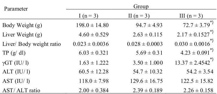
TABLE 1. Body and liver weight, liver/body weight ratio, TP and non-functional plasma enzymes: GT, AST, ALT, and AST/ ALT ratio
in experimental.
Parameter Group
I (n = 3) II (n = 3) III (n = 3)
Body Weight (g) 198.0 ± 14.80 94.7 ± 4.93 72.7 ± 3.79*)
Liver Weight (g) 4.60 ± 0.529 2.63 ± 0.115 2.17 ± 0.1527*)
Liver/ Body weight ratio 0.023 ± 0.0036 0.028 ± 0.0003 0.030 ± 0.0016*)
TP (g/ dl) 6.03 ± 0.321 5.69 ± 0.31 4.23 ± 0.091*)
GT (IU/ l) 1.63 ± 1.222 3.50 ± 1.00 0 13.37 ± 2.4542*)
ALT (IU/ l) 60.5 ± 12.28 54.7 ± 10.32 54.2 ± 3.54
AST (IU/ l) 118.0 ± 7.98 129.6 ± 16.75 122.5 ± 15.82
AST/ ALT ratio 2.00 ± 0.384 2.39 ± 0.189 2.26 ± 0.158
*)p<0.05 difference is statistically significant
Liver weight at the end of the experiment was significantly decreased in treated group compared to control (p=0.00); but Liver/ Body weight ratio was significantly increased in treated group compared to control (p=0.00). Varied factors may be involved in the enlargement of the liver, and up to this time only a few are known. The increase in the percentage of water, as found by McEwen and Haven, although only in limited amounts, seems to be important. It suggests the possibility of systemic changes in the cytoplasm. The suggestion of Yeakel that the increase in liver size induced by hypertrophy leading to an increased protein anabolism is still have to be proved. Histological investigations indicate that the increased weight of the liver may be explained in part by a mitotic activity of the liver.13The damage of the liver caused by iron overload and CCl4was evident by the alteration in serum marker enzymes concentration beside the clinical signs and histopathology, however, its exact mechanisms remain unclear.14Iron is essential for life but iron overload was shown to be toxic and potentially fatal. Hepatotoxicity is the most common finding in iron over-load because liver is the main recipient of the excess iron.12However, it is known that when iron is administered after the development of liver dysfunction, fibrosis of the liver is accelerated and increases the incidence of liver cirrhosis.15
The total protein after treatment with hepatocarcinogenic agent (FeSO43,5% and CCl4
0,1 ml/ kgBW) were significantly lower compared with those of the control group (p = 0.00). These proteins are important liver function marker. The concentration of plasma protein represents the nutritional status, which is one of the factors affecting the state of health of the animal.16Protein turnover is generally decreased with advanced liver disease, mostly so in hepatic coma patients where protein synthesis is only one third to one half of that observed in normal individuals.17The liver is considered to be the major source of plasma proteins, damage to this organ leads to decrease of total protein.18-20Since total protein, globulin and albumin are averagely responsive to total protein intake that the concentration of serum protein at any given time is a function of the nutritional status, water balance and other factors affecting the state of health of the animal.21
markers of serious hepatic dysfunction or damage.22 In most of liver diseases, both malignant and nonmalignant, GT estimation has been reported to be a sensitive but nonspecific indicator of the disease. Recent studies from this laboratory have shown a rise in activity of GT in patients with head and neck cancer, as well.23
Activity of ALT and AST have been widely used as sensitive laboratory parameters in clinical practice to evaluate the degree of liver injury. Alanine aminotransferase is an enzyme present in hepatocytes (liver cells), and it leaks into blood when liver cells are damaged.Alanine aminotransferase rises dramatically in acute liver damage (such as viral hepatitis and paracetamol overdose) and during liver inflammation. Aspartate aminotransferase is similar to ALT in that it is another enzyme associated with liver parenchymal cells. It is raised in acute liver damage and is also present in red cells and cardiac muscle. Observed values in this experiment showed there were no significant differences in the values of AST and ALT (p = 0.62, and 0.67 respectively). The serum values of these enzymes do not correctly reflect the degree of hepatic cell necrosis. Elevated activity of AST and ALT may be observed when cells containing these enzymes are injured or the permeability of cell membranes increases. Although serum levels of both ALT and AST are elevated when liver cells are injured, the degree of elevation is not parallel to the degree of injury.24The mechanism of the elevation is affected by many factors, such as etiology of the liver disease or severity of the liver cell necrosis. The ratio of AST to ALT is a useful parameter, which can predict the severity of liver disease.25 Observed values of AST/ALT ratio in experimental group was also not significantly different compared with those of the control group (p=0.26). When the ratio is over 2.0, alcoholic liver disease is strongly suspected. The discrepancy in serum AST and ALT remains controversial.24
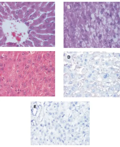
Histopathological changes
CONCLUSION
It was concluded that p-53 mutation was not detected in Rattus norvegicus rats that induced hepatocarcinogenic agent (FeSO4and CCl4) for 3 weeks and hepatic injury was still encountered in a pause of chemical hepatocarcinogen induced showed that the recovery was not complete.
ACKNOWLEDGMENT
The author would like to thank Totok Utoro, dr., DMedSc, S.PA(K) for his support to interpret the histological and immunohistochemical assessment.
REFERENCES
1. Anand BS. Cirrhosis of liver. West L Med 1999;171:110-15.
2. Aisen P, Enns C, Wessling-Resnick M. Chemistry and
biology of eukaryotic iron metabolism. Int J Biochem Cell Biol 2001;33:940–59.
3. Yajun Z, Hongshan C, Baoxi S, Dengbing Y, Jianhua S, Xinshun G, Li Y, Yi C. Translocation of Bax in rat hepatocytes cultured with ferric nitrilotriacetate. Life Sci 2005;76:2763–72.
4. Brittenham GM. Disorders of iron metabolism: iron
deficiency and overload. In: Hoffman R BE, Shattil SJ, Furie B, Cohen HJ, Silberstein LE, McGave P., ed. Hematology: Basic Principles and Practice 3rded. New
York: Churchill Livingstone, 2000:397-428.
5. Aisen P, Enns C, Wessling-Resnick M. Chemistry and
biology of eukaryotic iron metabolism. Int J Biochem Cell Biol 2001;33:940-59.
6. Kohgo Y, Ikuta K, Ohtake T, Torimoto Y, Kato J. Body iron metabolism and pathophysiology of iron overload. Int J Hematol 2008;88: 7-15.
7. Brattin WJ, Glende EA Jr, Recknagel RO. Pathological mechanisms in carbon tetracholoride hepatotoxicity. J Free Radic Biol Med 1985;1(1):27-38.
8. Pe´rez Tamayo R. Is cirrhosis of the liver experimentally produced by CCl4and adequate model of human cirrhosis? Hepatology 1983;3:112–20.
9. Darjono, Sleight SD, Stowe HD, and Aust SD. Vitamin A status, polybrominated biphenyl (PBB) toxicosis, and common bile duct hyperplasia in rats. Toxicol Appl Pharmacol 1983;71:184-93.
10. Murray RK, Granner DM, Mayes PA, and Rodwell VW.
Harper ’s Illustrated Biochemistry. 25 ed.
London:Prentice-Hall International Inc., 2003.
11. Benhar M, Engelberg D, Levitzki A. ROS,
stress-activated kinases and stress signaling in cancer. EMBO Rep 2002;3:420– 25.
12. Bonkovsky HL. Iron and the liver. Ame J Med Sci
1991;301(1):32-43.
13. Annaua E, Manginelli, Roth A. Increased weight and mitotic activity in the liver of tumor-bearing rats and mice. Cancer Res 1951:304-6.
14. Kumar PV, Sivaraj A, Elumalai EK, Kumar BS. Carbon tetrachloride-induced hepatotoxicity in rats - protective role of aqueous leaf extracts of coccinia grandis. Int J Pharm Tech Res 2009;1(4):1612-5.
15. Shiraishi T, Maekawa I, Komatsu T, et al. Experimental studies on the relation between hepatic fibrosis and iron deposition of the liver. Nippon Naika Gakkai Zasshi 1968;57: 211-9.
16. Edem DO. Haematological and histological alterations induced in rats by palm oil – containing diets. Eur J Sci Res 2009;32(3):405-18.
17. Matkowitz R, Hartig W, Junghans P, Slowig M, Bornhak H, Faust H et al. Protein turnover in liver failure -determination using the stable isotope 15N. Leber Magen Darm 1984; 14:83-9.
18. Egbal SA, Samia ABE, Iman ME, Inaam ELD.
Serobiochemical changes associated with highly pathogenic avian influenza in chickens. Res J Animal Vet Sci 2007;2:53-5.
19. Somi MH, Rahimi AO, Moshrefi B, Rezaeifar P, Maghami JG. Nutritional status and blood trace elements in cirrhotic patients. Hepatitis Monthly 2007;7(1):27-32.
20. Meshkibaf MH, Ebrahimi A, Ghodsi R, Ahmadi A.
Choronic effects of lamotrigine on liver function in adult male rats. Indian J Clin Biochem 2006;21(1):161-4. 21. Igwebuike JU, Anugwa FOI, Raji AO, Ehiobu NG, Ikurior
SA. Nutrient digestibility, haematological and serum biochemical indices of rabbits fed graded levels ofAcacia albidapods. J Agri Biol Sci 2008;3(4):33-40.
22. Atakisi E, Karapehlivan M, Atakisi O, Kontas T and Marasli S. Adenosine deaminase and biochemical liver function tests in the dermatophytic cattle. Bull Vet Inst Pulawy 2006;50:481-3.
23. Seth S , Ravi B, Randhawa HS, Chillar N, Lal H. Serum gamma glutamyl transpeptidase in breast cancer. Indian J Clin Biochem 1996;11(1):49-51.
24. Tameda M, Shiraki K, Ooi K, Takase K, Kosaka Y, Nobori T, et al. Aspartate aminotransferase-immunoglobulin complexes in patients with chronic liver disease. World J Gastroenterol 2005;11(10): 1529-31
25. Powell LW, Bassett MK, Halliday JW: Hemochromatosis: 1980 update. Gastroenterol 1980;78: 374-81.
26. Furness PN. The use of digital images in pathology.J Pathol1997;183:253–63.