Melanin, Energy and the Cell Diabetes Obes Int J
Melanin, Energy and the Cell
Herrera AS*
Human Photosynthesis Research Center, México
*Corresponding author: Arturo Solis Herrera, MD, PhD, Human Photosynthesis Research Center, Aguascalientes, México, E-mail: [email protected]
Abstract
In the 19th century, researchers first realized that metabolism can be viewed as a network of connected biochemical
reactions. Apparently, enzymes catalyze these reactions, accelerating the rate at which they take place; however, our
body is not merely a sack of enzymes. Several of the most prevalent diseases in modern society, as diabetes,
cardiovascular disease and obesity involve disruptions of metabolic processes. In patients with diabetes mellitus, this
results in an excessive amount of glucose in the blood. The first scientific article dating back to the 17th century, until
today; almost six million articles have been published about metabolism. In spite, new insights continuously
published, providing pieces of the puzzle about the mechanisms of metabolic processes; we cannot understand
metabolism if only study the individual parts of the network reactions. It is something like trying to understand the
complex functioning of our solar system with isolated photographs from the Moon, the Sun, or the planets.
To understand the metabolism, which means continuous change; It is necessary to study the network as an
ever-changing whole, including the energy needed. Any change requires energy, because energy is defined as everything
that produces a change. Therefore, cellular metabolism, involving continuous changes means that it requires the
presence of power on a continuous basis.
So far, glucose is considered as the energy source par excellence of eukaryote cell. But the models that have been
created based on this dogma, have resulted in the creation of databases whose metabolic pathways are controversial
by 97%.The lack of consensus between different metabolic pathway databases is substantive and cannot be explained
adequately.
We think that the discovery of the unsuspected intrinsic capacity of the melanin of transform the light, visible e
invisible, in chemical energy by means of the dissociation of the molecule of the water, as the chlorophyll in plants;
this is: producing chemical energy which is carried by the hydrogen (H2). It will bring the appropriate molecular logic
to implement more coherent, more consistent metabolic models. The discerning that the true role of glucose is only as
a source of carbon chains with which our body synthesizes the biomolecules that needs to operate, to maintain shape;
and the light visible and invisible as the true source of power, thanks to the melanin; means the birth of a new
biochemistry.
Keywords
: Energy; Cell; Melanin; Water Dissociation; Hydrogen; GlucoseReview Article
Volume 2 Special Issue 1
Received Date: March 04, 2017
Herrera AS . Melanin, Energy and the Cell. Diabetes Obes Int J 2017, 2(S1): cardiovascular, and neurological disease represent major global health issues.
Type 2 Diabetes is associated with both impaired insulin action at target tissues and impaired insulin release, and is evident early in the course of the disorder. Both type 1 (autoimmune) and type 2 of diabetes are associated with similar long-term complications. forgotten in the lapse of a few years, something that can do very well, because millions of years, has done millions of times.
It has been speculated much trying to explain this rare phenomenon, involving reactive species of oxygen (ROS), to mitochondrial dysfunction; the role of the potential of membrane, etc., but the ethiopathogeny of the diabetes continues without be understood, as it demonstrated by the ascending rhythm of the incidence and prevalence of diabetes mellitus worldwide, as example of metabolic disorder; especially in the countries in development.
Energy and the Cell
It is a long-lasting mystery how do living organism capture the energy available through two mainforms: (1) degradation of organic matter and (2) absorption of light (Photosynthesis). Thereafter, through a poorly understood energy coupling, the chemical energy released is harnessed to performance of useful work such as biosynthesis, membrane transport and movement. In Biochemistry, this is the most perplexing problem.
The Chemiosmotic theory of biological energy transduction proposed by Peter Mitchell in 1961 transformed bioenergetics from chemical to electrical [1]. Panoply of membrane functions was connected by way of ion currents. The molecular mechanisms that underlie energy transduction and results in transport of solutes and water into and out the cells, homeostatic regulation and signaling, and mechanical work; are still largely unknown. In spite Mitchell´s insights into the nature of biological energy coupling, it often seems that our appreciation of the Chemiosmotic theory remains
incomplete, even superficial. The mechanisms of integration of lifeless molecules into living systems are controversial; therefore, physiological and medical applications have been limited.
In the first half of past century, Fritz Lipmann [2] proposed the metabolic generation and utilization of phosphate energy. Thereby, the source of biological work could be ATP or some related phosphoryl donor, which participates chemically in the reaction that it supports.
Thereafter, the concept about function of great metabolic highways (respiration and photosynthesis) is to generate and maintain ATP supply, but no one knew how ATP is produced. And mitochondria related Mitchell´s Chemiosmotic theory based in bacteria, not in mitochondria; appeared. Briefly, the respiratory chain of redox catalysts is so arranged within and across the bacterial plasma membrane that, as electrons wend their way to oxygen, protons are translocated out of the cytoplasm.
Peter Mitchell applied his experience in bacteria to mitochondria. His theoretical model was that plasma membrane forms a closed vesicle that is relatively impermeable to protons (double membrane?), thereby, proton translocation generates an electrical potential across the membrane, with the interior negative; in time, a pH difference may also arise, with the interior alkaline. Protons at the external surface find themselves at a higher electrochemical potential than those of cytoplasm. Therefore, they are subject to a pull derived from both the pH gradient and the electrical potential, named as proton motive force (PMF); it pulls proton backs across the membrane, down the electrochemical gradient established by respiration.
The ATP synthase provides a pathway that allows protons to traverse the membrane. It is so articulated as to couple the downhill of protons to the uphill synthesis of ATP and Pi. Energy is conserved, not chemically but by the proton-motive force (PMF), or proton potential. To Mitchell, proton circulation could support also other kinds of membrane work. Any membrane-localized function can be coupled to the proton circulation whereas the appropriate molecular device allows passage to protons and harnesses their downhill flow to performance of work.
Herrera AS . Melanin, Energy and the Cell. Diabetes Obes Int J 2017, 2(S1): 000S1-004.
Copyright© Herrera AS.
The Unsuspected Intrinsic Property of
Melanin to Dissociate the Water Molecule,
as Chlorophyll in Plants
Suppose by a minute that the protons that mentions Mitchell, those subatomic particles that is found in the nucleus of any atom, with positive electric charge, equal but opposite to it of the electron, and whose mass is of only 1.673 x 10⁻²⁷ g, this is: something less that the mass of the neutron; and the hydrogen only has one proton; came from the dissociation of the water, and not from glycolysis; this is: thanks to the melanin.
Melanin intrinsic property to dissociate the water molecule, was found during an observational study about the three main causes of blindness: Age-related macular degeneration, diabetic retinopathy, and glaucoma. Our hypothesis work was try to find morphological characteristics of the optic nerve blood vessels that eventually could be useful as indicators of early disease. However, optic nerve in humans is so small, 1200 microns or 12 human hairs together.
Thereby image magnification needed to the
achievement of an adequate study of these minute blood vessels was substantive.
At those levels of magnification, besides
morphological characteristics of the optic nerve blood vessels, melanin was also visible, and in few weeks come up to be a variable in study, in addition to the blood vessels. The insistence of Nature in place melanin nearby optic nerve in 6000 studied patients from 1990-2002, draw powerfully our attention, due to nature just insist in important things (Figure 1).
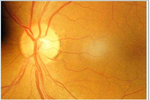
Figure 1: The photography of the ocular fundus, shows the optic nerve (blue arrow), the macula (yellow arrow), and the melanin in the temporary edge of the optic nerve (black arrow).
In the 6000 patients who comprised the study (1990-2002), which was descriptive in nature; all had melanin at the edge or nearby of the optic nerve.
Briefly, we had an initial axiom: The human´s optic nerve has melanin nearby always. As the studio went ahead, eventually a second axiom was identified: a greater amount of melanin, less number and size of blood vessels, and vice versa. And trying to explain the apparent anti-angiogenic effect of the melanin, after several years, the enigma was deciphered: the higher levels of oxygen usual in the pigmented tissues. What gave origin to another axiom: to greater amount of melanin, greater level of oxygen in the tissues, and vice versa.
Reviewing cell biology, we could not find a mechanism that could explain the high levels of oxygen in pigmented tissues, and in the case of the eye, specifically in the uveal tract, the difference becomes 34 or 37%, so it had to be a very efficient mechanism and that works consistently. But we could not find any molecule that is providing such a quantity of oxygen. The only option that remained to us was water that the eye contains, as it includes almost 90% of the eyeball.
Therefore, the vitreous body, with its great content of water was the ideal source of oxygen, but the water not donates oxygen for free, it is requiring first the dissociation of the water molecule, so the possibility of that the melanin could have that capacity appeared in our mind.
We review the literature looking for indirect data that support our hypothesis, and find them. For example, our prediction was: a greater amount of melanin, lower levels of hemoglobin, and vice versa. And thus was the difference between the people of fair skin and the people of dark skin reaches until 20 grams of hemoglobin by liter. Although the authors of the article it attributed to a bad nutrition [3], our explanation was that if the tissues had greater amount of melanin, then had greater capacity of dissociate the water, what ended in a greater availability of oxygen, thereby less need of hemoglobin.
After twelve years, we can have understood that melanin possesses the intrinsic property to dissociate the water molecule, this is: from liquid to gas, and vice versa. The reaction can be written as follows:
2H2O (Liquid)→ 2H2 (Gas) + O2 (Gas)→2H2O (Liquid) +
The main product of this reaction is molecular hydrogen (H2), the energy carrier by excellence in
Herrera AS . Melanin, Energy and the Cell. Diabetes Obes Int J 2017, 2(S1): 000S1-004.
Copyright© Herrera AS.
Therefore, we must rethink our concepts about the role of glucose in the human body. Glucose is not a source of energy, is a source of biomass. With atoms and carbon chains of carbohydrates, our body knows very well, since the beginning of time, the body synthesizes 99% of the bimolecular that make us up. Remember that all they have a backbone of chains of carbon. Our body as well-known glucose, which handles it very well, because it splits it very accurately, distorts it, combined with other elements, it adds other chains of carbon, etc.
But the energy, defined as all that that produces a Don't forget him. What happens is that when the levels of chemical energy that our body requires to function well, as it has done millions of years, millions of times, are not suitable, our body cannot do what it does very well. Levels of chemical energy needed by our body are the same that has taken over all the evolution, all the creation.
Then, how would be the Molecular
Biology?
Theoretically, about 98% of the inhaled oxygen is consumed by the mitochondria; and say theoretically because the blood cannot transport the atmospheric oxygen to the inside of the body and distribute it in the tissues [4]. If our organism no need mitochondria or its functions are different to Peter Mitchell conception, then we would have no need of the oxygen transfer machinery of the lungs, a long-lasting misconception in biology; neither red cells, hemoglobin; nor the circulatory system that delivers oxygen to tissues.
The organization of food intake, digestion and processing is not designed, primarily; to supply substrates destined for mitochondrial oxidation. Glucose is source of carbon atoms, carbon chains; which our body synthesizes 99% of our biomolecules destined to the replenishment of used compounds, but glucose cannot provide the energy that its own metabolism requires.
Physiology of higher organisms is not dictated by the demand of our mitochondria for a supply of oxygen, due to every cell has melanosomes, and the cell is highly energy in symmetric form, in all directions.
With the supply of energy provided by mitochondria as ATP, the evolution of higher organisms would not have been possible, not so melanin, because it can dissociate the water molecule both in and out eukaryotic cell.
Mitochondria are not essential to battle against entropy, necessary to maintain live. Homothermic
organisms characteristically have low or any
mitochondria. Thereby, mitochondria play a central role as a heat-generating mechanism in non-shivering thermo genesis in mammals, especially in the youngest [5], and probably as accurate control mechanism for
phosphate levels, compounds thermodynamically
stables, but biochemically unstable.
secretion of hormones and neurotransmitters;
processes central to sophisticated life, all them can be explained by the chemical energy that comes from melanin in form of H2 and e-.
Mitochondrial dysfunction has been implicated in all the major neurodegenerative diseases (Parkinson, Alzheimer, motoneuron disease -Lou Gehrig´s disease or amyotrophic lateral sclerosis). However, the body processes are astoundingly accurate, therefore when chemical energy levels (from melanin) are not the same the body have been along evolution, a generalized failure appears, and mitochondria is not an exception.
Herrera AS . Melanin, Energy and the Cell. Diabetes Obes Int J 2017, 2(S1): 000S1-004.
Copyright© Herrera AS.
Mitochondrion does not generate energy as electrons are passed from donors at lower to acceptors at higher redox potential, through various proteins complexes [6]. Bioenergetically it is not possible. Energy cannot be created or destroyed, simply is transformed. In other words: the energy that comes from melanin, in form of H2 and e-, are the base of mitochondria form and
function (Figure 3).
All diseases are multi-causals, therefore, when the mitochondria are damaged, in greater or lesser degree, also we can find data from alteration in other structures of the cell, due to when energy is altered, the fault is widespread. Recall that cell uses energy in many ways.
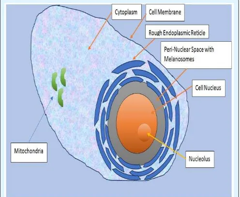
Figure 3: Schematic representation of the peri-nuclear space, the main location of the granules of melanin called melanosomes in mammals.
Peri-nuclear space is completely surrounded by the rough endoplasmic reticulum, probably to capture all the possible H2 and e- that melanin produces. Hydrogen
gas bubbles, that are not visible in solution; follows the Laws of simple diffusion, and gradually extending over cytoplasm all, due to H2 dos not combine with water,
reaching until the last corner of eukaryotic cell, i.e. mitochondria, Golgi apparatus, Lysosomes, Smooth endoplasmic reticule; cell membrane, etc.
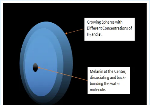
The location of the granules of melanin to the inside of the cell is strategic: in the peri-nuclear space. Energy melanin-releasing way of growing areas extends symmetrically in all directions, which, being surrounded by the granules of melanin, cell nucleus, the large intracellular organ, becomes a zone of high energy to the merge of growing energy spheres that melanin granules release constantly, day and night (Figure 4).
Figure 4: Drawing showing a segment of peri-nuclear space, the main location of melanin granules.
At the top is the rough endoplasmic reticulum, and lower left part represents the cell nucleus. It is no coincidence that the two organelles with increased metabolic activity of the cell, are so close to melanin, because that way they capture efficiently the energy that constantly emanates from it, both day and night.
Growing energy spheres, do so according to the laws of simple diffusion, so, as they deviate from the melanin and reach the cell membrane, its content of H2 and e -
decreasing the various cellular structures are capturing energy to molecular hydrogen. By the way, the best antioxidant that referred to is H2, because it is able to
reduce oxygen, forming water.
The size of the hydrogen molecule is very small, which allows that intracellular (Figure 5) permeates practically through any structure.
Herrera AS . Melanin, Energy and the Cell. Diabetes Obes Int J 2017, 2(S1): 000S1-004.
Copyright© Herrera AS.
Conclusion
From now in ahead, melanin has emerging as central player in the cell bioenergetic scheme.
Metabolic processes are constantly taking place in our body no matter whether we eat, sleep, or exercise. Food is not broken down to generate energy, instead to generate body building blocks.
Ascribed energy to the glucose, ATP or the mitochondria, given as results models too tangled, almost unrecognizable; as evidenced the fact that if we simply combine the various descriptions of the human metabolism provided by different databases available [7], and that despite having been implemented through the combined efforts of numerous groups of experts; It is impossible to obtain a consistent or at least integrated metabolic scheme since the conflicting information is not resolved or they may not be detected or eliminate errors.
Metabolic alterations in diabetes are not limited to single failure of insulin secretion or defective receivers. The problem is much more complex. Glucose found even in blood types, because we know that every cell in our body is thickly decorated by several layers of compounds derived from glucose, known as glucans. And each layer is different between itself and more still, because the neighboring cell has other different layers in different order.
The study of the metabolism of the glucose has proven to be a formidable challenge. It is easier to study proteins. But we now understand that the glucose only provides the building blocks of organic molecules, but does not provide the energy that requires its own metabolism.
Having understood that the energy needed by our body's metabolism comes to light and not food, our concepts should change radically; but at the same time, it opens new avenues for the study of the biology of the disease, which will allow overcoming the current slump, by placing the foundations of a new era in biochemistry.
Acknowledgements
This work was generously funded by Human Photosynthesis® Research Center.
References
1. Harold, Franklin M (2001) Gleanings of
Chemiosmotic eye. Bioessays 23(9): 848-855
2. Lipmann F (1941) Metabolic generation and
utilization of phosphate bond energy. Adv Enzymol 1: 99-162.
3. Gran, Stanley M. Smith, Nathan J, Clark, Diane C (1975) Lifelong Differences in Hemoglobin Levels Between Blacks and Whites. JAMA 67(2): 91-96.
4. Solis Herrera, Solis Arias A (2013) The Odyssey of Atmospheric Oxygen in their futile attempt to Reach the Interior of the Cell. Int J Health Res Innov 1(3): 37-52.
5. Duchen RM (2004) Roles of mitochondria in health
and disease. Diabetes 53(S1): S96-S102.
6. Sivitz WI, Mark AY (2010) Mitochondrial
dysfunction in diabetes: from molecular
mechanisms to functional significance and
therapeutic opportunities. Antioxidants and Redox Signaling 12(4): 537-577.