The cortico-basal ganglia-thalamocortical circuit with
synaptic plasticity. II. Mechanism of synergistic modulation
of thalamic activity via the direct and indirect pathways
through the basal ganglia
Isabella Silkis *
Neurophysiology of Learning Laboratory,
Institute of Higher Ner6ous Acti6ity and Neurophysiology of the Russian Academy of Sciences,117865,
Butlero6a5a str.Moscow Russia
Received 25 July 2000; accepted 28 August 2000
Abstract
A possible mechanism underlying the modulatory role of dopamine, adenosine and acetylcholine in the modifica-tion of corticostriatal synapses, subsequent changes in signal transducmodifica-tion through the ‘direct’ and ‘indirect’ pathways in the basal ganglia and variations in thalamic and neocortical cell activity is proposed. According to this mechanism, simultaneous activation of dopamine D1/D2 receptors as well as inactivation of adenosine A1/A2A receptors or muscarinic M4/M1 receptors on striatonigral/striatopallidal inhibitory cells can promote the induction of long-term potentiation/depression in the efficacy of excitatory cortical inputs to these cells. Subsequently augmented inhibition of the activity of inhibitory neurons of the output nuclei of the basal ganglia through the ‘direct’ pathway together with reduced disinhibition of these nuclei through the ‘indirect’ pathway synergistically increase thalamic and neocortical cell firing. The proposed mechanism can underlie such well known effects as ‘excitatory’ and ‘inhibitory’ influence of dopamine on striatonigral and striatopallidal cells, respectively; the opposite action of dopamine and adenosine on these cells; antiparkinsonic effects of dopamine receptor agonists and adenosine or acetylcholine muscarinic receptor antagonists. © 2001 Elsevier Science Ireland Ltd. All rights reserved.
Keywords:Basal ganglia; LTP; LTD; Dopamine, Adenosine; Movement disorders
www.elsevier.com/locate/biosystems
1. Introduction
The numerous movement disorders are ex-plained by functional model of the cortico-basal ganglia-thalamocortical motor loop (DeLong, 1990). In humans, lesions at the basal ganglia levels lead to hypokinetic and hyperkinetic dys-* Corresponding author. Tel.:+7-95-3344345; fax: +
7-95-3388500.
E-mail address:[email protected] (I. Silkis).
functions attributive to Parkinson’s and Hunting-ton’s disease. Diverse changes in signal transduc-tion through the basal ganglia can be caused by modifications in the efficacy of cortical inputs to the striatum, the entry structure of the basal ganglia. Plasticity of excitatory corticostriatal in-puts, such as long-term potentiation and depres-sion (LTP, LTD), involves convergent actions of glutamate, dopamine, adenosine and acetylcholine (Calabresi et al., 1992, 1998, 1999a). However, mechanism underlying different changes in signal transduction through the basal ganglia, produced by modulatory neurotransmitters, remains un-clear. The aim of this work has been to analyze the feasible mechanisms of variations of neuronal activity in the cortico-basal ganglia-thalamocorti-cal loop, triggered by modification of corticostri-atal synapses. Earlier suggested modification rules (Silkis, 2001) were used for this analysis. Accord-ing to these rules, an activation of dopamine D1 and D2, or adenosine A2A and A1, or muscarinic M1 and M4 receptors on striatal cells promotes the induction of LTP and LTD, respectively. Based on these rules we expected that modulatory neurotransmitters variously modify the neuronal interactions in the ‘direct’ and ‘indirect’ pathways through the basal ganglia, since different receptor types are expressed on striatal cells, originative these pathways.
2. Functional organization of the cortico-basal ganglia-thalamocortical ‘motor’ circuit
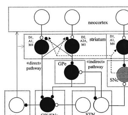
The main features and character of operation of the cortico-basal ganglia-thalamocortical ‘mo-tor’ loop are widely described (Alexander and Crutcher, 1990; DeLong, 1990; Parent and Hazrati, 1995a,b). Signals initiated in the precen-tral motor areas activate the inhibitory spiny cells in the putamen. This part of the striatum is one of the ‘input’ structures of the basal ganglia. Striatal cells inhibit ‘output’ neurons of the basal ganglia which are possess in the motor portion of the internal segment of the globus pallidus (GPi) and substantia nigra pars reticulata (SNr). This path-way is known as ‘direct’ (Fig. 1). Another group
of striatal cells inhibits the external segment of the globus pallidus (GPe) which inhibits excitatory cells of the subthalamic nucleus (STN) and in-hibitory cells of GPi. This pathway is known as ‘indirect’ (Fig. 1). One segment of STN is recipro-cally connected with GPe, while another projects to the output basal ganglia cells (Parent and Hazrati, 1995b). Possibly, the striatum more effec-tively reduce the activity of GPe than GPi, since striatal terminals form 85 and 31% of all in-hibitory contacts on the soma of GPe and GPi cells, respectively (Shink and Smith, 1995). GPe, which makes 47% of contacts on the soma of GPi cells (Shink and Smith, 1995) can strongly inhibit GPi. Cells of GPi/SNr inhibit the ventrolateral thalamus, which activates the precentral motor areas. Thalamic cells excite those spiny striatal neurons, that influence thalamic activity through GP/SNr (Funaki et al., 1998; Parent and Hazrati, 1995a). Terminals from ventrolateral nucleus
Fig. 1. The scheme of excitatory and inhibitory connections in the cortico-basal ganglia-thalamocortical loop. GPi and GPe, internal and external segments of the globus pallidus, respec-tively; SNr and SNc, substantia nigra pars reticulata and compacta, respectively; STN, subthalamic nucleus; D1and D2,
receptors sensitive to dopamine; A1and A2A, receptors
sensi-tive to adenosine; M1 and M4, muscarinic receptors sensitive
make synaptic contacts with cortical neurons, projecting to the striatum (Zin-Ka-leu et al., 1998). Thus, the thalamo-cortico-striatal loop is formed by monosynaptic connections. Striatoni-gral/striatopallidal cells, which are located in the matrix, express dopamine D1/D2 receptors (Loschmann et al., 1997). Neurons of striosomes, projecting to the dopaminergic cells of substantia nigra pars compacta (SNc) contain D1 receptors that are colocolized with muscarinic M4receptors (Harrison et al., 1996). Adenosine A1(A2A) recep-tors are specifically expressed on striatonigral (striatopallidal) cells, where they are colocolized with D1(D2) receptors (Ferre et al., 1997; Fuxe et al., 1998; Svenningsson et al., 1999). Muscarinic M1receptors are more often located on striatopal-lidal cells, while coexpression of D1, and M4 receptors on striatonigral neurons has been demonstrated (Wang and McGinty, 1996). Thus, the sign of modification (LTP or LTD) of synapses on spiny neurons giving rise to the direct and a indirect pathways through the basal gan-glia, depends on the activating D1/A1/M4 and D2/A2A/M1 receptors, respectively (Fig. 1). It is clear that in the absence of dopamine, adenosine and acetylcholine the cortex disinhibits the thala-mus through the direct pathway, while the indi-rect pathway is inhibitory (Fig. 1). Any persistent impact of modulatory neurotransmitters on the efficacy of corticostriatal synapses can lead to a shift in the balance between neural interactions in the direct and indirect pathways and subsequent variations in the thalamic and cortical functioning.
3. Synergistic changes in the basal
ganglia-thalamic transmission due to the opposite modification of cortical inputs to striatonigral and striatopallidal cells
According to earlier suggested modification rules (Silkis, 2001), an activation of dopamine D1 (D2) receptors leads to LTP (LTD) of excitatory cortical inputs to striatonigral (striatopallidal) cells and augmenting (lowering) GABA release into GPi/SNr (GPe). Actually, precisely this type of influence of D1, and D2 receptors on GABA
release by striatonigral and striatopallidal cells has been obtained (Hossain and Weiner, 1995; Ferre et al., 1996a; Harsing and Zigmond, 1997; Hooper et al., 1997; Wooten, 1997). It has also been shown that these changes lead to variations in neuronal activity in the direct and indirect pathways. We assume that the proposed role of dopamine in LTP and LTD induction can under-lie ‘excitatory’ and ‘inhibitory’ effect of dopamine on striatal cells, projecting into the GPi/SNr and GPe, respectively (DeLong, 1990). If an activation of A1 (A2A) receptors promotes LTD (LTP) in striatonigral (striatopallidal) cells (Silkis, 2001), then GABA release into GPi/SNr (GPe) can be reduced (strengthened) by adenosine. Indeed, A1 receptor activation antagonistically influenced GABAergic striatal input to the entopeduncular nucleus, the homologue of the non primates GPi (Ferre et al., 1996a). In contrast, an agonist of A2A receptors caused an augmentation of GABA release into the GPe (Corsi et al., 1999). This means that the sign of modification of each synapse depends on the difference between parameters of current and prior activation (Silkis, 1998). Apparently for this reason, both depression and potentiation of activity of SNr neurons have been observed after an injection of a selective D1, receptor agonist as well as D2 receptor agonist (Akkal et al., 1996).
In spite of suggested modification rules (Silkis, 2001) it seems paradoxical that the coactivation of D1 and D2 receptors resulted in striatal LTD (Calabresi et al., 1992). We assume that this effect is the sequence of reciprocal inhibition between striatal neurons (Fig. 1) that occurs through both GABAAand GABABreceptors (Shi and Rayport, 1994). Therefore, the efficacy of inhibitory con-nections can be modified (Silkis, 1998, 2000). An inducing LTP (LTD) in striatonigral (striatopalli-dal) cell caused by activation of D1(D2) receptors, can lead to the rising (lowering) the inhibitory influence on striatopallidal (striatonigral) cell. This should promote a decrease (enlargement) in postsynaptic Ca2+ and cAMP concentration and
receptor activation, has been strengthened by D1 receptor agonists (Ruskin et al., 1998). We assume that reciprocal inhibitory connections can also enhance the opposite modification of cortical in-puts to striatonigral and striatopallidal cell through the net interactions between A1 and A2A receptors, A1and D2receptors, A2Aand D1 recep-tors. Actually, it had been shown, that the effect caused by D1 receptor agonist has been strongly potentiated by inactivation of A2A receptors (Fenu et al., 1997; Le-Moine et al., 1997). Interac-tion between A2A and D1 receptors produced by transneuronal interactions has been obtained in behavioral experiments too (Ferre et al., 1997). Modifiable inhibitory reciprocal connections could be adjunct to the models of recurrent net-works with Hebbian learning such as the model for cortical and brainstem premotor circuits (Hua et al., 1999).
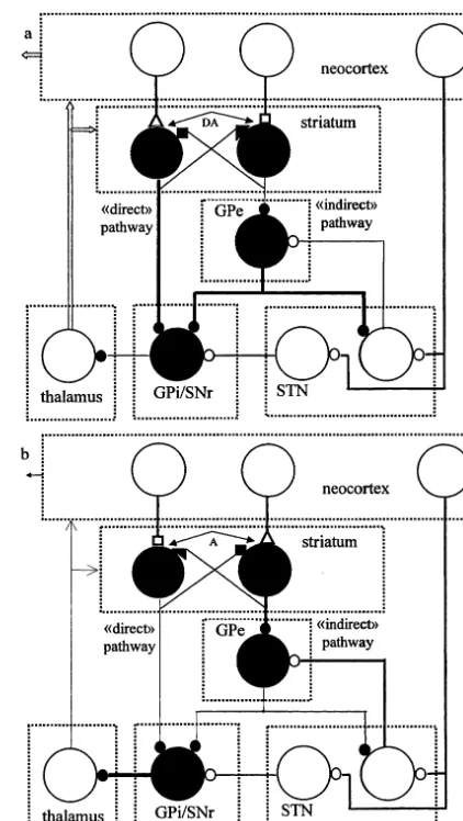
The presence (absence) of dopamine in the striatum can intensify (reduce) an inhibition of GPi/SNr as directly, by striatonigral cells in con-sequence of LTP (LTD) in cortical inputs, as indirectly by GPe neurons, which become more weakly (strongly) inhibited by striatopallidal cells due to LTD (LTP) of their cortical inputs (Fig. 2a). Thus, the synergistic decrease in the activity of output inhibitory neurons of the basal ganglia could be caused by dopamine. A change in GPi/ SNr activity caused by additional dopamine (deficit of dopamine) in the striatum must lead to a disinhibition (inhibition) of the thalamic and neocortical cell activity. Indeed, an increase and decrease in the amount of dopamine in the stria-tum resulted in strengthening and weakening tha-lamo-cortical inputs, respectively (Rodriguez and Gonzales-Hernandez, 1999). The presence (ab-sence) of adenosine in the striatum must lead to the opposite effects due to the promotion of LTD (LTP) on striatonigral cells and LTP (LTD) on striatopallidal cells (Fig. 2b). Since dopamine (adenosine) induced modification of corticostri-atal synapses synergistically influence the activity of output GPi/SNr cells, a conjunctive activation of D1and D2(A2Aand A1) receptors should cause more strong effects than those produced by acti-vation of one type of receptors. Actually, the selective agonist of D1 or D2 receptors produced
Fig. 2. Changes of synaptic transmission in the ‘direct’ and ‘indirect’ pathways through the basal ganglia in consequence of dopamine (a) and adenosine (b) induced modification of corticostriatal synapses. Small triangles and squares, potenti-ated and depressed synapses, respectively. Other designations are as in Fig. 1.
changes in firing rate of 23% or 17% of SNr neurons, respectively, while joint activation of both receptors resulted in changes of 70% of cells (Murer et al., 1997).
blockage of adenosine receptors together with ac-tivation of dopamine receptors on both striatoni-gral and striatopallidal cells will more strongly enhance the activity of the thalamus and neocor-tex. The same must be true for the joint activation of dopamine receptors and blockade of mus-carinic receptors. This is because M1– D2 recep-tors and M4– D1 receptors are coexpressed on striatopallidal and striatonigral cells, respectively. In line with modification rules (Silkis, 2001), do-pamine and muscarine oppositely influence the efficacy of a cortical input to a striatopallidal as well as to a striatonigral neuron, since an activa-tion of M1 and M4 receptors can promote the induction of LTP and LTD in these cells. Actu-ally, acetylcholine oppositely affects striatopalli-dal and striatonigral cells (Harrison et al., 1996; Wang and McGinty 1997).
Due to presence of excitatory thalamic afferents in the striatum (Fig. 1), an increase of thalamic activity can facilitate the firing rate of striatal cells. Additional excitation can promote LTP on striatonigral. cells, but impede LTD on striatopal-lidal neurons. The last effect must reduce thalamic and striatal activation. Thus, the excitation in the thalamo-basal ganglia-thalamic loop can be lim-ited. On the other hand, LTD induction on stria-topallidal cells, subsequent disinhibition of GPe neurons and augmented inhibition of STN cells can in turn decrease the excitation of GPe neu-rons (Fig. 2). Therefore, this local circuit can reduce inhibitory action of GPe not only on STN, but also on GPi/SNr, and thus decrease the disin-hibitory effect of dopamine on the thalamus through the indirect pathway. An activation of D1 receptors on spiny cells of striosomes and LTP induction must increase the inhibition of do-paminergic cells in SNc (Fig. 1) and cause a decrease of dopamine release in the striatum. The deficit of dopamine must impair corticostriatal LTP and decrease the inhibition of SNc. These effects can prevent the variation of dopamine concentration through a large range.
The tendency to bursting discharges and oscilla-tions has been found in corticostriatal and thala-mic cells (Magnin et al. 2000; Plenz and Aertsen 1996). Such synchronization must promote the modification of synapses in the striatum as well as
in the whole basal-ganglia-thalamocortical net, since it provides the fulfillment of the Hebbian rule (coincidence in the activity of pre- and post-synaptic neurons).
We have postulated that only receptors acti-vated by transmitter are modifiable (Silkis, 1998, 2000b). This postulate, which provides the fulfill-ment of the Hebbian rule and input specificity of LTP/LTD, is experimentally supported in differ-ent structures, including the basal ganglia (Cal-abresi et al., 1999b; Harsing and Zigmond, 1997; Matuszewich and Yamamoto, 1999). So, do-pamine and others neuromodulators, which are diffusely released into the striatum after arriving of excitatory signals from one or several cortical areas, can modulate the efficacy of those corticos-triatal synapses that have been simultaneously activated by others cortical areas. This effect may lead to the selection of signals and their ‘correc-tion’ depending on other ones. Such mechanism may underlie the one of the most important func-tions of the basal ganglia — the integration of information from different neocortical areas.
4. Correlation between effects emerging from proposed mechanism, and available data related to motor disorders and treatment of Parkinson’s and Huntington’s disease
receptors in caudate nucleus of the striatum in-creases (Augood et al., 1997). From the point of view of suggested mechanism, such fact should rise the probability of LTP induction on striatoni-gral cells and subsequent intensifying of a motor activity (hyperkinesia) through the direct path-way. It is known, that endogenous dopamine activates D2 receptors and does not activate D1, receptors (Harsing and Zigmond, 1997). We as-sume that LTD in striatopallidal cells, caused by D2 receptor activation, may promote a basic low-ering thalamic inhibition through the indirect pathway.
It follows from proposed mechanism, that the blockade of adenosine and/or acetylcholine sensi-tive receptors on striatal cells can facilitate LTP/ LTD induced by dopamine and thus reinforce disinhibition of thalamic and cortical activity. Ac-tually, a treatment of Parkinson’s disease by an-tagonists of adenosine and/or muscarinic receptors enhances the therapeutic effects caused by dopamine alone. Systemic administration of A1 receptor antagonist essentially potentiated a mo-tor activity caused by D1receptor agonist (Popoli et al., 1996), while A2A receptor antagonist, caf-feine, enhanced effects induced by D2 receptor agonist (Pollack and Fink, 1995). In the caudate nucleus A2A receptors are activated by endoge-nous adenosine. The model suggests that the pre-venting this activation can be useful in the treatment of hypokinesia. Indeed, the blockade of A2A receptors strengthened motor responses (Fuxe et al., 1998; Svenningsson et al., 1999), especially for animal with a low level of dopamine (Hauber et al., 1998), and caused a strong aug-mentation of a motor activity at small doses of L-dopa (Fenu et al., 1997). Moreover, A2A recep-tor antagonists administered alone produced an-tiparkinsonian effects that reached the level of efficacy of L-dopa but was less likely to elicit dyskinesias (Grondin et al., 1999).
As concern the antiparkinsonic effect of mus-carinic receptor antagonists, on the one hand, there are some proofs that it is linked to an excitatory action of cholinergic interneurons on those striatal cells that contain enkephalin (Alex-ander and Crutcher, 1990). These cells giving rise to the indirect pathway. On the other hand, it has
been shown, that antiparkinsonic effect is related to a blockage of those muscarinic receptors (M4 or M2 that inhibit the activation of adenylate cyclase, caused by D1receptor activation (Olianas and Onali, 1996). These receptors allow con-trolling the activity of striatal neurons originative the direct pathway. According to the current view, antagonistic interactions between A2A and D2 re-ceptors as well as interactions between A1 and D1 receptors in the basal ganglia could underlie a depressive influence of agonists of adenosine re-ceptors on motor activity (Ferre et al., 1996b). It follows from suggested mechanism that effects obtained on the macrolevel, are based on the opposite influence of adenosine and dopamine on cAMP concentration and protein kinase A activ-ity in both striatonigral and striatopallidal neu-rons. Antagonistic interactions between D1 and M4 receptors or D2 and M1 receptors could be also based on the opposite influence of dopamine and acetylcholine on the activity of protein ki-nases in striatal spiny cells.
Single-unit registration from GPe, GPi, STN and thalamus had shown that the oscillatory properties of individual cells, combined with the dynamic properties of the cortico-basal ganglia-thalamocortical network, are responsible for the parkinsonian symptoms (Magnin et al. 2000). The frequency of oscillations in the neuronal network depends on the parameters of inhibitory transmis-sion between its elements. Therefore, the shift in the dynamic properties of the net, specific to Parkinson’s or Huntington’s disease, can be artifi-cially corrected by diverse neurotransmitters, which modulate the strength of inhibitory connec-tions in the basal ganglia.
5. Conclusion
pro-mote LTP and LTD (LTD and LTP) of cortical inputs to striatonigral and striatopallidal cells, respectively. Subsequent simultaneous changes in signal transductions via the direct and indirect pathways through the basal ganglia can lead to a synergistic decrease (increase) in the activity of inhibitory neurons of the output nuclei of the basal ganglia and cause an essential disinhibition (inhibition) of thalamic and neocortical cell activ-ity. The proposed mechanism makes it possible to explain intensifying of a motor activity observed after joint administration of agonists of dopamine receptors and antagonists of adenosine and/or acetylcholine muscarinic receptors.
Acknowledgements
This work was partly supported by Russian Foundation of Fundamental Research, grant 98-04-48368.
References
Akkal, D., Burbaud, P., Audin, J., Bioulac, B., 1996. Re-sponses of substantia nigra pars reticulata neurons to intrastriatal D1 and D2 dopaminergic agonist injections in the rat. Neurosci. Lett. 213, 66 – 70.
Alexander, G.E., Crutcher, M.D., 1990. Functional architec-ture of basal ganglia circuits: neural substrates of parallel processing. Trends Neurosci. 13, 266 – 271.
Augood, S.J., Faull, R.L., Emson, P.C., 1997. Dopamine D1 and D2 receptor gene expression in the striatum in Hunt-ington’s disease. Ann. Neurol. 42, 215 – 221.
Berardelli, A., Noth, J., Thompson, P.D., Bollen, E.L., Curra, A., Deuschl, G., van Dijk, J.G., Topper, R., Schwarz, M., Roos, R.A., 1999. Pathophysiology of chorea and bradyki-nesia in Huntington’s disease. Mov. Disord. 14, 398 – 403. Calabresi, P., Centonze, D., Gubellini, P., Pisani, A., Bernardi, G., 1998. Blockade Of M2-like muscarinic receptors
en-hances long-term potentiation at corticostriatal synapses. Eur. J Neurosci. 10, 3020 – 3023.
Calabresi, P., Centonze, D., Gubellini, P., Bernardi, G., 1999a. Activation of M1-like muscarinic receptors is required for the induction of corticostriatal LTP. Neuropharmacology 38, 323 – 326.
Calabresi, P., Gubellini, P., Centonze, D., Sancesario, G., Morello, M., Giorgi, M., Pisani, A., Bernardi, G., 1999b. A critical role of the nitric oxide/cGMP pathway in corti-costriatal long-term depresion. J. Neurosci. 19, 2489 – 2499.
Calabresi, P., Maj, R., Mercuri, N.B., Bernardi, G., 1992. Coactivation of D1 and D2 dopamine receptors is required for long-term synaptic depression in the striatum. Neu-rosci. Lett. 142, 95 – 99.
Chase, T.N., Oh, J.D., Blanchet, P.J., 1998. Neostriatal mech-anisms in Parkinson’s disease. Neurology 51, S30 – S35. Corsi, C., Melani, A., Bianchi, L., Pepeu, G., Pedata, F., 1999.
Effect of adenosine A(2A) receptor stimulation on GABA release from the striatum of young and aged rats in vivo. Neuroreport 10, 3933 – 3937.
DeLong, M.R., 1990. Primate models of movement disorders of basal ganglia origin. Trends Neurosci. 13, 281 – 285. Fenu, S., Pinna, A., Ongini, E., Morelli, M., 1997. Adenosine
A2A receptor antagonism potentiates L-DOPA-induced turning behaviour and c-fos expression in 6-hydroxydo-pamine-lesioned rats. Eur. J. Pharmacol. 321, 143 – 147. Ferre, S., O’Connor, W.T., Svenningsson, P., Bjorklund, L.,
Lindberg, J., Tinner, B., Stromberg, I., Goldstein, M., Ogren, S.O., Ungerstedt, U., Fredholm, B.B., Fuxe, K., 1996a. Dopamine D1 receptor-mediated facilitation of GABAergic neurotransmission in the rat strioentopendun-cular pathway and its modulation by adenosine A1 recep-tor-mediated mechanisms. Eur. J. Neurosci. 8, 1545 – 1553. Ferre, S., Popoli, P., Tinner-Staines, B., Fuxe, K., 1996b. Adenosine A1, receptor-dopamine D1, receptor interaction
in the rat limbic system: modulation of dopamine D1
receptor antagonist binding sites. Neurosci. Lett. 208, 109 – 112.
Ferre, S., Fredholm, B.B., Morelli, M., Popoli, P., Fuxe, K., 1997. Adenosine-dopamine receptor-receptor interactions as an integrative mechanism in the basal ganglia. Trends Neurosci. 20, 482 – 487.
Funaki, S., Meguro, R., Abe, H., Norita, M., 1998. The organization of the thalamostriatal projection from the lateral posterior thalamic nuclear complex (LP) in the pigmented rat. Neurobiology Bp. 6, 273 – 294.
Fuxe, K., Ferre, S., Zoli, M., Agnati, L.F., 1998. Integrated events in central dopamine transmission as analyzed at multiple levels. Evidence for intramembrane adenosine A2A/dopamine D2 and adenosine A1/dopamine D1 recep-tor interactions in the basal ganglia. Brain Res. Rev. 26, 258 – 273.
Gibb, W.R., 1997. Functional neuropathology in Parkinson’s disease. Eur. Neurol. 38, 21 – 25.
Grondin, R., Bedard, P.J., Hadj Tahar, A., Gregoire, L., Mori, A., Kase, H., 1999. Antiparkinsonian effect of a new selective adenosine A2A receptor antagonist in MPTP-treated monkeys. Neurology 52, 1673 – 1677.
Harrison, M.B., Tissot, M., Wiley, R.G., 1996. Expression of m1 and m4 muscarinic receptor mRNA in the striatum following a selective lesion of striatonigral neurons. Brain Res. 734, 323 – 326.
Hauber, W., Nagel, J., Sauer, R., Muller, C.E., 1998. Motor effects induced by a blockade of adenosine A2A receptors in the caudate-putamen. Neuroreport 9, 1803 – 1806. Hooper, K.C., Banks, D.A., Stordahl, L.J., White, I.M.,
Re-bec, G.V., 1997. Quinpirole inhibits striatal and excites pallidal neurons in freely moving rats. Neurosci. Lett. 237, 69 – 72.
Hossain, M.A., Weiner, N., 1995. Interactions of dopaminer-gic and GABAerdopaminer-gic neurotransmission: impact of 6-hy-droxydopamine lesions into the substantia nigra of rats. J. Pharmacol. Exp. Ther. 275, 237 – 244.
Hua, S.E., Houk, J.C., Mussa-Ivaldi, F.A., 1999. Emergence of symmetric, modular, and reciprocal connections in re-current networks with Hebbian learning. Biol. Cybern. 81, 211 – 225.
Lawrence, A.D., Weeks, R.A., Brooks, D.J., Andrews, T.C., Watkins, L.H., Harding, A.E., Robbins, T.W., Sahakian, B.J., 1998. The relationship between striatal dopamine receptor binding and cognitive performance in Hunting-ton’s disease. Brain 121, 1343 – 1355.
Le-Moine, C., Svenningsson, P., Fredholm, B.B., Bloch, B., 1997. Dopamine-adenosine interactions in the striatum and the globus pallidus: inhibition of striatopallidal neurons through either D2 or A2A receptors enhances D1 receptor-mediated effects on c-fos expression. J. Neurosci. 17, 8038 – 8048.
Loschmann, P.A., Wullner, U., Heneka, M.T., Schulz, J.B., Kunow, M., Wachtel, H., Klockgether, T., 1997. Differen-tial interaction of competitive NMDA and AMPA antago-nists with selective dopamine D-1 and D-2 agoantago-nists in a rat model of Parkinson’s disease. Synapse 26, 381 – 391. Magnin, M., Morel, A., Jeanmonod, D., 2000. Single-unit
analysis of the pallidum, thalamus and subthalamic nu-cleus in Parkinsonian patients. Neurosci. 96, 549 – 564. Matuszewich, L., Yamamoto, B.K., 1999. Modulation of
GABA release by dopamine in the substantia nigra. Synapse 32, 29 – 36.
Murer, M.G., Riquelme, L.A., Tseng, K.Y., Cristal, A., San-tos, J., Pazo, J.H., 1997. D1-D2 dopamine receptor inter-action: an in vivo single unit electrophysiological study. Neuroreport 8, 783 – 787.
Olianas, M.C., Onali, P., 1996. Antagonism of striatal mus-carinic receptors inhibiting dopamine D1 receptor-stimu-lated adenylyl cyclase activity by cholinoreceptor antagonist used to treat Parkinson’s disease. Br. J. Phar-macol. 118, 827 – 828.
Parent, A., Hazrati, L.N., 1995a. Functional anatomy of the basal ganglia. I. The cortico-basal ganglia-thalamo-cortical loop. Brain Res. Rev. 20, 91 – 127.
Parent, A., Hazrati, L.N., 1995b. Functional anatomy of the basal ganglia. II. The place of subthalamic nucleus and external pallidurn in basal ganglia circuitry. Brain Res. Rev. 20, 128 – 154.
Plenz, D., Aertsen, A., 1996. Neural dynamics in cortex-stria-tum co-cultures II. Spatiotemporal characteristics of neu-ronal activity. Neuroscience 70, 893 – 924.
Pollack, A.E., Fink, J.S., 1995. Adenosine antagonists potenti-ate D2 dopamine-dependent activation of Fos in the stria-topallidal pathway. Neuroscience 68, 721 – 728.
Popoli, P., Gimenez-Llort, L., Pezzola, A., Reggio, R., Mar-tinez, E., Fuxe, K., Ferre, S., 1996. Adenosine A1 receptor blockade selectively potentiates the motor effects induced by dopamine D1 receptor stimulation in rodents. Neurosci. Lett. 218, 209 – 213.
Rodriguez, M., Gonzales-Hernandez, T., 1999. Electrophysio-logical and morphoElectrophysio-logical evidence for a GABAergic ni-grostriatal pathway. J. Neurosci. 19, 4692 – 4694. Ruskin, D.N., Rawji, S.S., Walters, J.R., 1998. Effects of full
D1 dopamine receptor agonists on firing rates in the globus pallidus and substantia nigra pars compacta in vivo: tests for D1 receptor selectivity and comparisons to the partial agonist SKF 38393. J. Pharmacol. Exp. Ther. 286, 272 – 281.
Shi, W.-X., Rayport, S., 1994. GABA synapses formed in vitro by local axon collaterals of nucleus accumbens neurons. J. Neurosci. 14, 4548 – 4560.
Shink, E., Smith, Y., 1995. Differential synaptic innervation of neurons in the internal and external segments of the globus pallidus by the GABA- and glutamate-containing terminals in the squirrel monkey. J. Comp. Neurol. 358, 119 – 141. Silkis, I.G., 1998. The unitary modification rules for neural
networks with excitatory and inhibitory synaptic plasticity. Biosystems 48, 205 – 213.
Silkis, I.G., 2000a. Unitary postsynaptic mechanisms of LTP and LTD in the neocortex, hippocampus and cerebellum. In: Miller, R., Ivanitsky, A.M., Balaban, P.M. (Eds.), Complex Brain Functions: Conceptual Advances in Rus-sian Neuroscience. Harwood Academic Publishers, Lon-don, UK, pp. 21 – 51.
Silkis, I.G., 2000b. Interrelated modification of excitatory and inhibitory synapses in three layer olivary-cerebellar neural network. Biosystems 54, 141 – 149.
Silkis, I, 2001. Synaptic plasticity in the cortico-basal ganglia-thalamocortical circuit. i. Modification rules for excitatory and inhibitory synapses in the striatum, Biosystems. In Press.
Svenningsson, P., Le Moine, C., Fisone, G., Fredholm, B.B., 1999. Distribution, biochemistry and function of striatal adenosine A(2A) receptors. Progr. Neurobiol. 59, 355 – 396. Wang, J.Q., McGinty, J.F., 1996. Muscarinic receptors regu-late striatal neuropeptide gene expression in normal and amphetamine-treated rats. Neuroscience 75, 43 – 56. Wang, J.Q., McGinty, J.F., 1997. The full D1 dopamine
receptor agonist SKF-82958 induces neuropeptide mRNA in the normosensitive striatum of rats: regulation of D1/D2 interactions by muscarinic receptors. J. Pharmacol. Exp. Ther. 281, 972 – 982.
Wooten, G.F., 1997. Functional anatomical and behavioral consequences of dopamine receptor stimulation. Ann. N.-Y. Acad. Sci. 835, 153 – 156.