Vol. 7, no. 45-48, 2012
ISSN 1312-7586
INTERNATIONAL JOURNAL
OF CONTEMPORARY MATHEMATICAL
SCIENCES
Editorial Board
A. Amirteimoori (Iran) D. Perrone (Italy)
K. S. Berenhaut (USA) J. M. Rassias (Greece)
G. Bosi (Italy) A. Schulze-Halberg (Mexico)
Ching-Ter Chang (Taiwan) K. P. Shum (China)
Wenbin Guo (China) L. Tamassy (Hungary)
D. Hong (USA) M. Tang (USA)
W. Klostermeyer (USA) L.A. Zadeh (USA)
F.H. Li (USA) L. Zamboni (USA)
Chih-Min Lin (Taiwan) X. Zhou (USA)
Managing Editor: Emil Minchev
International Journal
of Contemporary Mathematical Sciences
Aims and scopes : The International Journal of Contemporary Mathematical
Sciences publishes refereed, high quality original research papers in all
areas of pure and applied mathematics.
Call for papers : The authors are cordially invited to submit papers to the
Managing Editor: Emil Minchev. Manuscripts submitted to this journal will
be considered for publication with the understanding that the same work has
not been published and is not under consideration for publication elsewhere.
Instruction for authors : The manuscript should be prepared using LaTeX or
Word processing system, basic font Roman 12pt size. The papers should be in
English and typed in frames 14 x 21.6 cm (margins 3.5 cm on left and right
and 4 cm on top and bottom) on A4-format white paper or American format
paper. On the first page leave 7 cm space on the top for the journal's
headings. The papers must have abstract, as well as subject classification
and keywords. The references should be in alphabetic order and must be
organized as follows:
[1] D.H. Ackley, G.E. Hinton and T.J. Sejnowski, A learning algorithm for
Boltzmann machine, Cognitive Science, 9 (1985), 147-169.
[2] D.O. Hebb, Organization of Behaviour, Wiley, New York, 1949.
Editorial Office:
Hikari Ltd, P.O. Box 15, Ruse 7005, Bulgaria
Managing Editor: Dr. Emil Minchev, Pres. of Hikari Ltd
e
–
mail: [email protected]
Int. Journal of Contemp. Math. Sciences, Vol. 7, 2012, no. 45
–
48
Contents
K. A. Hasan, M. F. Hama,
Complex dynamics behaviors of a discrete
prey-predator model with Beddington-DeAngelis functional response 2179
F. T. Dikkartin Ovez,
The effect of the 4MAT model on student's algebra
achievements and level of reaching attainments 2197
A. Bakhtvar, R. Safakish,
Property for graph of some commutative-
transitive finite rings 2207
K. S. Gehlot, The generalized K-Mittag-Leffler function 2213
M. K. Azarian,
Identities involving Lucas or Fibonacci and Lucas numbers
as binomial sums 2221
B. Widodo,
The influence of hydrodynamics on pollutant dispersion in the
river 2229
A.A.N. Gunawan, Ir. Suhariningsih, K.S.P. Triyono, B. Widodo,
Determina-
tion of physical parameter model for the photo film mammographic X-ray
results on the breast cancer histology classification 2235
M.A. Hussian,
Inverse joint moments of multivariate random variables 2245
H. S. Shukla, B. N. Prasad, O. P. Pandey,
Exponential change of Finsler
metric 2253
A. Al-Saket,
On estimating a lower bound for the Laplacian spectral radius
of triangle-free graphs 2265
S. S. Thakur, D. S. Yadav,
A related common fixed point theorem in fuzzy
2-metric spaces 2277
Chang-An Tian,
A new Hardy-Hilbert's inequality 2285
V. M. Goudar, K. S. Ashalatha, Venkatesha, M. H. Muddebihal,
On the
geodetic number of line graph 2289
S. K. Datta, S. Tamang, Further results related to the generalised growth
properties of analytic functions in a unit disc 2297
K. K. Dash, R. S. Ota, S. Dash,
Sequence t-balancing numbers 2305
Z. Suhail, G. M. Habibullah, Extended Bateman polynomials 2311
O. P. Vinocha, J. S. Bhullar, B. S. Brar, On generator Cauchy matrices of
GDRS/GTRS codes 2317
E. Dupree, B. Mathes,
Singular values of k-Fibonacci and k-Lucas Hankel
matrices 2327
S. Currarini, M. Marini, On the effect of premia and penalties on optimal
portfolio choice 2341
Guang Shi,
Stratification orthonormal bases for complete random inner
product modules 2345
O. Gonzalez-Gaxiola, J. A. Santiago, An
α
-Mellin transform and some
applications
Int. J. Contemp. Math. Sciences, Vol. 7, 2012, no. 45, 2235 - 2244
Determination of Physical Parameter Model for the
Photo Film Mammographic X-Ray Results on the
Breast Cancer Histology Classification
Anak Agung Ngurah Gunawan1, Ir. Suhariningsih 2,
K. S. P. Triyono 3 and Basuki Widodo 4
1
Department of Physics, University of Udayana at Bali, Indonesia [email protected]
2
Department of Physics, University of Airlangga at Surabaya, Indonesia [email protected]
2
Department of Medical, University of Airlangga at Surabaya, Indonesia [email protected]
4
Department of Mathematics, Sepuluh Nopember Institute of Technologi at Surabaya, Indonesia
Abstract
The breast cancer is tendentious issues, primarilly on public health problem. To overcome these issues, the scientists have been developed various methods for early detection of breast cancer. The breast cancer have been detected, potentially using the physical parameter of the x-ray mammographic images. To find out the type of breast cancer histology have been developed with a fine needle biobsi method. These studies will have been classifying histological type of breast cancer by using the characteristics of gray-level image structure are referred to as the physical parameter of the mammographic x-ray images. These parameters had been analized using logistic regression methods. The method can be assist radiologist to minimize misinterpretation on these images. The results showed that the physical parameters of the x-ray mammographic images can be classify histological types of breast cancer with a sensitivity of 86.36%.
Determination of physical parameter model 2236
1. INTRODUCTION
The intensity of radiation on breast cancer, fractionally there are absorbed and partly transmitted. The intensity of the transmitted radiation related on the density of breast cancer. Solidity of breast cancer density can be increase the amount of radiation absorbed and decrease transmitted intensity on the other. These transmition radiations will be recorded by mammographic films in gray- level values (0-255) are composed as matrix form. Intensity values of each image pixels of the breast cancer featured in varies related on the type cancer which known as gray-level image structure. The more malignant the cancer pixel intensity values are not uniform (entropy). Besides the lack of uniformity there is also the sharpness of the structural variations (contrast), the structural uniformity (angular second moment), the local homogeneity (inverse difference moment), the linear dependence (correlation), the nature of authenticity (mean), the density ( deviation). Value of gray-level image structure features or it is called very good physical scale used to classify the types of breast cancer histology.
Breast cancer is a public health majority issues in the world. Mammographic X-ray is the primary imaging method used to detect breast cancer based on its sensitivity and high resolution, that helps early detection [9]. However, mammographic not reveal the network and has a lower sensitivity for dense breast [13] and thus used in the breast are often equipped Computer Aided Diagnosis (CAD).
Determination of physical parameter model 2237
2. MATERIALS AND METHODS
A. Data Acquitions
This study was approved by the ethics committee of health research at the Dr Soetomo Surabaya Hospital, focused on number 07 / Panke KKE/I/2012 and the patient informed consent are signed alltogether. All the mammographic images are collected from the data base computer at radiodiagnostic Dr. Soetomo Surabaya Hospital, these images of mammographic labelling Kodak brand, type 6900 laser imager dryview data prints of mammographic films and its placed in the direct view 975 CR. The reason importing mammographic image data from a computer data base caused theese images is not deformed by the external scratch influences. Images saved in bmp format and sampled with a matrix size of 5 cm x 5 cm.
B. Radiation Interaction in The Breast Cancer Mater.
If the x-ray enters a material in this case breast cancer, can result in ionization, but ionization is produced mainly through the process of secondary ionization. Thus, the x-rays interact with breast cancer just a few pairs of primary ions are formed. Primary ions were subsequently conducted in order to obtain the secondary ionization ion pair more than which had been formed in the primary ionization process.
When x-rays (electromagnetic waves) into the breast tumor, the radiation intensity will decrease, while the energy remains unchanged. The relationship between the radiation intensity of a material can be written,
With I, I0, µ, and L is the intensity of each of the transmitted radiation, the
radiation intensity at first, the linear absorption coefficient of materials, and material thickness.
There are three main processes that can occur when x-rays pass through breast cancer, namely photoelectric effect, Compton scattering and pair production. The third process is the next release electrons that can ionize other atoms in the material. Opportunities for interaction between the x-ray with breast cancer, is determined by the linear absorption coefficient (µ). Because the absorption intensity of the electromagnetic wave through three main processes, the value of µ is also defined by opportunities for all three processes, ie µf for photoelectric, µc for Compton scattering and pair production µpp. Total absorption coefficient (µt) of the three coefficients are
µt = µf + µc + µpp
C. The physical scale film mammographic X-ray photos.
There are ten films that have physical scale on the x-ray mammographic images is [1]:
Determination of physical parameter model 2238
! !
for y
r≠ y
qDetermination of physical parameter model 2239
With YQ, yr, d, are respectively pixel gray-level value of unity, the value of the
pixel gray-level and the distance between pixels of unity with the second pixel. H (YQ, yr, d) is the second-order histogram illustrating the distribution of likelihood
of occurrence of a pair of gray-level.
High uniformity will show a lower structural variation. Conversely if the value is low it can be concluded that the sign of possible events related microcalsification greater. (YQ - yr) 2 is the unequal size. These measurements provide evidence of
how sharp variations in the structure of the image [1]. D.
Logistic mapping function
Review the following probability function:
) and
where the dependent variable that is bound to free variables
,
and
linearly independent with X
jthat is
Where Y, is output category, eg:
y=0, normal category, y = 1, The category is rather normal, and so on, y=k, particular
category.
This form is multinomial, or multiple linear rate.
Review the logistic function (logit) the following:
Further
For example the category Y = Z
k,
Note: Use of functional ln (natural logarithm related to qualitative mapping (Entropy) to
qualitative (histological types of breast cancer), which is not satisfy the normal Gaussian,
statistically
or
to obtain:
,
Determination of physical parameter model 2240
And
as a multinomial logistic regression of statistical model.
eg for
, it will be found in all categories
,
to
,
And because the fulfillment of all categories / infiltrating into force,
so that
E.
Linear Regression Multinomial Function as Outcome of The Histological Type
Review the following linear regression:
Z
kis the outcome / impact of a number of
Z
k0is the intersection / intersection of the axis, or the initial value of outcome,
For the tribe
is a nuisance parameter / variable-free number
the rank of 1 (one)
or linear.
Determination of physical parameter model 2241
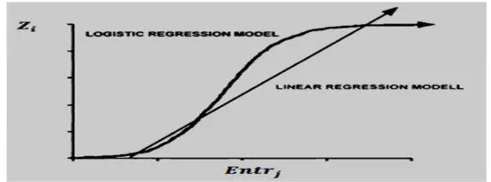
There are illustrated as bellows:
Figure 1. Linear Regression Model
3. RESULTS AND DISCUSSION
Figure 2 (a) and (c) are characteristic lobuler type of infiltrating carcinoma (ILC) and the type of infiltrating Ductal Carcinoma (IDC), while (b) and (d) is the excerpts of each ILC and IDC with a size of 5 cm x 5 cm which will be analyzed to make mathematical models.
(a) (b) (c) (d)
Figure 2. (a) ILC, (b) Excerpts of ILC with a size of 5 cm x 5 cm. (c) IDC. (d) Excerpts of IDC with size 5 cm x 5 cm.
Formulating mathematical models that can classify infiltrating ca mamma, then do skraning variable physical quantities movies x-ray results of
mammography images using the method of logistic regression results as follows.
Determination of physical parameter model 2242
Z := -1836.934 + 6888.371* E[1] -14247.818* E[2] + 6611.320* E[3] + - 4079.380* E[4] + 5428.171* E[5] + 14.284* E[6] + 1427.621* E[7] -1537.421* E[8] -648.404* E[9] + 6.619* E[10] + 2647.499* MA[1] + 305.296* MA[2] - 81529.334*MA[3] -19958.633* MA[4] + 9211.990* MA[5] + 3650.557* MA[6] + 1058157.031* MA[7] - 2563702.464*MA[8] + 2349513.476*MA[9] -
747689.772*MA[10] -177.402*MN[1] + 225.654*MN[2] – 0. 253*MN[3] -
1.094*MN[4] + 38.244* MN[5] - 0.023*MN[5] + 13.896* MN[7] – 300.025*
MN[8] + 90.543* MN[9] + 110.452*MN[10] -1588.560*EH[1] + 8028.265* EH[2] -193.301*EH]3] + 4998.507*EH[4] -7983.892*EH[5] -19.248*EH[6] + 215.800*EH[7] -24539.684*EH[9] + 23227.280* EH[10] + 19212.830*MAH[1] + 1581.604*MAH[2] + 42634.038*MAH[3] -119589.405* MAH[4] -
227899.575* MAH[5] + 308931.535* MAH[6] + 137648.993*MAH[7] + 12686.232* MAH[8] -226984.576* MAH[9] + 48797.966* MAH[10] -63.103* MHD[1] -14.829* MHD[2] + 240.890* MAH[3] -606.835* MHD[4] - 90.952*MHD[5] + 573.267* MHD[6] + 3.386* MHD[7] + 113.840* MHD[8] + 7.225* MHD[9] -232.076* MHD[10];
Probabilitas_ILC :=
;
Probabilitas_IDC = 1
–
probabilitas_ILC;
IDC and ILC VALUE RANGE
Variabel Fisis Film
IDC
ILC
Entropy
1.05095
–
5.01892
3.69954
–
4.17982
Anguler Secound Moment
0.00008
–
0.74840
0.00008
–
0.00028
Mean
10.20608
–
158.26904
113.7815
–
132.54800
Entropy of Hdiff
0.68777
–
1.92526
1.47797
–
1.90791
Anguler Secound Moment of Hdiff
0.01402
–
0.400670
0.01479
–
0.04042
Determination of physical parameter model 2243
4. CONCLUSION
Research has been done by formulating a mathematical equation model for classifying types of breast cancer histology. The results obtained from clinical cases can be made the following conclusions.
1) The optimum physical film to mengkalsifikasi histological types of breast cancer is the entropy, angular secound Moment, Mean, Entropy of Hdiff, angular secound Moment of Hdiff, Mean of Hdiff.
2) Determination of the amount of physical models the movie x-ray mammographic images in the determination of histological type of breast cancer could be increase performance in diagnosing breast cancer with a sensitivity of 86.36%.
REFERENCES
[1] Atam P. Dhawan, Yateen Chitre, Christine Kaiser-Bonasso, and Myron
Moskowitz, “Analysis of mammographic microcalcifications using gray-
level image structure features,” ieee transactions on medical imaging, vol. 15, no 3, pp. 246-259, june 1996.
[2] Bhagwati Charan Patel, Dr. G.R.Sinha,” Early detection of breast cancer using self similar fractal method,” International Journal of Computer Applications
(0975 – 8887),Vol10– N.4, pp. 39-42, November 2010.
[3] Dheeba.J, Wiselin Jiji.G,” Detection of microcalcification clusters in
mammograms using neural network, “International Journal of Advanced
Science and Technology,Vol. 19, pp. 13-22, June, 2010.
[4] Dr.H.B.Kekre, Saylee M. Gharge and Tanuja K. Sarode,” Image segmentation
of mammographic images using kekre’s proportionate error technique on
probability images,” International Journal of Computer and Electrical Engineering, Vol.2, No.6, pp. 1048-1052, December, 2010.
[5] Humberto Ochoa, Osslan Vergara, Vianey Cruz, Efrén Gútierrez,” A hybrid system based on a filter bank and a successive approximations threshold for microcalcifications detection, “ journal of computers, vol. 4, no. 8, pp. 691 – 696, August 2009.
.[6] L.S.S.Reddy, Ramaswamy Reddy, CH.Madhu & C. Nagaraju, “A novel
image segmentation technique for detection of breast cancer,” International Journal of Information Technology and Knowledge Management, Volume 2, No. 2, pp. 201-204, July-December 2010.
[7] Mohanalin, Beenamol, Prem Kumar Kalra, Nirmal Kumar,” A novel
Determination of physical parameter model 2244
[8] Oh Whi-Vin, Kim KwangGi, Kim Young-Jae,” Detection of
microcalcifications in digital mammograms using foveal method, “ J Kor
Soc Med Informatics, Vol. 15(1), pp. 165 – 172, 2009.
[9] Po-Hsiang Tsui, Yin-Yin Liao, Chien-Cheng Chang, Wen-Hung Kuo, King-
Jen Chang, and Chih-Kuang Yeh, “Classification of benign and malignant
breast tumors by 2-d analysis based on contour description and scatterer characterization,” IEEE Transactions on Mmedical Imaging, vol. 29, no. 2, pp. 513-521 February 2010.
[10] R. Krishnamoorthy, N. Amudhavalli, M.K. Sivakolundu,” Identification of
microcalcifications with orthogonal polynomials model,” Interna tional
Journal of Engineering Science and Technology, Vol. 2(5), pp. 1204-1210,
2010.
[11] S. Bouyahia, J. Mbainaibeye, N. Ellouze,” Wavelet based microcalcifications
detection in digitized mammograms,” ICGST-GVIP Journal, ISSN 1687-
398X, Vol. 8, pp.23-31, January 2009.
[12] S.Saheb Basha, Dr.k.Satya Prasad,” Automatic detection of breast cancer
mass in mammograms using morphological operators and fuzzy c –means
clustering,” Journal of Theoretical and Applied Information Technology, pp. 704-709, 2009.
[13] T. M. Kolb, J. Lichy, and J. H. Newhouse, “Occult cancer in women with dense breasts: Detection with screening US-diagnostic yield and tumor characteristics,” Radiology, vol. 207, pp. 191–199, 1998.
[14] Yufeng Zheng,“ Breast Cancer detection with gabor features from digital
mammograms, ” algorithms,ISSN 1999-4893, pp. 44-62, 2010.