Diagnostic Performance Addition of Abnormal P Wave Dispersion on Moderate Risk Duke Treadmill Score Criteria in Detecting Severe Stenosis of Coronary Arteries in
Stable Angina Pectoris Patients
Widodo Darmo Sentono, Erika Maharani, Irsad Andi Arso
Department of Cardiology and Vascular Medicine, Faculty of Medicine Universitas Gadjah Mada – Dr. Sardjito Hospital, Yogyakarta, Indonesia
Abstract
Background: Moderate risk Duke treadmill score (DTS) in threadmill test (TMT), needs additional diagnostic tool to increase its sensitivity of detecting severe coronary artery stenosis in patients with stable angina pectoris. A P wave dispersion (PWD) during TMT currently has a signifi cant relationship with DTS value and can increase the sensitivity in identifying an ischemia. This study aimed to investigate whether the addition of PWD in moderate risk DTS can improve the prediction of the severity of coronary artery stenosis in patients with stable angina pectoris.
Methods: This study is a diagnostic test with cross-sectional design. The subjects were patients who had undergone TMT and coronary angiography at the Dr. Sardjito General Hospital Yogyakarta. The outcomes were sensitivity and specifi city of abnormal PWD in moderate risk DTS patients for predicting the severe stenosis in those with stable angina pectoris as compared with moderate risk DTS and normal PWD.
Results: The additional of PWD at moderate risk DTS on 64 subjects had a sensitivity of 86.7%, a specifi city of 44.1%, a positive predictive value of 57.8%, a negative predictive value of 79.0%, a positive likelihood ratio of 1.6, a negative likelihood ratio of 0.3 and a prevalence of 46.9%, with a prediction accuracy of 64.1%.
Conclusion: The additional of abnormal PWD in moderate risk DTS had a sensitivity of 86.7% and a specifi city of 44.1% for severe coronary artery stenosis in patients with angina pectoris.
Keywords: P wave dispersion, moderate risk Duke treadmill score, severe coronary stenosis.
Introduction
Currently, treadmill exercise test (TMT) is still one of the modalities for diagnostic and risk stratifi cation measurement in coronary heart disease. Examination that combine surface electrocardiogram (ECG) and exercise, heart rate also blood pressure response is a simple and affordable modality compared to other modalities such as echocardiography and nuclear test, with the expense of lower sensitivity and specifi city. Meta-analysis study showed that TMT had 68% sensitivity, 77% specifi city with positive predictive value 73%. 1,2,3,4,5
Duke treadmill score (DTS) is a scoring system that was developed from multivariate analysis variable in TMT and clinical performance and was designed to give direct assessment to the prognosis of TMT. 6 DTS was initially used
as a stenosis predictor in 1995 and published by American Heart Association in 18th June 1998.7
ST segment depression is still used as the main variable in diagnosing ischemia, although many
studies showed that using multiple indicator from TMT were superior in increasing diagnostic and probability.8
In TMT, the increased duration of P wave also stated as a predictor of ischemia with sensitivity of 64.3%, specifi city 86.5% and positive predictive value of 57.8%.9 The abnormality of P
wave duration correlated with the increasing of pressure and enlargement of atria due to volume overload caused by diastolic dysfunction. All of these processes was triggered by myocardial ischemia and also correlate with conduction disturbance in atrium. Silva et al. (2011) showed that there was a positive correlation between duration alteration and P wave dispersion with DTS obtained from TMT in stable angina pectoris patients.10 Stable angina pectoris is a clinical
syndrome, which is marked by chest discomfort during activity or emotional stress, and alleviate with rest or consumption of nitroglycerin.11
in moderate risk TMT group, the use of more sensitive modality exercise test such as exercise perfusion imaging or coronary angiography as a gold standard of diagnosing ischemia are preferred.12 Nevertheless, those modalities are
currently not vastly available in all regions along with operator ability and a greater cost.
Based on all those facts that adding P wave dispersion (PWD) to a moderate risk DTS would yield higher sensitivity and specifi city for predicting severe stenosis in stable angina pectoris that underwent TMT, we conducted a diagnostic study to observe adding PWD to a moderate risk DTS in predicting severe coronary stenosis in stable angina pectoris patients.
Methods
This was a cross-sectional study that used retrospective data obtained from medical record in RSUP Dr. Sardjito Yogyakarta up to March 2015. Subjects were stable angina pectoris patients that underwent TMT in RSUP Dr. Sardjito Yogyakarta outpatient clinic with moderate risk DTS that had undergone coronary angiography and met the inclusion criteria. Inclusion criteria were patients that were diagnosed as stable angina pectoris, had undergone TMT with moderate risk DTS and coronary angiography and also had complete medical records. Exclusion criteria are (1) patients with atrial fi brillation or fl utter in resting ECG, (2) patients with pacing rhythm, and (3)patients with uninterpretable waveform due to artifacts.
Independent variable in this sudy was PWD abnormality in moderate risk DTS. Dependent variable was severe coronary stenosis. Attributed variables were age, gender, diabetes mellitus, hypertension, smoking history, dyslipidemia, and family history of coronary heart disease.
Study protocol
Subjects were collected using outpatient clinic registry data to fi nd patients with positive TMT test with moderate risk DTS that had undergone coronary angiography. Complete data were then crossed reference with medical record data to obtain coronary heart disease risk factors. Patients with incomplete data were not included in the study. This study did not do direct observation to patients.
The measurement of PWD was obtained from ECG recording during TMT that were stored in
2 treadmill test equipments. The measurement of PWD was done in resting stage and in fi rst minute recovery stage soon after heart rate was below 140 x/minutes. The measurement of PWD that was obtained from GE healthcare CASE™ Version 6.6 treadmill equipment made in Finland, 2011, used an internal software which positioned cursor in exact starting point and enPWDoint of P wave in the display monitor that was magnifi ed four times. The measurement of PWD that was obtained from Quinton® Q-Stress® Cardiac Science treadmill equipment made in USA, 2011, was done manually after four times magnifi cation.
The measurement of PWD was done by one single observer that had been tested using Kappa validity test. The observer was blinded to the clinical data and coronary angiography results. The measurement of coronary lesion was done by single observer that had been tested using Kappa validity test. The observation was done by re-assessing coronary angiography using video recording. The observer was blinded to the clinical data and coronary angiography results.
Baseline characteristic was displayed and analyzed using T test for numerik data and 2x2 cross table for categorical data. Hypothesis test with 2x2 cross table was used to analyze sensitivity, specifi city and positive and negative predictive value of abnormal PWD in moderate risk DTS to severe stenosis. All statistical analysis was done using SPSS 15.0.
Ethical issue of the study
This study was a non experimental, retrospective study and there were no intervention that had been done to the patients. This study was conducted after the approval from Faculty of Medicine UniversitasGadjahMada / RSUP Dr. Sardjito Medical and Health Research Ethical Committee.
Results
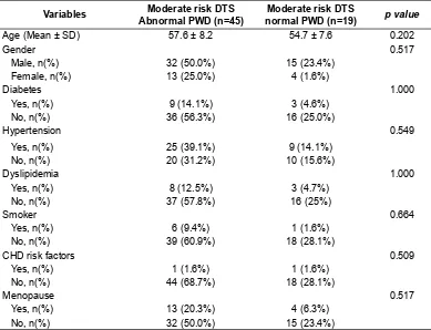
Study subjects were then divided into two groups which are group with abnormal PWD and group with normal PWD. Cut-off point for abnormality were PWD ≥12 milliseconds (ms). Mean PWD was 18.99 ± 11.87 ms. Baseline characteristic of the study subjects could be seen in table 1.
Mean age of patients with abnormal PWD was 57.6 ± 8.2 years, and 54.7 ±7.6 year in patients with normal PWD. There was no signifi cant difference between two groups based on the T test result (p= 0.202). Subjects were consisted of 47 male and 17 female. From 47 male subjects there were 32 subjects (50%) with abnormal PWD dan 15 subjects (23.4%) with normal PWD. From 17 female subjects there were 13 subjects (25.0%) with abnormal PWD and 4 subjects (1.6%) with normal PWD. There was no signifi cance association between gender and PWD based on the hypothesis test result (p=0.517).
There were 14.1% subjects with diabetes had abnormal PWD and 4.7% with normal PWD (p=1.000). Hypertensive subjects with abnormal PWD were 39.1% and 14.1% with normal PWD (p=1.000). Smoker subjects with abnormal PWD were 9.4% and 1.6% with normal PWD
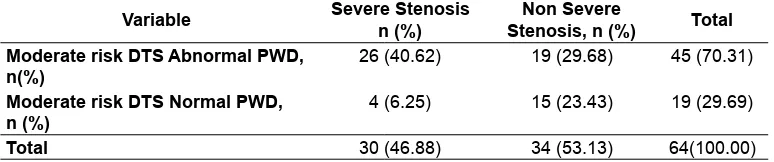
(p= 0.664). There were 1.6 % subjects with family history of CHD had abnormal PWD and 1.6% with normal PWD (p=0.509). Menopause subjects with abnormal PWD were 20.3% and 6.3% with normal PWD (p=0.424). There were no correlation between all of the baseline variables and subjects PWD. Table 2 showed the sensitivity and specifi city test on PWD to severe stenosis after 2x2 hypothesis analysis.
Based on 2x2 table, sensitivity that was obtained 86.7% with specificity 44.1% and positive predictive value 57.8% with 79%. Ratio of positive probability was 1.55 and ratio of negative probability was 0.3. Prevalence or pretest probability was 46.9% and predictive accuracy 64.1%.
Discussion
Diagnostic study from observation of PWD abnormality during TMT was expected to add its sensitivity, specifi city and prediction in moderate risk DTS. Therefore, it could be used as an alternative examination other than coronary angiography that is still becoming a gold standard.13,14,15 All of
this diagnostic study result (sensitivity, specifi city, Table 1. Baseline characteristics
Variables Abnormal PWD (n=45)Moderate risk DTS normal PWD (n=19)Moderate risk DTS p value
Age (Mean ± SD) 57.6 ± 8.2 54.7 ± 7.6 0.202
Gender 0.517
Male, n(%) 32 (50.0%) 15 (23.4%)
Female, n(%) 13 (25.0%) 4 (1.6%)
Diabetes 1.000
Yes, n(%) 9 (14.1%) 3 (4.6%)
No, n(%) 36 (56.3%) 16 (25.0%)
Hypertension 0.549
Yes, n(%) 25 (39.1%) 9 (14.1%)
No, n(%) 20 (31.2%) 10 (15.6%)
Dyslipidemia 1.000
Yes, n(%) 8 (12.5%) 3 (4.7%)
No, n(%) 37 (57.8%) 16 (25%)
Smoker 0.664
Yes, n(%) 6 (9.4%) 1 (1.6%)
No, n(%) 39 (60.9%) 18 (28.1%)
CHD risk factors 0.509
Yes, n(%) 1 (1.6%) 1 (1.6%)
No, n(%) 44 (68.7%) 18 (28.1%)
Menopause 0.517
Yes, n(%) 13 (20.3%) 4 (6.3%)
positive and negative prediction value also ratio of positive and negativeprobability) had signifi cance statistical value. The calculation of sensitivity was 86.7%, specifi city 44.1%, positive predictive value
57.8% with 79.0%.
Sensitivity is defi ned as the percentage of subject that is suffering from the disease in the abnormal test group. Specifi city is defi ned as the percentage of subjects that is not suffering from the disease in the normal test group.5 Sensitivity
and specifi city in PWD, with high sensitivity and low specificity, meant that abnormal PWD in moderate risk DTS could be use for diagnosing severe coronary stenosis. In other hand, we could not point the cause of PWD abnormality was only due to severe coronary stenosis. Abnormality of PWD could be caused by other event which meant that severe coronary stenosis in abnormal PWD had probability of 86.7% meanwhile the probability of non severe stenosis with a normal PWD was 44.1%.
In this study, 46.9% subjects were patients with severe coronary artery stenosis. These were a high prevalence number. The prevalence of severe coronary stenosis in moderate risk DTS was reported 31%.7 Based on the discriminating value
in sensitivity and specifi city, this study focused on the sensitivity due to severe coronary stenosis as a dependent variable. This had a worse outcome due to a high yearly mortality rate. Other choice was the low specifi city that was related to the pathomechanism in P wave dispersion event.
A P wave dispersion during TMT was related to the left ventricle dysfunction caused by ischemia that triggered instability and heterogeneity of atrium conduction other than atrium ischemia itself.10,16 Ischemia study that was used with
deployment of angioplasty balloon could cause enhancement of left atrium pump, either in left artery descendent or left artery circumfl ex.17 Abnormality
in P wave also related to the disruption of left ventricular fi lling or diastolic dysfunction. In this mechanism, the event was not specifi cally caused
by ischemia.13 Non-ischemic diastolic dysfunctions
could also cause abnormal PWD. Gelzinis (2014) reported that non-ischemic condition that could be the etiology of diastolic dysfunction were systemic hypertension, obesity, diabetes mellitus, older age, renal insufficiency, thyrotoxicosis, aorta stenosis, restrictive cardiomyopathy, idiopathic cardiomyopathy, hypertrophy obstructive cardiomyopathy, infi ltrative diseases, amiloidosis, hemochromatosis, sarcoidosis, constrictive pericarditis, and pericardial effusion.18 Due to the
abundance causes of diastolic dysfunction, we conclude that to increase the specifi city, efforts must be done to eliminate non ischemic diastolic dysfunctions.
High positive and negative predictive value in this research signified that abnormal PWD could not conclude severe coronary stenosis but there were higher chance of non severe stenosis in normal PWD. This was in accord with the hypothesis value of high sensitivity and low specifi city. We could use extrapolation with Fagan normogram to estimate changes in diagnostic value.
Study Limitation
The limitation of this study could be cause by some factors in measurement protocol. Measurement used 2 different treadmill equipments that used different softwares and measurement methods. This could cause bias in measurement. Other factors that could affect the result was the diffi culty in determining the starting point of P wave. This was due to dynamic ECG image during exercise which caused the wave and ECG complex becoming blurred and caused absurd value. Thoroughness were very important for a valid measurement.
Difference in recovery duration was also become a limitation. A different recovery time in each subject depended on the time to achieve heart rate <140 x/minute. This caused the measurement Table 2. Sensitivity and specifi city test on PWD to severe stenosis
Variable Severe Stenosisn (%) Stenosis, n (%)Non Severe Total Moderate risk DTS Abnormal PWD,
n(%) 26 (40.62) 19 (29.68) 45 (70.31)
Moderate risk DTS Normal PWD,
n (%) 4 (6.25) 15 (23.43) 19 (29.69)
of PWD was unequal in each subject. Other study limitation was the use of retrospective data. Due to the nature of retrospective data, the exact cause-effect relationship could not be determined. Retrospective data made independent variable could not be adjusted to the need of dependent variable and the data modifi cation could not be done.
Further study would be needed to increase specifi city diagnostic by eliminating confounding factors of PWD during TMT. Special attention would also be needed in measurement bias to ensure validity of diagnostic value. A prospective study is preferred to determine cause-effect relationship due to the ability to eliminate confounding variable.
Conclusion
Addition of abnormal P wave dispersion in moderate risk DTS had sensitivity 86.7% and specificity 44.1% to predict severe coronary stenosis in patients with stable angina pectoris.
References
Diamond, GA, Forrester, JS. 1979. Analysis of 1.
prob ability as an aid in the clinical diagnosis of coro nary-artery disease. N Engl J Med, 300:1350.
Detrano R, Janosi A, Steinbrunn W, Pfi sterer 2.
M, Schmid JJ. 1991. Algorithm to predict triple-vessel/left main coronary artery disease in patients without myocardial infarction. Circulation. 83:III-89 –III-96.
Morise, AP, Diamond, GA. 1995. Comparison 3.
of the sensitivity and specifi city of exercise electrocar diography in biased and unbiased populations of men and women. Am Heart J, 130:741-747.
Flecher GF, Flipse T, Kligfi eld P, Malauf J, 4.
1998. Current status of ECG stress test testing. Curr Probl Cardiol, 23:353-423. G i b b o n s R
1. J , B a l a d y G J , B r i c k e r JT, Chaitman BR, Fletcher GF, Froelicher VF, Mark DB, McCallister BD, Mooss AN, O’Reilly MG, Winters WL, Gibbons RJ, Antman EM, Alpert JS, Faxon DP, Fuster V, Gregoratos G, Hiratzka LF, Jacobs AK, Russell RO, Smith SC. 2002. ACC/ AAHA 2002 guideline update for exercise testing: summary article. A report of the
American College of Cardiology/American Heart Association task force on practice guidelines. Circulation, 40:1531-1540. Mark DB, Hlatky MA, Harrell FE Jr, Lee KL, 2.
Califf RM. 1987. Exercise treadmill score for predicting prognosis in coronary artery disease. Ann Intern Med, 106:793-800. Shaw LJ, Peterson ED, Shaw LK, Kesler 3.
KL, DeLong ER. 1998. Use of a prognostic treadmill score in identifying diagnostic coronary disease subgroups. Circulation, 98:1622-1630.
Ellestad MH, Savitz S, Bergdall D, Teske J. 4.
1977. The false positive stress test. Multivariate analysis of 215 subjects with emodynamic and angiographic and clinical data. Am J Cardiol, 40:681.
Maganis JC, Drimmer DA, Rojo FB, Gamie 5.
SH, Selvester RSH, Ellestad MH. 2013. Enhanced recognition of ischemia by three variable analysis of the exercise stress test. Journal of Electrocardiology (46) : 644–648. Silva RM, Cunha LBS, Teixeira LRM, Dias 6.
MSA. 2011. Correlation of P-wave duration and treadmill exercise testing with Duke score. The Cardiology, 6:20-24.
Fox
7. K, Garcia MA, Ardissino D, Buszman P, Camici PG, Crea F, Daly C, De Backer G, Hjemdahl P, Lopez-Sendon J, Marco J, Morais J, Pepper J, Sechtem U,Simoons M, Thygesen K, Priori SG, Blanc JJ, Budaj A, Camm J, Dean V, Deckers J, Dickstein K, Lekakis J, McGregor K, Metra M, Morais J, Osterspey A, Tamargo J, Zamorano JL. 2006. Guidelines on the management of stable angina pectoris: executive summary: The Task Force on the Management of Stable Angina Pectoris of the European Society of Cardiology. Eur Heart J. 27:1341-1381. Fletcher GF, Ades PA, Kligfi eld P, Arena R, 8.
Balady GJ. 2013. Exercise standards for testing and training: a scientifi c statement from the American Heart Association. Circulation, 128:873-934
Shettigar UR, Barry WH, Hultgren HN. 1977. 9.
P wave analysis in ischaemic heart disease, An echocardiographic, haemodynamic, and angiographic assessment. Brit Heart J, 39:894-899.
Myrianthefs MM, Ellestad MH, Startt-Selvester 10.
and correlation with angiographic fi ndings. Am J Cardiol, 68:1619-1624.
Myrianthefs MM, Shandling AH, Startt-Selvester 11.
RH. 1992. Analysis of the signal-averaged P-wave duration in patients with percutaneous coronary angioplasty-induced myocardial ischemia. Am J Cardiol, 70:728-732.
Turgut O, Yalta K, Yılmaz MB. 2007. 12.
Effect of coronary collateral circulation on P-wave dispersion. Türk Aritmi, pacemaker veelektrofi zyolojidergisi, 5:158-165.
Stefanadis C, Demellis J, Toutozas P, Tsiamis 13.
E. 1999. Effects of pacing-induced and
balloon coronary occlusion ischemia on left atrial function in patients with coronary artery disease. J Am Coll Cardiol, 33:687-696. Gelzinis TA. 2013. Review: New insights 14.
into diastolic dysfunction and heart failure with preserved ejection fraction. Semin Cardiothorac Vasc Anesth. 21:208-217. Agoritsas T, Courvoisier D, Combescure C, 15.