Indo. J. Chem., 2007, 7 (3), 350-353 350
ISOLATION OF ACTINOMYCIN D FROM MARINE DERIVED
Streptomyces
Abdullah Rasyid1* and Kyoko Adachi2
1
Research Center for Oceanography, Indonesian Institute of Sciences Jl. Pasir Putih No. 1 Ancol Timur, Jakarta14430 Indonesia 2
Marine Biotechnology Institute,3-75-1 Heita, Kamaishi, Iwate 026-0001 Japan
Received 4 September 2007; Accepted 1 October 2007
ABSTRACT
The bioactive substance producing strain, a marine bacterium A5S-46 was found in the antibacterial screening. This strain was isolated from the artificial sponge set in the coastal seawater at Iriomote islands (Japan). The crude extract from culture broth of the strain A5S-46 was assayed to the antibacterial activity test against the seven kinds of bacterial test strains. The active substance accumulated in the both bacterial cells and culture supernatant. Based on the 1H NMR and the LC/PDA/MS data, the bioactive substance was identified to be actinomycin D.
Keywords: Marine bacteria, Actinomycin D, Antibacterial, Anticancer
INTRODUCTION
During the last 10 years marine organisms have provided a large number of new natural products. Interesting compounds have mainly been derived from macro organisms such as sponges, ascidians, corals and bryozoans. The number of secondary metabolites microorganisms is smaller, but rapidly increasing. Because of the enormous difficulties involved in harvesting products from marine animals, and the fact that some of the bioactive compounds are produced by associated bacteria, the advantages of sustainable production of bioactive metabolites by bacteria or fungi, under the protection of natural resources, seem to be very attractive for the future [1].
Marine natural products have been found to be an important source of drugs and drug leads. These natural products as secondary metabolites and enhance survival fitness and may serve as chemicals weapons used against bacteria, fungi, viruses and small or large animals. Most of the natural products of interest to the pharmaceutical industry are secondary metabolites and several such compounds, derived from marine invertebrates, have been in clinical trials as experimental anti-cancer drug [2].
There is growing interest in marine natural products or marine secondary metabolites. This field of research receives the attention of investigators in various fields such as marine biology, marine ecology, biochemistry, chemistry and pharmacology. In the industrialized countries about 25% prescription drugs contain active principles that are still extracted from higher plants. Natural products, have been, and still are, an inexhaustible source of drug leads as well as drugs. Natural products are, along with combinatorial chemistry, at the forefront of research in the search for new therapeutic agents [2].
For the purpose of the discovery of the bioactive compounds lead to drugs, the antibacterial screening is useful based on the easy handling and high sensitivity. Moreover, the antibacterial compounds sometimes have another bioactivities such as antifungal, anticancer, immunosuppressant, neuroleptic actions. In the present paper, we tried to find the antibacterial compounds produced by the marine bacteria against the seven test strains containing five marine bacteria.
EXPERIMENTAL SECTION
Materials
All chemicals containing Nutrient Broth, Marine Broth were obtained commercially. Seawater was taken at Kamaishi Bay in Japan. Test strains shown in Table 1 were purchased from each microbial collection such as ATCC.
Instrumentation
NMR spectra were measured by a Unity INOVA 750 NMR (Varian Co. Ltd.). LC/PDA/MS was carried out by a LC-MS system consisted with a LCQ Advantage ion trap mass spectrometer (Thermo Electron Co. Ltd.) and a NanoSpace SI-2 HPLC/PDA (Shiseido).
Procedure
Isolation of bacteria by the special media
A piece of organism was dipped into the sterilized seawater (5 mL) contained in 15 mL of centrifuge tube. After homogenizing, the suspended solution was diluted 10-fold and 100-fold with sterilized seawater. These stock solutions were used for the isolation of the microorganisms. Sand and mud were collected into 15 mL of centrifuge tube. After standing, the supernatant
Abdullah Rasyid & Kyoko Adachi
Indo. J. Chem., 2007, 7 (3), 350-353 351
was used with dilution. The 50 µL aliquots of the sample solutions were spread on the isolation plates on the day when samples were collected. The special medium, namely 1/10NA (1/10 diluted Nutrient Agar), was used in
this research for the selection of Firmicutes and
Actinobacteria which could often produce active compounds. The 1/10 NA is consisted of Nutrient Broth (Difco, 0.8 g), agar (15 g) and distilled water (1 L).
For bacteria, after several days, each distinct strain of bacteria forming colonies on the isolation plates was isolated to new plate by disposable needle. For the storages, each bacterial strain was harvested with the sterilized loop and suspended in seawater containing 10% of glycerol. These bacterial solutions were stored at -80 °C.
Taxonomy
Genomic DNA was purified by using Genomic-tip and buffer set (Qiagen). The 16S rRNA gene fragment was amplified by using universal primers corresponding
to positions 341 as forward primer (Escherichia coli
numbering system). The partial sequencing of the 16S rRNA gene was done by MBI researches. The 16S rRNA gene sequence was compared with the bacterial sequence data stored in DDBJ database by using BLAST algorithm [3].
The marine bacterium A5S-46 was collected by Marine Biotechnology Institute researchers by the artificial sponge method [4] at Iriomote islands waters, in Japan. By 16S rRNA gene sequence analysis, A5S-46
strain was identified as a Streptomyces sp.
Sample preparation for the screening
The isolated strains were cultured in 4 mL of a liquid medium containing distilled water, 37.4 g of Marine Broth (Difco) and 3.3 g of tryptophan per liter of solution. The culture tube was incubated for 4 - 6 day at 30 °C on a shaker. After harvest, each culture was freeze-dried and extracted with 2 mL of 60 % ethanol.
Isolation and structural determination of active substances
The bacterial strain A5S-46 was incubated in 300 mL Marine Broth (Difco) medium at 30 ºC for 5 days on a rotary shaker (100 rpm). The culture broth was centrifuged, for the separation of supernatant and pellet. The supernatant and pellet were extracted with ethyl
acetate and acetone respectively. The organic solvent layer was continued to the bioactivity test and column chromatographic separation on a silica gel column with the elution of chloroform-methanol solvent system. The active fractions were followed by the further purification using reversed-phase chromatography with a ODS column, the mobile phase of 20-100 % methanol.
The structural determination of the isolated compounds was carried out by NMR and LC/PDA/MS. Analysis of LC/MS for the active fraction was conducted using column Inertsil ODS-2 (1.5 mm I.D. x 250 mm) and 70% aqueous acetonitrile as a solvent, at 0.1mL/min of flow rate. Separation profile of HPLC was detected at UV 220 nm and followed by the erectorspray ionization (ESI) MS with a positive TIC (Total Ionization Chromatograph) mode.
Antibacterial test
The antibacterial test against terrestrial and marine bacteria on a Nutrient Agar plate or a Marine Agar (Difco) plate was tested by the paper disk method. The samples were dissolved in methanol or 60
% ethanol and the 15 µLof the solution was applied to
a disk of 6 mm in diameter. All bacteria were incubated at 30 ºC. The tested bacteria are shown in Table 1.
RESULT AND DISCUSSION
Collection of Marine Bacteria
By using 1/10NA as a selection medium, about 80 strains were isolated from many kinds of samples collected at Iriomote islands. Fourteen isolates different in colony-type were selected for the taxonomy based on 16S rRNA gene sequences. As shown in Table 2,
most isolates were Actinobacteria and Firmicutes as
our expectation.
Screening for antibacterial activity
We screened the antibacterial activity of the isolated bacteria against the seven test strains. Many strains have antibacterial activities but pattern were different (Table 3). For the comparison, authentic antibiotics were tested in the same manner. The strain A5S-46 was selected for the further investigation because of its highest activity among the tested strains.
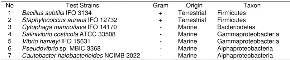
Table 1. Test strains in the antibacterial test
No Test Strains Gram Origin Taxon
1 Bacillus subtilis IFO 3134 + Terrestrial Firmicutes 2 Staphylococcus aureus IFO 12732 + Terrestrial Firmicutes 3 Cytophaga marinoflava IFO 14170 - Marine Bacteriodetes
4 Salinivibrio costicola ATCC 33508 - Marine Gammaproteobacteria
5 Vibrio harveyi IFO 15631 - Marine Gammaproteobacteria
6 Pseudovibrio sp. MBIC 3368 - Marine Alphaproteobacteria 7 Cautobacter halobacterioides NCIMB 2022 - Marine Alphaproteobacteria
Indo. J. Chem., 2007, 7 (3), 350-353 352
Table 2. The taxon of the isolated marine derived bacteria
Strain No. Sample No. Sample class Blast top % Taxon
A5S-01-1 05IRI002 alga AY964602.1 Bacillus sp. P01 99 Firmicutes
A5S-03-1 05IRI002 alga AM162319.1 Paenibacillus sp. P30 99 Firmicutes
A5S-06-1 05IRI008 driftwood AB023353.1 Promicromonospora sukumoe 100 Actinobacteria
A5S-09-1 05IRI008 driftwood AY999890.1 Streptomyces cuspidosporus strain AS 4.1686 99 Actinobacteria
A5S-11-1 05IRI016 sediments DQ289997.1 Bacillus pumilus clone B557 100 Firmicutes
A5S-18-1 05IRI035 sediments AY833099.1 Bacillus simplex isolate Ph18 99 Firmicutes
A5S-20-1 05IRI035 sediments DQ490381.1 Bacillaceae bacterium KVD-1700-08 99 Firmicutes
A5S-31-1 05IRI046 ascidian CP000157.1 Erythrobacter litoralis HTCC2594 98 Alphaproteobacteria
A5S-34-1 05IRI047 sediments AF319539.1 Microbacterium nematophilum 98 Actinobacteria
A5S-46 05IRI055 artificial sponge AY999720.1 Streptomyces coerulescens strain ISP 5146 99 Actinobacteria
A5S-60 05IRI041 alga AF319539.1 Microbacterium nematophilum 98 Actinobacteria
A5S-62 05IRI042 ascidian AY314782.1 Streptomyces glauciniger strain FXJ14 98 Actinobacteria
A5S-66 05IRI044 sponge DQ316786.1 Brevibacillus sp. Y2 99 Firmicutes
A5S-70 05IRI053 artificial sponge AY835926.1 Nocardioides kribbensis strain KSL-6 97 Actinobacteria
Table 3. The results of antibacterial test
Diameter of clear zone (mm) Strain No.
1 2 3 4 5 6 7
A5S-01-1 - - -
A5S-03-1 - - - - 12 - -
A5S-06-1 - - - - 10 - -
A5S-09-1 - - - - 12 - -
A5S-11 14 33 35 - - 11 15
A5S-18-1 - 32 42 - 15 9 -
A5S-20-1 - 12 - - 14 - -
A5S-31-1 - - - - 15 - 14
A5S-34-1 - - 35 - - - -
A5S-46 24 21 14 - 13 20 21
A5S-60 17 - - - - 13 -
A5S-62 - 12 - - 12 14 -
A5S-66 14 18 - - 14 - -
A5S-70 - - - - 13 - -
Polymyxin B (200 ppm) 24 24 - 10 - - -
Penicillin G (200 ppm) 8 13 - - - 26 -
Chloramphenicol (200 ppm) 19 14 -`` 17 13 - -
Kanamycin (200 ppm) - - -
Kanamycin (2000 ppm) 25 31 - - 12 - -
CCCP+(200 ppm) - - -
CCCP+(2000 ppm) 34 30 32 36 14 33 38
+
CCCP ( carbonyl cyanide m-chlorophenylhydrazone)
Purification and activity
The active compound produced by the marine bacterium A5S-46, which was eluted by the 80% methanol through the SepPak-C18 (ODS column), showed antibacterial activity against six strains among the seven antibacterial test strains (Table 3). The
diameter of clear zones against Bacillus subtilis isbigger
than others. Whereas the fraction A5S-46 has no activity
against to Salinivibrio costicola. This antibacterial activity
pattern is different from the authentic antibiotics.
For the easy selection of the active strains, samples are dispensed on an agar plate inoculated with an indicator microorganism, and after incubation a clear
circular zone of growth inhibition surrounding the sample is visible against partially opaque background of growth. The approach employed for most indicator organisms is very similar although the details of inoculums, medium petri dish size, sample application, and zone of inhibition measurement can very widely. In recent years, many different pieces of equipment have become commercially available to mechanize and automate the agar diffusion assay, thereby greatly increasing the number of samples that can be processed [5]. However, this methods sometimes show the error data because the clear circular zone of growth inhibition were appear because of the lack of
Indo. J. Chem., 2007, 7 (3), 350-353
Abdullah Rasyid & Kyoko Adachi
353
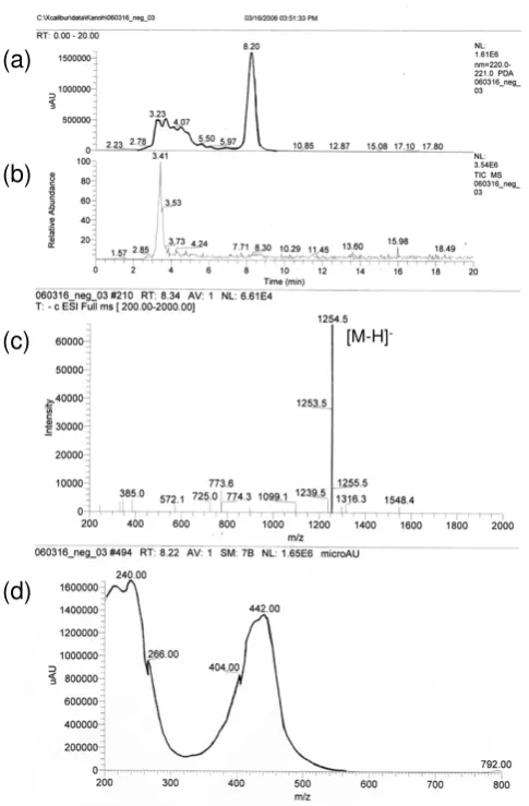
[M-H] -(a)
(b)
(c)
(d)
Fig 1. LC/PDA/MS profile of the active compound isolated from strain A5S-46 (a) HPLC chromatogram at 220 nm (b) Total ion chromatogram of MS (negative mode) (c) Mass spectrum (negative mode) and (d) UV spectrum
theneutients. Therefore, we used the agar diffusion assay with the paper disk, which was applied by the extract of the bacterial culture broth.
Structural determination of the active substance
By the LC/PDA/MS (Fig 1) and 1H NMR (Fig 2), the
active compound was identified to be Actinomycin D (Fig 3). This compound was first isolated by another species of Sterptomyces. Actinomycin D is one of the older chemotherapy drugs which has been used in therapy for many years. It is most commonly used in the treatment of cancer, such as kidney cancer, soft tissue sarcoma, bone sarcoma, etc.
CONCLUSION
The bioactive substance produced by marine bacterium A5S-46 was identified to be Actinomycin D.
Fig 2. 1H NMR spectrum of the active compound
isolated from strain A5S-46 (in CD3OD)
O N
O NH2
O O
O N
N H N N
O
O
O O
O
NH O
N
HN
N N
O
O
O
HN O O
Fig 3. The chemical structure of Actinomycin D
(C62H86N12O16, Mol.wt.: 1255.5)
This compound showed antibacterial activity against six antibacterial test strains and the diameter of clear
zones against Bacillus substilis is bigger than others.
ACKNOWLEDGEMENT
Grateful thanks are extended to Japan International Cooperation Agency (JICA) and staff of Marine Biotechnology Institute, Japan, who provide laboratory and other research facilities.
REFFERENCES
1. Wagner-Dobler,I., Winfried B., Siegmynd L.,
Marinus M., and Hartmut L.,In Advances in
Biochemical Engineering / Biotechnology, Ed. Th. Scheper, Springer-Verlag, Berlin Heiderberg, 2002, Vol. 74, 208-238
2. Gudbjarnason, S., 1999, Rit Fiskideildar,16,
107-110
3. Altsuchul, S.F., Gish, W., Miller, W., Myers, E.W.,
and Lipman, D.J., 1990,. J. Mol. Biol., 215,
403-410
4. Yasumoto-Hirose, M., Nishijima, M., Ngirchechol, M.K., Kanoh, K., Shizuri, Y., and Miki, W., 2006,
Mar. Biotechnol. (NY), 8(3), 227-237
5. Hunter-Cevera, J.C., M.E. Fonda and Angela B.
1986. In Arnold L. (eds.). Manual of Industrial