Corresponding author: [email protected]
Prolonged Kidney Ischemia-Reperfusion
Injury Associates with Inflammation,
Vascular Remodelling, and Myofibroblast
Formation
Nur Arfian*, Hilma Kholida Ats-tsani, Pratiwi Indah Sayekti, Dwina Agrila Lakabela, Amelia, Toni Febriyanto, Hana Rutyana Putri Antonio, Dian Prasetyo Wibisono, Dwi Cahyani Ratna Sari
Departement of Anatomy, Faculty of Medicine, Universitas Gadjah Mada, Yogyakarta,
Indonesia
DOI: http://dx.doi.org/10.19106/JMedSci005001201801
ABSTRACT
Prolonged kidney ischemia-reperfusion injury (IRI) is the important risk factor for leading to chronic kidney disease (CKD). Persistent hypoxia and inlammation are considered as the main pathogenesis of chronic injury, followed by myoibroblast expansion and ibrosis process. Tubular injury, cell proliferation, and vasoconstriction, as acute compensatory responses, are restored in chronic phase. The aim of the study was to investigate the relation between inlammation, vascular remodeling, and myoibroblast formation as response to ischemia injury after prolonged kidney ischemia-reperfusion (I/R). Fifteen male Swiss mice aged 3-4 months were used as kidney I/R injury model after bilateral pedicle renal clamping. Rats were divided into 3 groups with ive rats in each group i.e. control group (sham operation/SO), acute I/R model (IR1), and chronic I/R model (IR12). PAS staining was used for scoring tubular injury. Fibrosis was assessed using
ABSTRAK
Perpanjangan cedera iskemik-reperfusi ginjal (kidney ischemia-reperfusion injury/IRI) merupakan fakor risiko penting terjadinya penyakit ginjal kronis (chronic kidney disease/
CKD). Inlamasi dan hipoksia berkepanjangan diduga merupakan pathogenesis utama cedera kronik, diikuti ekspansi mioibroblas dan kejadian ibrosis. Cedera tubulus, proliferasi sel dan vasokontriksi sebagai respon balik akut terjadi pada fase kronik. Tujuan penelitian ini adalah mengkaji hubungan antara inlamasi, remodelling vaskular dan pembentukan mioibroblas sebagai respon cedera iskemik setelah perpanjangan iskemik/reperfusi (I/R) ginjal. Lima belas mencit Swiss jantan berumus 3-4 bulan digunakan sebagai model cedera setelah dilakukan penjepitan bilateral pedicle renal. Tikus dibagi menjadi tiga kelompok dengan 5 ekor setiap kelompok yaitu kelompok sham operation (SO), kelompok model IR akut (IR1) dan kelompok model IR kronis (IR12). Pengecatan PAS digunakan untuk menilai cedera tubulus. Terjadinya ibrosis diukur menggunakan pengecatan imunologi merah
sirius dan α-SMA untuk ekspansi mioibroblas. Pengecatan imunologi PCNA dan CD68 digunakan untuk mengidentiikasi proliferasi sel dan iniltrasi makrofag. RT-PCR dilakukan untuk mengkaji ekspresi MCP-1, HIF-1α dan ppET-1 yang diukur dengan program ImageJ. Data dianalisis menggunakan ANAVA satu jalan dan uji Kruskal-Wallis dengan tingkat signiikansi 0,05. Kenaikan secara nyata terjadi pada skor cedera tubulus (p<0,05) dan sel positif PCNA (0,05) pada kelompok IR1 dibandingkan SO, selain itu terjadi kenaikan HIF-1α pada kelompok IR12. Jumlah makrofag (p<0,01) dan ekspresi MCP-1 (p<0,05) meningkat secara nyata pada kelompok IR1 dan IR12 dibandingkan kontrol. Ketebalan dinding arteri meningkat (p0,05) diikuti penurunan area lumen vascular (p<0,05) dan kenaikan ekspresi ppET-1 (p<0,01) pada kelompok IR1 dan pulih secara nyata (p<0,05) pada kelompok IR12. Fraksi daerah ibrosis dan ekspansi mioibroblas meningkat nyata secara bertahap dari IR1 ke IR12 (p<0,01). Dapat disimpulkan, perpajangan cedera I/R ginjal menginduksi hipoksia dan respon inlamasi berkelanjutan yang menyebabkan pembentukan mioibroblas dan penurunan respon pemodelan kembali vaskular.
Keywords: ischemia-reperfusion injury - kidney – inlammation - vascular remodelling - myoibroblast.
INTRODUCTION
Kidney ischemia-reperfusion injury (IRI) is sudden temporary impairment of blood low
to the kidney, which is characterized by blood supply restriction to kidney and followed by restoration of blood low and re-oxygenation (perfusion).1 Kidney IRI is a major cause of
acute kidney injury (AKI) and 70% of AKI progress to chronic kidney disease (CKD).2
CKD is the chronic consequence of ischemia injury and thought to be related to glomerulo-interstitial ibrosis and persistent kidney dysfunction.3
The pathophysiology of kidney IRI is complicated. There are 3 stages of tissue
response to ischemia injury, that are initiation, extension, and maintenance.4 In early period or
initiation phase, microvascular damage causes hypoxia in corticomedullary junction which is characterized by obstruction, inlammation, and coagulopathy. Then persistent hypoxia and inlammatory responses stimulate extension phase in 24 hours after initiation phase.4
Loss of tubular brush border, exfoliation, and tubular obstruction are found in this period.4
Inlammation has the important role in early stage. Chemokines are major mediators of the inlammation that regulate pro-inlammatory cytokine, adhesion molecule expression, leukocyte activation and iniltration to the tissue.1 Inlammatory mediators, reactive oxygen species (ROS), intracellular adhesion molecule (ICAM-1), and P-selectin can promote leukocytes and neutrophil iniltration into post-ischemic tissue.1 The iniltration of leukocyte,
including macrophages, may play an important role in development of kidney injury which is facilitated by chemotactic factors and/or adhesion molecules.5 Monocyte
chemoattractant protein-1 (MCP-1) is a potent chemokine that stimulates migration of monocyte into the intimate layer of arterial walls and organs.5
Enhancement of leucocyte-endothelial interaction can cause endothelial injury, then leads to decreasing blood low to the tissue which aggravate ischemia.1,6 Hypoxia tissue
stimulates hypoxia-inducible factor-1 (HIF-1) expression. Then HIF-1 activates transcription of vascular endothelial growth factor (VEGF), which plays an essential role in angiogenesis.6 Kidney injury causes tubular system damage, followed by rapid cell proliferation. The proliferation is an acute compensatory mechanism of injury, which is characterized by differentiation of tubular epithelial cell.7
Those responses are associated with initial phase of injury. Cell proliferation can be represented by the expression of proliferating cell nuclear antigen (PCNA). PCNA is a
monoclonal antibody which is expressed dominantly on S phase and essential for DNA replication.8
Imbalance between vasodilator and vasoconstrictor mediator causes reducing renal perfusion in AKI.2 Endothelial dysfunction is
responsible for reducing renal blood low by impaired dilator capacity, which is attributed to
reduce production of nitric oxide.9 Endothelial
nitric oxide synthase (eNOS) has important role in preservation of medullary blood low in response to renal vasoconstrictor, such as angiotensin II. However, following renal injury, eNOS function is impaired which is demonstrated by a loss of responses to acetylcholine and bradykinin.9 Endothelial cells also secrete endothelin-1 (ET-1), a potent vasoconstrictor. Through vasoconstriction effect, ET-1 induces reduction of renal blood low and glomerular iltration rate,2 which causes oliguria. In maladaptive response of maintenance phase, ROS can causes interstitial cells expansion and extracellular matrix production by inhibit tubular epithelial cell proliferation.10 Fibrogenesis process
and kidney interstitial ibrosis are shown in interstitial area expansion which is the main characteristic of progressive kidney disease.11 This study was conducted to elucidate the kidney tissue response to prolonged injury, mainly about tubular injury appearance, inlammatory process, vascular changes, and myoibroblast formation.
MATERIALS AND METHODS
Animal
water ad libitum before used. Protocol of this study has been approved by the Medical and Health Research Ethic Committee of the Faculty of Medicine, Universitas Gadjah Mada, Yogyakarta.
Kidney ischemia/reperfusion injury model
The mice were administrated general anaesthesia with intraperitoneal injection of pentobarbital sodium (0.1 mg/g BW, Somnopentyl®). Kidney IRI model were
performed by clamping both of renal pedicles, using non-traumatic vascular clamp (Hammacher®) for 30 minutes. Then, both clamps were released and followed by reperfusion. The incision site then closed using silk surgical thread 3/0 (One Med®).
Kidney harvesting
IR1 group was sacriiced in day-1 after operation, while IR12 groups after day 12 after operation. Prior to abdomen and thorax opened, mice were anaesthetized with intraperitoneal injection of pentobarbital sodium (60 mg/kg BW, Somnopentyl®).
Perfusion of the organ was done from left ventricle, using 0.9% NaCl solution. Both perfused kidneys were harvested, one kidney was kept into RNA later® for RNA extraction and the rest was ixated into 4% PFA in PBS were deparafinized and rehydrated using xylene and alcohol serial. Specimens were then stained with sirius red (SR) for measuring ibrosis interstitial fraction area and periodic acid-schiff (PAS) to determine tubular injury.
For immunohistochemical staining, after deparafinized and rehydrated, followed antigen retrieval, blocking peroxidase using H
2O2 3% in PBS solution, and then blocking
non-speciic antigen using background sniper. The slides were incubated with α-SMA (1:400, Sigma, A2547), CD68 (1:400, Abcam, ab125212), and PCNA (1:200, Abcam, ab29) as 1st antibodies, TrekAvidin-HRP, 2nd
antibody anti rabbit Trekkie Universal Link (Biocare Medical®), then diaminobenzidine
tetrahydrochloride (DAB). α-SMA antibody immunostaining was used for measuring myoibroblast expansion, CD68 antibody for counting macrophage cells, and PCNA for assessing cell proliferation in kidney injury. Quantiication was measured from 15 ields for each sample with 400x magniication, using ImageJ software.
Tubular Injury and Interstitial Fibrosis Fraction-area quantiication
Tubular injury score was assessed by PAS staining, which was determined using semi quantitative scoring system in 15 ields for each specimen with 200x magniications. The variables of scoring are tubular atrophy and dilatation, loss of brush border, accumulation of inlammatory cells, and intraluminal cast. Scale of the lesion are from 0 to 4: 0, normal; 1, mild, injury <25%; 2, moderate, injury 25-50%; 3, severe, injury 50-75%; 4, extensive damage, injury >75%. Fibrosis fraction-area was stained using Sirius red, which was quantiied using ImageJ software on 15 non-overlapping ields and expressed in percentage (%).
Vascular remodeling
and then assessed using ImageJ software. The arteries were measured vessel area (diameter of outer layer), lumen area (diameter of inner layer), vessel perimeter, lumen perimeter, wall area (the difference between vessel area and lumen area), and central perimeter (the average of vessel and lumen perimeter). Wall thickness calculation is from the ratio between wall area and central perimeter.
Reverse transcriptase PCR (RT-PCR) RNA was extracted using Genezol solution (Genezol®, Cat. No. GZR100), followed
by RNA concentration quantiication using sphectophotometry. cDNA was synthesized using Rever Tra Ace® (Toyobo, Japan, Cat.
No. TRT-101) and random primer (Toyobo, Japan, Cat. No. 3801). Reverse transcriptase PCR was done for assessing the expression of following genes: HIF-1α forward
AGCTTCTGTTATGAGGCTCACCATC3’,
reverse AATGTCAAGATCACCAGCAC-3’), MCP-1 (forward 5’-CTTCTGGGCCTG
CTGTTCA-3’, reverse 5’- CTTCTGG
GCCTGCTG TTCA-3’), ppET-1 (forward
5’-GCCACAGACCAGGCAGTTAGA-3’, r e v e r s e5’ - A C C A G C T G C T G AT A
GATACACTTC-3’), GAPDH (forward
5’-TTGCTGTTGAAGTCGCAGGAG-3’,
reverse 5’-TGTGTCCGTCGTGGAT
CTGA-3’) were used as reference. The gene expressions were quantiied using densitometry analysis (ImageJ software) and GAPDH gene was used to normalized the gene expressions (housekeeping gene).
Statistical analysis
Data were presented as mean ± standard error of mean (SEM) for PCNA, HIF-1α, MCP-1 levels, ibrosis fraction-area, myoibroblast expansion, vascular remodelling (wall thickness and lumen area), and ppET-1 expression. For tubular injury score and macrophage cell counting were presented as median (min-max) data. Median data were analyzed using non-parametric test, Kruskal-Wallis, and then each group was compared using post-hoc Mann-Whitney. While the rest data, which have normal data distribution, were analyzed using one-way ANOVA test, followed by post-hoc LSD test. The level of statistical signiicance was p<0.05.
RESULTS
Kidney IRI induced tubular injury, cell proliferation, and inlammation
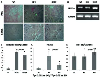
FIGURE 1. A. Microscopic picture of PAS and PCNA staining to show tubular injury and cell prolif
-eration; B-C Quantitative analysis of tubular injury score and PCNA positive cell count; D-E Electrophoresis band and RT-PCR measurement of HIF-1α.
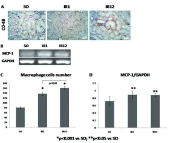
This study used MCP-1 and macrophage as representation of inlammation process. MCP-1 is a regulator of macrophage migration and iniltration. Therefore, increase of MCP-1expression in IR1 group was followed by
FIGURE 2. A. Representative picture of CD-68 immunostaining; B. Electrophoresis band of MCP-1 and GAPDH; C-D Quantitative analysis of macrophage cell count and RT-PCR measurement of MCP-1
Vascular remodelling of intrarenal artery
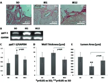
Vasoconstriction response in acute phase is related to the expression of ppET-1 gene. It was found that there were histological vascular changes of intrarenal artery (FIGURE 3.A). One day post-exposure, the vessel was constricted. It was proved by increase of wall thickness as well as decrease of vascular lumen
FIGURE 3. A. Histological changes of intrarenal artery are evaluated using sirius red staining; B-C. Electrophoresis band and RT-PCR measurement of ppET-1; D-E. Quantita
-tive analysis of intrarenal artery on wall thickness and lumen area.
Fibrosis and myoibroblast expansion
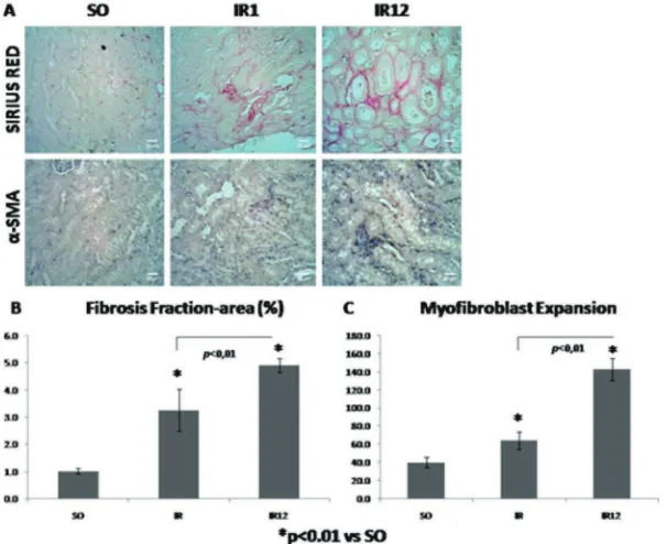
Fibrosis area was stained in purplish-red by sirius red staining (FIGURE 4.A). The widest ibrosis fraction-area was shown from IR12 group (4.927%), then followed by IR1 group (3.260%), and SO group (1.021%) (FIGURE 4.B). These inding was similar to
FIGURE 4. A. Sirius red and α-SMA IHC staining to show ibrosis and myoi
-broblas expansion; B-C. Quantitative analysis of ibrosis fraction-area and myoibroblast expansion
DISCUSSION
Kidney IRI effect on cell proliferation and inlammation response to tubular injury
The principle of kidney IRI is sudden temporary impairment of blood low to the kidney.1 There is injury/regeneration process
in kidney injury which is characterized by impairment of cell polarity, cytoskeleton integrity, and loss of renal tubular brush border which can induce apoptosis and necrotic process.12,13 Brush border and cell debris can
cause intraluminal obstruction which form an intraluminal cast in the distal tubules. These obstructions can promote the dilatation and atrophy of proximal tubules in acute kidney injury.14 Therefore, those characteristics
were used for assessing tubular injury score. After 7 days post injury, there is a repairing process by the differentiation tubular cell and
restoration of renal function and structure.7 In
this study, IR1 group, as an acute response, was impaired signiicantly compared to SO and IR12 group (FIGURE 1.A-B). It was correspond with study that was done by Basile
et al.9 reduction of tubular injury score in IR12
group was affected by repairing mechanism of tubular cells which was restored incompletely (FIGURE 1.B).
Bonventre & Dufield15 study showed that
replace the necrotic/apoptotic cells.16 PCNA
positive cell of IR1 group was more expressed than in chronic period (IR12) (p< 0.001, FIGURE 1.C). It meant that the proliferation rate in chronic injury was reduced. Decreasing of cell proliferation is caused by repair process which is began from day-7 after IRI.7 Furthermore, there is a maladaptive
repair process that decrease cell proliferation by discontinuing G2/M phase, then PCNA, which is dominantly expressed in S phase, will be decreased.16
Persistent hypoxia in I/R injury can causes sustainable injury and increase of HIF-1α expression.17 HIF is activated by low
oxygen condition and induces widespread changes in gene expression.17 Many of genes
whose expression is increased by HIF are expected to improve the cellular capacity when oxygen supply is reduced.17 Therefore,
activation of HIF may improve the survival of ischemic cell and also promote adaptive changes, such as increased angiogenesis.18
HIF appear in 10 minutes after ischemia, the most optimal period is 2 hour post-ischemia, and will be decreased in 8-24 hours after the optimum phase.18 HIF-1α expression of IR1 group was not increased, it was estimated that the level was decline after 24 hours post-exposure, due to HIF-1α is degraded rapidly (FIGURE 1.E). Several study revealed that HIF has protective effect in acute phase of ischemia injury. Paradoxically, in prolonged period of hypoxia, HIF-1α will be increased. Higgins et al.19 found that the ablation of
HIF-1α gene can prevent ibrosis tubulointerstitial expansion, through mesenchyme-epithelial cell transition. Moreover, Haase20 showed
that the prolonged HIF signal activation can stimulate ibrosis and persistent destruction of tissue. When HIF is stable and not degraded, it can lead to enhancement of pro-ibrotic gene transcription, connective tissue growth
factor (CTGF).20 Hence, the highest level
of HIF-1α was expressed in IR12 group signiicantly (p<0.05) (FIGURE 1.E). It is due to maladaptive response of repair process, so that in certain condition the level of HIF is associated with chronic injury.
Inlammatory response is the main role in pathogenesis of kidney injury. It can affect on acute and chronic (maladaptive amelioration) phase.21 When kidney is
exposed to ischemia injury, the epithelial cells will be change, the barrier and endothelial integrity can be damaged.9 This process
produce proinlammatory cytokine and chemotactic, such as TNF-α, MCP-1, IL-8, IL-6, TGF-β, RANTES and ephitelial neutrophil-activating protein 78 (ENA-78), which activate inlammatory cells, including macrophage.21 Enhancement of MCP-1 is associated with the presence of macrophage.5
Sutton et al.4 reported that in initial phase (less
than 24hour) cytokine and chemokine are increased, including MCP-1. MCP-1 can be produced by vascular smooth muscle cells.22
Ischemia condition will stimulate endothelial dysfunction, followed by inlammatory cells iniltration. As a compensated mechanism, smooth muscle tone will be increased, then induces the activation of vascular smooth muscle to produce MCP-1.22 Therefore,
MCP-1 level will be increased and macrophage iniltration will be stimulated. Increase of MCP-1 in exposed group (IR1 and IR12) is signiicant (p<0.05, FIGURE 2.D). This enhancement was followed by increase of macrophage cell signiicantly (p<0.05), which is observed by CD68 immunostaining (FIGURE 2.C).
Contrarily, when the repair response is incomplete, it will induce maladaptive process, such as ibroblast proliferation, excessive extracellular matrix deposition, and inlammatory response will be persistent,21
so that proinlammatory chemokine still produced. It can be found in MCP-1 expression and macrophage level of IR12 group, which still increased (FIGURE 2.C-D).
Kidney IRI effect on vascular remodelling
Renal ischemia affects the renal vascular and tubules. There is morphological and structural changing of renal tubules post-exposure, while auto regulation disturbance and vasoconstriction are the vascular response to ischemia. Furthermore endothelial dysfunction, stimulated by ROS, also play role in vascular maladaptive response.14 One
of endothelial reaction to any type of injury, including ischemia injury, is remodelling of vascular wall.14 This mechanism involves
cell growth, cell death, cell migration and degradation or cellular matrix production.9
These changes eventually result in intimal accumulation of smooth muscle-like cells and extracellular matrix, medial smooth muscle degeneration, and adventitial ibrosis.23 The
histopathological changes can be observed by thickening of vascular wall and narrowing of lumen area, which increase vascular resistance. Based on the result, there were enhancement of vascular thickness and narrowing of lumen area after one day exposure, then restored after day12 (FIGURE 3.A). Those alteration between IR1 and IR12 group was signiicant statistically (p<0.05, FIGURE 3.D-E). The changes that occurred in IR12 group are considered as restoration mechanism in maintenance phase of ischemia injury.4
Vasoconstriction response in ischemia injury is inluenced by the presence of ET-1,
mediated by ETAR.2 Endothelial cells secrete
ET-1 as a response to endothelial injury, caused by ischemia.1 ET-1 can stimulate
hypertrophy, migration, and proliferation of vascular smooth muscle cell by transduction signal.24 Therefore, ET-1 can promote the
thickening and narrowing of intrarenal vessel, caused by smooth muscle cell proliferation. ET-1 expression is proportional to the vascular remodelling mechanism. It is consistent with this study, ET-1 expression was increased signiicantly (p<0.05) in IR1 group as well as enhancement of wall thickness and narrowing of lumen area (FIGURE 3.C-E). Those expressions then decrease in chronic group, it signiied the presence of repairing process. It was correspond to the previous study conducted by Arian et al.2 that reduction of
the remodelling level is associated with ET-1 deletion in IRI model.
Kidney IRI effect on myoibroblast formation
It has been explained that there is amelioration mechanism which is characterized by cell differentiation and restoration of kidney function. When the injury process is extended and the restoration mechanism is incomplete, it will progress to chronic injury which is observed by the presence of ibrosis in tubulointerstitial area.12 In acute
periode, tubular injury can be compensated by adaptive amelioration process, through inconsiderable ibrosis formation and tubular cell proliferation as compensation to maintain the kidney structurally.4 Kidney tissue is
intact, but the function is reduced. Reduction of kidney function is caused by replacement of ibrotic tissue which is loss of elasticity, proliferation capacity, and differentiation ability.25 However, adaptive mechanism
supported by persistent inlammatory process.4
This mechanism is stimulated by hypoxia environment which activate proinlamatory cytokines, proibrotic, growth factor.13
Tubulointerstitial ibrosis was illustrated on IR1 and IR12 group signiicantly (p<0.05, FIGURE 4.B). Purplish-red colored area in sirius red staining (FIGURE 4.A) indicates the presence of type I and II collagen.25 The
widest ibrosis area was found in IR12 group (4.927%), represented chronic injury in IRI model (FIGURE 4.B). It was correspond to study conducted by Skrypnyk et al.26
and Varrier et al.27 that reported the highest
level of ibrosis was presence in chronic condition and there is TGF-β1 expansion which stimulate myobibroblast activation. The activated myoibroblast will produce and degrade a matrix, then stimulate connective tissue formation in tubulointerstitial area. α-SMA expression, as spesiic marker, is used for indicating the presence of myoibroblast.28
It was found enhancement of α-SMA expression in IR1 and IR12 (p<0.05, FIGURE 4.C). Signiicantly, IR12 group was the most expressed between goups (p<0.05) (FIGURE 4.C).
Fibrosis fraction-area and myoibroblast expansion are more progressive on IR12, it is related to ROS which is originated from inlammatory metabolism.26 ROS can
promotes death cell of tubular system and stimulates proliferation factor in interstitial tubular cell.4 Kim et al.10 showed that
enhancement of interstitial cell proliferation parallel to ROS level in kidney tissue. It is proved by increasing of α-SMA, FSP1, dan protein NADPH oksidase-2 expression.10
The excessive proliferation and expansion of extracellular matrix, followed by apoptotic tubular cell without regeneration mechanism, will accelerate the progressivity of injury.9,10
CONCLUSION
Prolonged IRI leads to chronic injury via persistent hypoxia and inlammatory response, signiied by myoibroblast formation in tubulointerstitial area. Vascular remodelling and cell proliferation response are reduced in long-term period of injury.
ACKNOWLEDGEMENTS
The authors thank Mr. Mulyana for animal-maintenance support and the Ministry of Research, Technology, and High Education, Republic of Indonesia for research grant through Excellence High Education Institution Research (Penelitian Unggulan Perguruan Tinggi) program. This manuscript
had been used for inishing undergraduate program of Pratiwi Indah Sayekti, Dwina Agrila Lakabela, Amelia, Toni Febriyanto, Hana Rutyana Putri Antonio.
REFERENCES
1. Malek M, Nematbakhsh M. Renal Ischemia/ Reperfusion Injury; from Pathophysiology to treatment. J Renal Inj Prev 2015; 4(2):20-7. http://dx.doi.org/10.12861/jrip.2015.06 2. Arian N, Emoto N, Vignon-Zellweger N,
Nakayama K, Yagi K, Hirata K. ET-1 Deletion
from endothelial cells protects the kidney
during the extension phase of ischemia/ reperfusion injury. Biochem Biophys Res Commun 2012; 425(2):443-9. http://dx.doi. org/10.1016/j.bbrc.2012.07.121
3. Le Clef N, Verhulst A, D’Haese PC, Vervaet BA. Unilateral renal ischemia-reperfusion
as a robust model for acute to chronic
kidney injury in mice. PLoS ONE 2016;
11(3):0152153.
h t t p : / / d x . d o i . o rg / 1 0 . 1 3 7 1 / j o u r n a l .
4. Sutton TA, Fisher CJ, Molitoris BA. Microvascular endothelial injury and dysfunction during ischemic acute renal failure. Kidney Int 2002; 62(5):1539-49. h t t p : / / d x . d o i . o r g / 1 0 . 1 0 4 6 / j . 1 5 2 3 -1755.2002.00631.x
5. Sung FL, Hu TY, Au-Yeung KK, Siow YL, Karmin O. Enhanced MCP-1 expression during ischemia/reperfusion injury is mediated by oxidative stress and NF-kB. Kidney Int 2002; 62(4):1160-70.
http://dx.doi.org/10.1111/j.1523-1755.2002.
7. Basile DP, Donohoe DL, Roethe K, Mattson DL. Chronic renal hypoxia after acute ischemic injury: effects of L-arginine on hypoxia and secondary damage. Am J Physiol Renal Physiol 2003; 284(2):338-48.
http://dx.doi.org/10.1152/ajprenal.
00169.2002
8. Caskurlu T, Kanter M, Erboga M, Erboga, ZF, Ozgul M, Atis G. Protective effect of nigelia sativa on renal reperfusion injury in rat. Iran J Kidney Dis 2016; 10(3):135-43.
9. Basile DP, Yoder MC. Renal endothelial
dysfunction in acute kidney ischemia
reperfusion injury. Cardiovasc Hematol Disord Drug Taergets 2014; 14(1):3-14. https://doi.or g/10.2174/1871529X1401140724093505 10. Kim J, Jung KJ, Park KM. Reactive oxygen
species differently regulate renal tubular
epithelial and interstitial cell proliferation
after ischemia and reperfusion injury. Am J Physiol Renal Physiol 2010;
298(5):F1118-29.
http://dx.doi.org/ 10.1152/ajprenal.
00701.2009
11. Strutz F, Zeisberg M. Renal ibroblast and myoibroblast in chronic kidney disease. J
Am Soc Nephrol 2006; 17(11):2992-8. http://dx.doi.org/10.1681/ASN.2006050420 12. Venkatachalam MA, Grifin KA, Lan R, Geng
H, Saikumar P, Bidani AK. Acute kidney injury: a springboard for progression in chronic kidney disease. Am J Physiol Renal Physiol 2010; 298(5):1078-94.
h t t p : / / d x . d o i . o r g / 1 0 . 1 1 5 2 /
ajprenal.00017.2010
13. Bonventre JV, Yang L. Cellular
pathophysiology of ischemic acute kidney injury. J clin invest 2011; 121(11):4210-21. http://dx.doi.org/10.1172/JCI45161
14. Schrier RW, Wang W, Poole B, Mitra A. Acute renal failure: deinitions, diagnosis, pathogenesis, and therapy. J Clin Invest 2004;
114(1):5-14.
http://dx.doi.org/10.1172/JCI22353
15. Dufield JS, Bonventre JV. Kidney tubular epithelium is restored without replacement with bone marrow-derived cell during repair after ischemic injury. Kidney Int 2005;
68(5):1956-61.
h t t p : / / d x . d o i . o r g / 1 0 . 1111 / j . 1 5 2 3 -1755.2005.00629.x
16. Canaud G, Bonventre JV. Cell cycle arrest and
the evolution of chronic kidney disease from
acute kidney injury. Nephrol Dial Transplant 2015; 30(4):575-83.
http://dx.doi.org/10.1093/ndt/gfu230
17. Hill P, Shukla D, Tran MG, Aragones J, Cook
HT, Carmeliet P, et al. Inhibition of hypoxia
inducible factor hydroxylases protect against renal-ischemia reperfusion injury. J Am Soc Nephro 2008; 19(1):39-46.
http://dx.doi.org/10.1681/ASN.2006090998 18. Rosenberger C, Heyman SN, Rosen S, Shina
A, Goldfarb M, Griethe W, et al. Up-regulation
of HIF in experimental acute renal failure:
evidence for a protective transcriptional
response to hypoxia. Kidney Int 2005; 67(2):
http://dx.doi.org/10.1111/j.1523-1755.2005. 67110.x
19. Higgins DF, Kimura K, Iwano M, Haase VH. Hypoxia-inducible factor signaling in the development of tissue ibrosis. Cell Cycle 2008; 7(9):1128–32.
http://dx.doi.org/10.4161/cc.7.9.5804
20. Haase VH. Regulation of erythropoiesis by hypoxia-inducible factors. Blood Rev 2013;
27(1):41-53.
http://dx.doi.org/10.1016/j.blre.2012.12.003 21. Akcay A, Nguyen Q, Edelstein CL. Mediators
of inlammation in acute kidney injury. Mediators inlamm 2009; 2009:137072. http://dx.doi.org/10.1155/2009/137072 22. Granger DN, Senchenkova E. Inlammation
and the microcirculation. In: Colloquium series on integrated systems physiology: from molecule to function. San Rafael: Morgan & Claypool Life Sciences. 2010; 1-87.
http://doi.org/10.4199/c00013ed1v01
y201006isp008
23. Ponticelli C. Ischaemia-reperfusion injury: a major protagonist in kidney transplantation. Nephrol Dial Transplant 2014; 29(6):
1134-40.
http://dx.doi.org/10.1093/ndt/gft488
24. Bouallegue A, Daou GB, Srivastava AK. Endothelin-1-induced signaling pathways in vascular smooth muscle cells. Curr Vasc Pharmacol 2007; 5(1):45-52. http://doi. org/10.2174/157016107779317161
25. Akamatsu T, Arai Y, Kosugi I, Kawasaki
H, Meguro S, Sakao M, et al. Direct
isolation of myoibroblasts and ibroblasts from bleomycin-injured lungs reveals their
functional similarities and differences.
Fibrogenesis Tissue Repair 2013; 6(1):15. http://dx.doi.org/10.1186/1755-1536-6-15 26. Skrypnyk NI, Harris RC, de Caestecker MP.
Ischemia-reperfusion model of acute kidney
injury and post injury ibrosis in mice. J Vis Exp 2013; 78:6-11.
http://dx.doi.org/10.3791/50495
27. Varrier M, Forni LG, Ostermann M. Long-term sequelae from acute kidney injury:
potential mechanisms for the observed poor
renal outcomes. Crit Care 2015; 19(1):102. http://dx.doi.org/10.1186/s13054-015-0805-0 28. Kramann R, Dirocco DP, Maarouf OH,
Humphreys BD. Matrix producing cells in ch ronic kidney disease: origin, regulation, and activation. Curr Pathobiol Rep 2013;
1(4):1-17.