Int. J. Trop. Vet. Biomed. Res. Vol. 1 (2) : 6-14; November 2016
www.jurnal.unsyiah.ac.id/IJTVBR E-ISSN : 2503-4715
Administration of Centella Leaf Extract (Centella asiatica (L.) Urban) for Decreasing cAMP
Responsive Element Modulator (CREM) Expression in Testicular Seminiferous Tubule of Male Rats (Rattus norvegicus)
Susi Darmayanti1, Sri Wahyuni2,3, Muslim Akmal4, Tongku N. Siregar5, Sugito6
1Postgraduate Study Program of Veterinary Public Health, Faculty of Veterinary Medicine, Syiah Kuala
University, Banda Aceh
2Laboratory of Veterinary Anatomy, Faculty of Veterinary Medicine, Syiah Kuala University, Banda Aceh 3Laboratory of Veterinary Research, Faculty of Veterinary Medicine, Syiah Kuala University, Banda Aceh 4Laboratory of Veterinary Histology, Faculty of Veterinary Medicine, Syiah Kuala University, Banda Aceh 5Laboratory of Veterinary Reproduction, Faculty of Veterinary Medicine, Syiah Kuala University, Banda Aceh
6Laboratory of Veterinary Clinics, Faculty of Veterinary Medicine, Syiah Kuala University, Banda Aceh
E-mail for correspondence: [email protected]
Abstract
The objective of this study was to determine the effect of centella leaf extract administration on decreased of the molecule cAMP responsive element modulator (CREM) expression in the testicular seminiferous tubules of male rats (Rattus norvegicus). Eight rats, aged 3.5 months with 150-250 grams of body weight (BW) were used in this study. All rats were divided randomly into four groups as if K0 as a control group whereas K1, K2, and K3 were given the centella leaf extract with doses 125, 250, and 500 mg / kg body weight respectivelly that given once daily for 30 days. At the end of the treatment, rats were sacrificed and their testes were collected and subsequently fixed in buffered neutral formalin (BNF) 10% as fixative solution for histological preparation. The CREMs expressions were detected using immunohistochemical methods. The results showed that the number of CREM expression in the seminiferous tubules significantly differ (P <0.05) between K0 and the treatment group (K1, K2, and K3). Conclusion, the administration of centella leaf extract with of the dose 125, 250, and 500 mg/kg BW can decreased CREM expression spermatids of testicular seminiferous tubules in male rat.
Key words: centella leaf extract, seminiferous tubules, spermatogenesis, CREM, Rattus norvegicus immunohistochemical staining
Background
The use of medicinal plants for healing and preventing disease is the oldest treatment techniques in the world. Every country, including Indonesia has a wide range of plants that can be used as a medicine. It has 940 species of plants, many of which are known having medicinal properties and have been used in traditional way for generations by the people of Indonesia (Masyhud, 2010).
One of the many plants that can be used as a medicinal plant is Centella asiatica (L.) Urban). It is a tropical plant that related to the family of Apiaceae which is found in Southt Asia, South Africa, and Madagascar and has long been used as traditional medicine both in fresh or dried state. It has
many chemical compounds such as triterpenoids, essential oils, flavonoids, polysaccharides, polyne-alkene, amino acids, fatty acids, sesquiterpenes, alkaloids, sterols, carotenoids, tannins, chlorophyll, pectin, and inorganic compounds (Zheng and Qin 2007).
7
antiviral (Brinkhaus et al., 2000). According to Anggraeni (2013), the compounds that are found in the leaves of centella and play a role in inhibiting the formation of kidney stones are potassium, sodium, and flavonoid. Flavonoids can improve urination and glomerular filtration rate (Adha ,2009). The improving in glomerular filtration rate can cause nephrotoxic substances released quickly from the kidney due to the increasing in activation of urination (Guyton and Hall, 1997).
However, the active substance of centella leaf can also cause side effects that lead to the decreasing of both the development of spermatogenic cell and the quality of spermatozoa in the testes (Solihati et al., 2013). Research that has been done by Hasanah (2009) showed that the use of centella leaf extract at a dose of 125 mg / kg body weight can decrease spermatogenic cells level like spermatogonia and spermatocytes. It is reinforced by Toras (2014) who reported that centella leaf extract can reduce concentrations of testosterone in white male rats. According to Sihombing (2014), the effect of centella leaf extract can decrease the development rate of spermatid cells in mice.
At molecular level, the quality of sperm is strongly influenced by the expression of cAMP-responsive element modulator (CREM) in seminiferous tubules which is an important molecular regulator that has responsibility to the formation of spermatozoa (Walker and Habener, 1996). In addition, CREM play a role in initiating the maturation and the development of the spermatid cells (Blendy et al., 1996). It is believed to be a key factor in the process of spermiogenesis. CREM transcription factor can regulate the expression of some post meiosis genes, such as transition protein and protamine (Sassone-Corsi, 1998). Protamine is a major protein on spermatozoa that has a low molecular weight which is responsible for protecting deoxyribonucleic acid (DNA) in spermatid cells (D'Auria et al., 1993).
In this study, we aimed to investigate the effect of Centella leaf extract administration on cAMP responsive element
modulator (CREM) expression in the testicular seminiferous tubules of male rats.
Materials and Methods
Isolation Lactic Acid Acid Bacteria
This study used eight male rats which were divided into 4 treatment groups (K0, K1, K2, and K3), each groups consisted of two replication. A group of K0; a group without the Centella leaf extract administration, while K1, K2, and K3 were group that injected by 125, 250, and 500 mg / kg of Centella leaf extract , respectively, one time in the morning for 30 days. Rat testes were collected and were immersed in 10% BNF solution for histological observation.
Preparation procedure of centella leaf extract
Centella leaf extract prepared by maceration method. It has been done before by Toras (2014) and Sihombing (2014). Centella leaf were sorted and then washed with water. They were dried without exposed to direct sunlight for three days before soaked in ethanol 70% for 48 hours and then were filtered. It was concentrated using rotary evaporator to obtain thick extract.
Preparation procedure of histological tissue
Testes that have been fixed in BNF 10% were immersed in 70% alcohol (stopping point) to make histological tissue. It prepared according to Kiernan's methode (1990) which starts from dehydration process using alcohol 70%, 80%, 90%, 95% and absolute alcohol respecively prior to clearing process in wich the tissues were immersed 3 times in xylol and liquid paraffin. The next step was embedding which testes were immersed in liquid paraffin and molded into paraffin blocks (blocking). Furthermore, testes were slice up
to 5μm using microtome and were placed on
J. Trop. Vet. Biomed. Res.2:6-14
8
Immunohistochemistry staining methode
Immunohistochemistry staining was done using avidin biotin peroxidase complex (ABC) method. The First step of the methode was coloring process that begins with deparaffinization and rehydration. In the process, Histological tissues are immersed in xylol and absolute alcohol 3 times, respectively. It followed by alcohol 95%, 90%, 80%, 70%, and finally by tap water and distilled water. Furthermore, Antigen retrieval process was performed by heating the tissue in microwave for 15 minutes followed by immersing them in 0.3% hydrogen peroxide diluted in methanol for 15 min at room temperature.
After the incubation, tissues were washed using distilled water and PBS pH 7.4 3 times. 10% normal goat serum was added and the tissues were incubated at 37 ° C for 60 minutes then washed using PBS 3 times for 10 minutes. The next step was incubation process in which the tissues were incubated in a refrigerator at 4 ° C overnight after the addition of primary antibody (CREM antibody). The tissues were left to reach room temperature then rinsed with PBS 3 times. Secondary antibody (0.02% anti-rabbit IgG) was added to the tissues and were incubated at 37 ° C for 30 minutes then rinsed again with PBS for 3 times.
Combination of 10 µL avidin and 10 µL biotin diluted in 1 ml PBS was dropped on the tissues and were incubated at 37 ° C for 30 minutes, then washed with PBS 3 times. To visualize the result, the tissues were dropped by 0.03% diaminobenzidine (DAB) then incubated at room temperature for 15 minutes while observed under a microscope. After staining reaction is formed. The tissues were washed using distilled water and counterstained with hematoxylin at room temperature, then washed again using distilled water. Brown color was formed on the tissue which is the positive reaction of CREM expression. The Final step of staining procedure is dehydration, clearing, and mounting using Entellan®. Observation of the tissue was done using a light microscope (Olympus
BX41TF, Japan) with 40 times
magnification of objective lenses.
Data Analysis
The observation of CREM
expression was focused on seminiferous tubules stage IV, VI, X, and XIII including round spermatids, elongating spermatids and elongated spermatids in testicular tissue of white male rats (Rattus norvegicus). Data were analyzed using analysis of variance (ANOVA) then followed by Duncan test.
Results and Discussion
Effect of Centella asiatica leaf extract administration on the decreased of CREM expressions in seminiferous tubules
Administration of centella leaf extract 125, 250, and 500 mg/kg BW decreased CREM expression in spermatids of testicular seminiferous tubules (stage IV, VI, X, XIII) in K1, K2, K3 compared by K0. The statistical value of CREM expression in seminiferous tubules are presented in Table 1.
Table 1. Average ± SD CREM
expression in spermatid cells of
seminiferous tubules after administration of Centella leaf extract for 30 days.
Grou
a,b. Different superscripts in the same
column indicated that CREM expression has significant differences quantity (P <0.05).
9
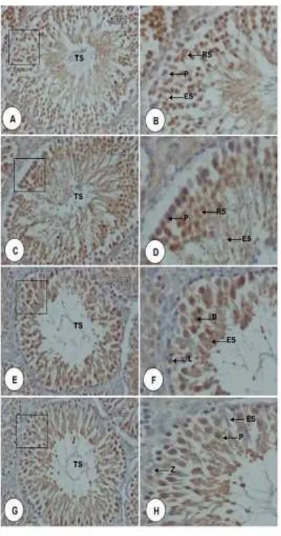
and the treatment groups. Althought K0 vs K1 and K2 vs K3 showed no significant differences (P> 0.05), Duncan test revealed significant differences between K0 vs K2, K3 and K1 vs K2 and K3. CREM expressions in the seminiferous tubules were indicated by the formation of brown color on spermatid cells that was showed in Figure 6.
Figure 6. Micrographs of CREM expression in spermatid cells of seminiferous tubules rat testicular tissue. A. Stage IV, C. Stage VI, E. Stage X, and G. Stage XIII. B, D, E, dan F were an inset of A,C, E, dan G. Seminiferous tubules (TS), round spermatids (RS), elongated spermatids (ES), pachyten (P), elongating spermatids (ES), diakinase (D), leptoten (L), and zigoten (Z). Staining
of Immunohistochemistry, 40 x
magnification (A, C, E, and G) and 100 x magnifications (B, D, E, and F).
The reduction of CREM expression in seminiferous tubules associated with the active compounds activity contained in Centella leaf extracts, especially saponins and tannins. Antifertility compounds in medicinal plants have two different effect such as cytotoxic and hormonal effects which inhibit the derivative rate of spermatogonia by interfering with hormonal system balance. Saponins and tannins are cytotoxic for tumor cells, both of which have cytotoxic effect on the seminiferous tubules of spermatogenic cells like spermatids and spermatozoa (Herdiningrat, 2002). Tannins are able to bind proteins and ions contained in sperm membrane which disrupt tyrosine activity and phosphorylation process that causes sperm morphology became abnormal (Zhou et al., 2013)
According to Hartini (2011), another possibility is affected by flavonoids activity. Flavonoids can inhibit aromatase, an enzyme that convert androgens into estrogen, to increases testosterone. Testosterone in high level lead to a negative feedback mechanism (feedback) on hypothalamus and pituitary. Testosterone will inhibit hypothalamus to produce GnRH. Thus, GnRH level will decrease and inhibit anterior pituitary to produce FSH and LH.
Steroid has responsibility in inhibiting secretion of FSH and LH (Raqifa et al.,2013). The decline in LH concentrations can affect the activity of the Leydig cells to synthesize testosterone, which results in the reduction of testosterone
production (Zhang, 2003). The
administration of Centella leaf extract in male rats can decrease testosterone level (Toras, 2014). Inhibition of FSH, LH and testosterone can disrupt spermatogenesis.
J. Trop. Vet. Biomed. Res.2:6-14
10 cAMP (cyclic adenosine monophosphate) -
protein kinase A (PKA) pathway (Walker and Cheng, 2005). In addition, FSH can stimulate Sertoli cells to produce androgen binding protein (ABP) and inhibin. ABP has an important role in transporting high level of testosterone to lumen seminiferous tubules. In fact, testosterone will not be able to enter tubules lumen without ABP. Meanwhile, inhibin is responsible for inhibiting the formation process of FSH (Akbar, 2010).
The inhibition of LH secretion would disrupt signaling LH in Leydig cells to produce testosterone. It will also disrupt MAP kinase pathway and calcium pathway in Sertoli cells (Walker and Cheng, 2005). The disruption of FSH and testosterone signaling pathway in Sertoli cells will affect the phosphorylation of cAMP element binding protein (CREB) and cAMP responsive element modulator (CREM). It is also reported by Macho et al., (2002) which stated that the inhibition of CREM activation caused a complete block toward the process of spermiogenesis in mice. Thus, it will cause failure in round spermatids formation. Sihombing (2014) reported that the administration of Centella leaf extract can reduce the number of spermatid cells (round spermatids, elongating spermatids and elongated spermatids) which eventually lead to infertility in male rats (Blendy et al., 1996).
Wistuba et al., (2002) reported that malfunctioning or CREM deficiency will cause infertility in mice as a result of spermiogenesis disruption in round spermatid phase. Spermatogenesis process will take place properly, if the hypothalamus, pituitary gland and gonads in a normal condition. However, in this study, the relationship between FSH and testosterone pathway disruption with the administtration of centella leaf extract were not fully understood.
Conclusion
The administration of centella leaf extract with serial doses 125, 250 and 500 mg/kg body weight (BW) can reduce CREM expression. The most efective dose to
decrease CREM expression in spermatid cells of seminiferous tubules in white male rats (Rattus norvegicus) is 500 mg/kg BW.
Acknowledgments
This study was funded by the Rector of syiah kuala university under the
Chairman of Program Kreativitas
Mahasiswa Penelitian (PKMP) Syiah Kuala University.
References
Adha, A.C., (2009). Pengaruh Pemberian Ekstrak Etanol Daun Alpukat
(Persea americana Mill.) terhadap
Aktivitas Diuretik. Skripsi. Fakultas Kedokteran Hewan Institut Pertanian Bogor. Bogor.
Akbar, B. (2010). Tumbuhan dengan Kandungan Senyawa Aktif yang Berpotensi Sebagai Bahan Antifertilitas. Adabia Press UIN, Jakarta.
Anggraeni, S. (2013). Uji Aktifitas Penghambatan Batu Ginjal (Anti Nefrolitiasis) Ektrak Etanol dari Herbal Pegagan (Centella asiatica
(L.) Urban) pada Tikus Putih Jantan. Skripsi. Fakultas Kedokteran dan Ilmu Kesehatan. Jakarta.
Blendy, J.A., K.H. Kaestner., G.F. Weinbauer., F. Nieschlag, and G. Schutz. (1996). Severe impairment of spermatogenesis in mice lacking the CREM gene. Nature. (380): 162-165.
Brinkhaus B., M. Lindner., D. Schuppan, and E.G. Hahn. (2000). Chemical, pharmacological and clinical profile of the East Asian medical plant Centella asiatica. Phytomedicine. (7): 48-427.
D’Auria, G., L. Paolillo, R. Sartorio, and S.
11
Guyton, A., and J. Hall. (1997). Buku Ajar Fisiologi Kedokteran. Edisi ke-9. Diterjemahkan oleh Setiawan, I. EGC, Jakarta.
Hartini. (2011). Pengaruh Dekok Daun Jambu Biji Merah (Psidium guajava L.) terhadap Jumlah Kecepatan Dan Morfologi Spermatozoa Tikus Putih Jantan (Rattus norvegicus) Tesis. Program Studi Ilmu Biomedik. Universitas Andalas Padang. Hasanah, I.W. (2009). Pengaruh Ekstrak
Daun Pegagan (Centella asiatica) terhadap Spermatogenesis Mencit
(Mus musculus). Skripsi. Jurusan
Biologi Fakultas Sains dan Teknologi UIN Maulana Malik Ibrahim. Malang.
Herdiningrat, S. (2002). Efek pemberian infus buah manggis muda (Garcinia
mangostana Linn) terhadap
spermatozoa mencit (Mus musculus).
Majalah Andrologi Indonesia.
10:130.
Kiernan, J.A. (1990). Histological and Histochemical Methods: Theory and Practice. 2nd ed. Pergamon Pr, England.
Macho, B., S. Brancorsini, G.M. Fimia, M. Setou, N. Hirokawa, and P. Sassone-Corsi. (2002). CREM-dependent transcription in male germ cells controlled by a kinesin. Science. (298): 2388–2390.
Masyhud. (2010). Tanaman Obat Indonesia. http://www.dephut. go.id/indexphp? =id /node/54. (12 Januari 2011). Orhan, I. E. (2012). Centella asiatica (L.)
Urban: From Traditional Medicine to Modern Medicine with
Neuroprotective Potential.
Evidence-Based Complementary and
Alternative Medicine Volume 2012, Article ID 946259, 8 pages.
Raqifa., A. Ramadhan, dan D. Tureni (2013). Pengaruh Pemberian Ekstrak Buah Terung Belanda (Solanum
Bataceum) Terhadap Morfologi Dan
Motilitas Spermatozoa Mencit (Mus
Musculus) Galur Ddy. e-Jipbiol. (1):
50-56.
Sassone-Corsi, P. (1998). CREM: a master-switch governing male germ cell differentiation and apoptosis.
Seminars in Develop. Biology, (9):
475-482.
Sihombing, W. (2014). Efek Ekstrak Daun Pegagan (Centella asiatica (L). Urban) terhadap Perkembangan Sel Spermatid Tikus (Rattus norvegicus). Skripsi. Fakultas Kedokteran Hewan Universitas Syiah Kuala. Banda Aceh.
Solihati, N., P. Purwantara, I. Supriatna, dan A. Winarto (2013). Perkembangan sel-sel spermatogenik dan kualitas sperma pascapemberian ekstrak pegagan (Centella asiatica). JITV
18(3): 192-201.
Toras, M. (2014). Pengaruh Pemberian Ekstrak Daun Pegagan (Centella
asiatica)terhadap Konsentrasi
Testosteron pada Tikus Putih Jantan
(Rattus norvegicus). Skripsi.
Fakultas Kedokteran Hewan Universitas Syiah Kuala. Banda Aceh.
Walker, W.H., and J.F. Habener. (1996). Role of transcription factors CREB and CREM in cAMP-regulated transcription during
spermatogenesis. Trends Endoc.
Meta. (7): 133-138.
Wistuba, J., S. Schlatt, C. Cantauw, V.V. chönfeldt, E. Nieschlag, and R. Behr. (2002). Transplantation of wild-type spermatogonia leads to complete spermatogenesis in testes of cyclic
3’,5’-adenosine monophosphate
response element modulator-deficient mice. Biol. Reprod.
(67):1052-1057.
Zhang, F.P. (2003). The low gonadotropin independent constitutive production of testicular testosteron is sufficient to mainten spermatogenesis. PNAS.
(23): 13692-13697.
J. Trop. Vet. Biomed. Res.2:6-14
12 Zhou, B., Z. Qiu, G. Liu, C. Liu, and J.