The LDL receptor is the major pathway for
b
-VLDL uptake by
mouse peritoneal macrophages
Ste´phane Perrey
a, Shun Ishibashi
a,*, Tetsuya Kitamine
a, Jun-ichi Osuga
a,
Hiroaki Yagyu
a, Zhong Chen
a, Futoshi Shionoiri
a, Yoko Iizuka
a, Naoya Yahagi
a,
Yoshiaki Tamura
a, Ken Ohashi
a, Kenji Harada
a, Takanari Gotoda
a,
Nobuhiro Yamada
baDepartment of Metabolic Diseases,Faculty of Medicine,Uni6ersity of Tokyo,7-3-1Hongo,Bunkyo-ku,Tokyo113-8655,Japan bDepartment of Endocrinology,Metabolism and Atherosclerosis,Institute of Clinical Medicine,Uni6ersity of Tsukuba,1-1-1Tennodai,Tsukuba,
Ibaraki305-8575,Japan
Received 9 August 1999; received in revised form 7 February 2000; accepted 2 March 2000
Abstract
In order to determine the contribution of the low density lipoprotein receptor (LDL-R) to the removal of apoB-containing native lipoproteins by macrophages, we compared the uptake ofb-VLDL in peritoneal macrophages (MPM) from wild type mice and mice lacking the LDL-R. ThedB1.006 g/ml lipoproteins obtained from apoE deficient mice fed a high fat diet were poorly degraded by macrophages and caused only a slight formation of CE in macrophages from both types of mice. On the other hand,
dB1.006 g/ml lipoproteins obtained from LDL-R deficient mice fed a high fat diet,b-VLDL with apoE, were avidly taken up by and markedly stimulated CE formation in wild type macrophages, but not in macrophages lacking the LDL-R. The degradation of125I-labeled-apoE-containingb-VLDL by wild type MPM was poorly inhibited by unlabeled human LDL, and
b-VLDL without apoE had no effects. In conclusion, we propose that the in vitro uptake of native apoE-enriched lipoproteins by murine macrophages is primarily mediated by the LDL receptor and not by other apoE-recognizing receptor systems such as: the LDL receptor related protein, the VLDL receptor or the triglyceride-rich lipoprotein receptor. © 2001 Elsevier Science Ireland Ltd. All rights reserved.
Keywords:Foam cell formation; Knock-out mice; Receptor
www.elsevier.com/locate/atherosclerosis
1. Introduction
An early event in the development of an atheroscle-rotic lesion is the intimal accumulation of lipid-laden foam cells which originate from either monocyte-macrophages or vascular smooth muscle cells [1]. It is believed that foam cells are generated by cellular
up-take of lipoproteins through pathways independent of the low density lipoprotein receptor (LDL-R), because massive accumulation of foam cells is found even in atherosclerotic lesions of patients who completely lack the LDL-R, i.e. homozygous familial hypercholes-terolemia (see Refs. [2,-4] for review). Furthermore, the LDL-R is barely detectable in lesions from patients
Abbre6iations: apo, apolipoprotein; BSA, Bovine Serum Albumin; CE, cholesterol ester; CHO, Chinese Hamster Ovary; DMEM, Dulbeco modified Eagle’s medium; EDTA, Ethylenediaminetetra-acetic; FCS, fetal calf serum; FH, familial hypercholesterolemia; LDL, low density lipoproteins; LDL-R(− / −), LDL-R deficient; LPDS, lipoprotein deficient serum; LPL, lipoprotein lipase; LRP, low density lipoprotein receptor-related protein; MPM, mouse peritoneal macrophages; PAGE, polyacrylamide gel electrophoresis; PBS, phosphate buffered saline; PMSF, phenylmethylsulphonylfluoride; SDS, Sodium Dodecyl Sulphate; TGRLP, triglyceride-rich lipoproteins; TLC, thin layer chromatography; WHHL, Watanabe Heritable Hyperlipidemic; VLDL, very low density lipoprotein; b-VLDL, b-migrating very low density lipoprotein; b-VLDL-E(+),dless than 1.006 g/ml lipoproteins obtained from the LDL receptor knockout mice fed an atherogenic diets;b-VLDL-E(+),d
less than 1.006 g/ml lipoproteins obtained from apolipoprotein E knockout mice fed an atherogenic diets. * Corresponding author. Tel.: +81-3-3815-5411 (ext. 33113, 33129); fax: +81-3-5802-2955.
E-mail address:[email protected] (S. Ishibashi).
with the LDL-R [5,6]. In support of this notion, numer-ous receptors, which recognize and take up denatured lipoproteins have been identified in macrophages. These include scavenger receptor A [7,8], CD36 [9], Fc recep-tor [10], macrosialin [11] and others [12]. Furthermore, modified lipoproteins have been demonstrated in atherosclerotic lesions [13,14].
In the development of atherosclerosis, lipoprotein retention precedes lipoprotein denaturation [15]. There-fore, the mechanisms by which lipoproteins deposited in the extracellular milieu are eliminated before they are denatured are no less important than are those of foam cell formation. Receptor systems, which mediate the uptake of native lipoproteins, account for the elimina-tion by surrounding cells of native lipoproteins, which are retended in the extracellular matrix. The LDL-R is expressed in macrophages and mediates the uptake of
LDL,b-VLDL and apoE-richb-migrating VLDL [16 –
19] The LRP is widely expressed in various tissues including macrophages [20 – 22] and potentially medi-ates the uptake of lipoproteins that are rich in apoE or LPL [23 – 25]. Finally, the VLDL receptor (LR8) also
binds tob-VLDL [26,27]. In addition to these receptors
belonging to the LDL receptor gene family, other re-ceptors such as lipolysis-stimulated receptor (LSR) [28] and triglyceride-rich lipoprotein receptor (TGRLP re-ceptor) [29,30] have been postulated to be involved in the uptake of triglyceride-rich lipoproteins, although their molecular identity is yet to be determined. Among these receptors for native lipoproteins, LRP [3,31] and the VLDL receptor [32], but not the LDL-R [3,4], have been demonstrated to be present in atherosclerotic le-sions, implicating their involvement in atherosclerosis.
b-VLDL is an atherogenic lipoprotein that is found
in the plasma of animals, which are fed a high
choles-terol diet. In vitro, b-VLDL has been shown to cause
the conversion of macrophages into foam cells [16]. Murine models of both LDL-R [33] and apoE defi-ciency [34,35] have been created by targeted disruption of these genes in embryonic stem cells, providing valu-able tools for the detailed analysis of the pathways involved in the uptake of native lipoproteins [36,37]. In the current study, we made use of these animals to re-examine the roles of the LDL-R and apoE in cellular
cholesterol homeostasis in mouse peritoneal
macrophages (MPM) from wild-type and LDL-R knockout mice.
2. Materials and methods
2.1. cDNA
A plasmid containing mouse LDL-R cDNA [33] was
digested with EcoRI, religated and used as a template
to synthesize an antisense riboprobe spanning from
nt233 to nt801 of the cDNA sequence. Polyclonal anti-body against the human LDL-R was a generous gift from Drs Ho, Goldstein and Brown.
2.2. Animals
Generation of the mutated mice lacking the LDL-R [33] and apoE [34] was described in the indicated references. These animals were allowed access to food and water ad libitum. Two diets were used: (i) a normal chow (MF from Oriental Yeast, Tokyo); and (ii) an atherogenic diet which contained 1.25% cholesterol 7.5% cocoa butter, 7.5% casein, and 0.5% cholic acid (Oriental Yeast, Tokyo). The wild-type mice had a genetic background similar to that of the other two mutant mice; hybrids of C57BL6 and 129Sv.
2.3. RNase protection assay
The 33P-labeled riboprobes for the LDL-R and b
-actin were synthesized from linearized plasmid tem-plates by reverse transcriptase using MAXI scripts™ (Ambion, Austin, TX). The specific activity of the
b-actin riboprobe was one tenth that of the LDL
receptor riboprobe. An RNase protection assay was performed using RPAII™ (Ambion, Austin, TX) ac-cording to the manufacturer’s instruction.
2.4. Macrophage collection
One ml of 5% thioglycollate broth was injected into the peritoneal cavities of mice aged between 2 and 6 months. After 4 days, the peritoneal cavities were lavaged with 10 ml of ice-cold saline. The cells were washed three times with PBS and resuspended in
DMEM to give a concentration of 106cells/ml. A total
of 1 ml/well and 10 ml/dish were used for plating in 12
well-plates and in 55 mm2 dishes (Corning),
respec-tively. After incubation at 37°C for 2 h, the non-adher-ent cells were removed by washing three times with pre-warmed PBS and the adherent cells were incubated
with DMEM containing 5 mg/ml of LPDS for 36 h at
37°C in an atmosphere of 5% CO2 and 95% air unless
otherwise stated.
2.5. Immunoblot analysis
MPM from five mice were scraped in 1 ml of ice-cold Buffer A containing 20 mM Tris – HCl, pH 8.0, 1 mM
CaCl2, 150 mM NaCl and a cocktail of protease
in-hibitors (1 mM PMSF, 1 mM o-phenanthroline, 1 mM
leupeptin, 5mg/ml aprotinin and 1mg/ml pepstatin) and homogenized in a Potter – Elvehjem homogenizer. The
homogenate was centrifuged at 2000×gfor 10 min and
the resulting supernatant was centrifuged at 100 000×g
B containing 250 mM Tris – maleate pH 6.0, 2 mM
CaCl2 and 1% (v/v) Triton X-100 supplemented with
the mixture of protease inhibitor. Resuspended pellets
were centrifuged at 100 000×g for 60 min. The
super-natant, the Triton X-100 soluble fraction, was stored at
−70°C for up to 2 weeks. Thirty micrograms of
protein was subjected to 7% SDS-polyacrylamide gel electrophoresis. After transfer to a nitrocellulose mem-brane (Hybond ECL™, Amersham), the LDL receptor was detected with the polyclonal antibody using the ECL Western blotting system (Amersham) according to the manufacturer’s procedure.
2.6. Lipoproteins
Mice were fed an atherogenic diet for 2 weeks, and blood was collected from the retro-orbital venous plexus into tubes containing EDTA after an overnight
fast. The dB1.006 g/ml lipoproteins were isolated by
ultracentrifugation at a density of 1.006 g/ml for 16 h.
b-VLDL containing apoE (b-VLDL-E(+)) and b
-VLDL without apoE (b-VLDL-E(−)) were prepared
from the LDL-R deficient mice and apoE knock-out mice, respectively. Blood was drawn from healthy
vol-unteers and LDL (d=1.019 – 1.063 g/ml) and LPDS
(d\1.21 g/ml) were prepared by step-wise
ultracen-trifugation as described [38]. After re-cenultracen-trifugation, the lipoprotein and LPDS solutions were dialyzed against 2 l of 2 mM sodium phosphate, pH 7.4, 150 mM NaCl,
0.01% EDTA and 0.01% NaN3. After dialysis against
saline, protein concentrations were determined using a BCA™ protein assay reagent kit (Pierce, IL), Total cholesterol, free cholesterol, triglycerides and phospho-lipids were determined using the determiner TC-555, determiner L FC, determiner TG-S555 and determiner L PL (Kyowa Medex, Tokyo, Japan), respectively.
Lipoproteins were radioiodinated by the iodine
monochloride method as described [39]. LDL were acetylated as described [40]. These lipoproteins were used within 1 week after preparation.
2.7. Uptake and degradation of the radioiodinated lipoproteins
The cells were incubated with a medium containing radioiodinated lipoprotein with or without unlabeled
lipoproteins and 5 mg/ml of BSA for 5 h. The medium
was removed and the radioiodinated lipoproteins de-graded by the cells were measured according to Basu et al. [40].
2.8. Cholesterol ester formation assay
Cholesterol ester (CE) formation from [1-14
C]oleate was determined essentially as described [40]. Briefly, the cells were incubated in a medium containing
lipo-proteins, the [1-14C]oleate-albumin complex and 5 mg
/ ml of BSA at 37°C for 24 h. Cells were washed twice
with PBS containing 2 mg/ml BSA and once with PBS.
CE was extracted with hexane-isopropanol (3:2) to
which [3H]cholesteryl-oleate and unlabeled oleic acid
were added as the internal standard and carrier, respec-tively. The organic phase was evaporated to dryness
under flowing N2. Cholesterol [1-14C]oleate was
sepa-rated on silica coated TLC plates (c5583, Merck,
FRG) using a solvent system composed of heptane/
ethyl ether/acetic acid (90:30:1; v/v/v), their position was identified using a BAS2000 phosphoimager (Fuji Film, Tokyo, Japan). The spots corresponding to CE were scraped and their radioactivities were quantified by liquid scintillation. Cellular protein was dissolved in 0.1 N NaOH and its concentration was determined by the Lowry method [41].
2.9. Cellular cholesterol mass measurement
Lipids were extracted from cultured monolayers of MPM by hexane-isopropanol. TC and FC were deter-mined by fluorometric microassay according to a modified method of Heider and Boyett [42]. Cellular protein was dissolved in 0.1 N NaOH and its concen-tration was determined by the Lowry method [41].
2.10. Statistics
A two-tailed Student’s t-test was used to assess the
significance of difference between two groups.
3. Results
3.1. Lipoproteins
The results of the SDS/PAGE analysis of the
apolipoprotein composition of the lipoproteins used for the experiments are shown in Fig. 1(A). ApoB-100, apoB-48 and apoE were the major apolipoprotein
con-stituents of the b-VLDL-E(+) and the apoB-48 and
apoB-100 concentrations were similar. In contrast, the
b-VLDL-E(−) contained apoB-48, apoA-IV and
apoA-I, but lacked apoE. Upon agarose gel
elec-trophoresis (Fig. 1(B)), b-VLDL-E(+) hadb-mobility,
whileb-VLDL-E(−) migrated faster and had mobility
between the b and pre-b positions, suggesting that
b-VLDL-E(−) was charged more negatively than b
-VLDL-E(+). The ratios of free cholesterol, cholesterol
ester and phospholipids to the proteins of b
-VLDL-E(+) were similar to those of b-VLDL-E(−) (Table
1). The ratio of triglycerides were higher in b
-VLDL-E(+) however this difference was not statistically
3.2. Expression of the LDL-R
RNase protection assays were performed to confirm the expression of the LDL-R mRNA (Fig. 2). After treatment with LPDS for 36 h, a band corresponding to the LDL-R was observed in the wild type MPM (614 bp). The mRNA for the disrupted
LDL-R was detected in the LDL-R(− / −) MPM (401
bp) and was markedly up-regulated (between 50- and 100-fold) compared to the band for the wild-type mRNA.
Immunoblot analysis was performed to estimate the LDL-R protein expression in the wild type MPM (Fig. 3). Without treatment with LPDS, the LDL receptor protein was not detectable. After treatment with LPDS for 36 h, LDL receptor expression was substantially induced.
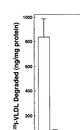
3.3. Degradation of 125I-b-VLDL-E(+) and 125I-b-VLDL-E(−) by MPM
The cellular degradation of 125I-b-VLDL-E(+) and
125I-b-VLDL-E(−) were compared in MPM obtained
from the LDL-R(− / −) and wild type mice (Fig. 4).
The MPM possessing functional LDL-R degraded
sub-stantial amounts of 125I-b-VLDL-E(+). In contrast,
the degradation of 125I-b-VLDL-E(+) in LDL-R(− /
−) MPM was less than 10% of that in wild type MPM.
In wild type MPM, 125
I-b-VLDL-E(−) were
signifi-cantly less degraded than 125
I-b-VLDL-E(+) and the
level was comparable to that of 125
I-b-VLDL-E(+)
degraded by LDL-R(− / −)MPM. There was no
dif-ference in the degradation of 125I-b-VLDL-E(
−)
be-tween the two types of cells. These results indicate that both the LDL-R on the cells and apoE on the
lipo-Fig. 1. Biochemical characteristics ofb-VLDL-E(+) andb-VLDL-E(−).b-VLDL-E(+) andb-VLDL-E(−) were isolated by ultracentrifugation at a density of 1.006 g/ml from plasma obtained from LDL-R deficient and apoE deficient mice which had been fed an atherogenic diet for 2 weeks. Twenty-five micrograms of the lipoproteins were delipidated with ethanol/ether and subjected to 5 – 15% gradient polyacrylamide SDS gel electrophoresis (A). Proteins were visualized with Coomassie Brilliant Blue R250. Rainbow marker (Amersham) was used to calibrate the molecular weight. Lane 1,b-VLDL-E(+); lane 2,b-VLDL-E(−). One microliter of the concentrateddB1.006 g/ml lipoproteins were subjected to agarose gel electrophoresis (B). Lipids were visualized with Fat Red 7B. The migratory positions are indicated. Lane 3,b-VLDL-E(+); lane 4,b-VLDL-E(−).
Table 1
Lipid composition ofb-VLDL used in the cell studiesa
Lipid/protein ratio Lipoprotein
TC FC CE TG PL
1.0190.19 0.6790.11
b-VLDL-E(+) 3.1190.73 2.4390.63 1.3390.73
0.9590.27 0.8190.21 3.1790.95
b-VLDL-E(−) 3.9891.14 0.2490.01
Fig. 2. RNase protection assay for the LDL receptor mRNA. MPM were incubated with DMEM containing 5 mg/ml of LPDS. Ten micrograms of total RNA was used for RNase protection assay for mouse LDL receptor andb-actin. Seven percent polyacrylamide gel was used for separation of the protected probes. The sizes of the protected RNA are 614, 401 and 245 bp, for the wild-type, mutated LDL receptor andb-actin, respectively.
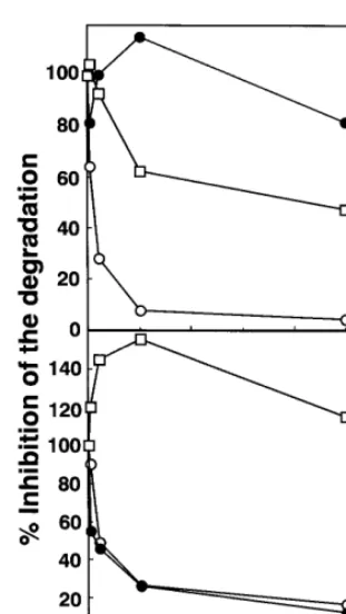
5). The degradation of125I-b-VLDL-E(
+) by wild type
MPM was almost completely inhibited by excess
amounts of unlabeled b-VLDL-E(+), while even
125-fold excess of b-VLDL-E(−) had no inhibitory effects
on its degradation (Fig. 5(A)). These results indicate
that the degradation process for b-VLDL-E(+)
pri-marily depends upon the presence of apoE. As ex-pected, 125-fold excess of human LDL reduced the degradation by 50%. This confirms that human LDL have lower affinity for receptors expressed by MPM
thanb-VLDL-E(+) [17]. On the other hand, the
degra-dation of 125
I-b-VLDL-E(−) by wild type MPM,
which was substantially lower than that of 125
I-b
-VLDL-E(+), was displaced by both unlabeled b
-VLDL-E(−) and b-VLDL-E(+) to a similar extent,
but not by LDL, suggesting that the degradation
pro-cess forb-VLDL-E(−) involves a molecule(s) common
to bothb-VLDL-E(−) andb-VLDL-E(+) (Fig. 5(B)).
Fig. 4. Cellular degradation of125I-b-VLDL-E(+) and125I-b -VLDL-E(−) in MPM. MPM were prepared from mice as described in Section 2. After incubation with DMEM containing 10% (v/v) FCS for 36 h, the cells were incubated with DMEM containing 5 mg/ml of LPDS for 36 h. A total of 2mg/ml of125I-b-VLDL-E(+) (open bar) and125I-b-VLDL-E(−) (hatched bar) were incubated with and with-out 100 mg/ml of unlabeled lipoproteins for 5 h. Data are the mean9S.D. of four wells for the wild type MPM, and the mean of two wells for the LDL-R(− / −) MPM. The total degradation by the wild type macrophages was inhibited by 85% with an excess amounts of unlabeled b-VLDL-E(+) (data not shown), indicating that a specific endocytic pathway is involved in the uptake and degradation of the125I-b-VLDL-E(+).
Fig. 3. Western blot analysis for LDL receptor protein. MPM were treated with DMEM containing either 10% (v/v) FCS (1) 5 mg/ml of LPDS (2) or for 36 h. The membrane fractions were prepared and 30 mg were subjected to 7% polyacrylamide SDS gel electrophoresis. Immunoblot analysis was performed as described in Section 2. Rain-bow marker (Amersham) was used to calibrate molecular weight.
proteins are required for the specific uptake and
degra-dation of dB1.006 lipoproteins. The amounts of 125
I-LDL degraded by wild type MPM was two-fold larger than that of125I-b-VLDL-E(−) by the same cells (data
not shown). Yet, the LDL-R(− / −) MPM did not
degrade any human125I-LDL above background levels
and this degradation could not be displaced by excess unlabeled LDL (data not shown).
Fig. 5. Inhibition of the degradation of125I-b-VLDL-E(+) (A) and 125I-b-VLDL-E(−) (B) by unlabeledb-VLDL-E(+),b-VLDL-E(−) and LDL in MPM. A total of 2mg/ml of125I-b-VLDL-E(+) (A) and 125I-b-VLDL-E(−) (B) were incubated without and with 2, 10, 50, 250mg/ml of unlabeledb-VLDL-E(+) (open circle),b-VLDL-E(−) (closed circle) and LDL (square) for 5 h. Data are presented as the means of duplicate wells. The amounts of125I-b-VLDL-E(+) and 125I-b-VLDL-E(−) degraded without excess unlabeled lipoproteins were 351 and 64 ng/mg, respectively.
3.4. Cholesterol ester formation in MPM stimulated by lipoproteins
CE formation stimulated by LDL, acetyl LDL, b
-VLDL-E(+) or b-VLDL-E(−) was compared
be-tween wild type and LDL-R(− / −) MPM (Fig. 6).
b-VLDL-E(+) substantially stimulated CE formation
in wild type MPM, but not in LDL-R(− / −) MPM. In
LDL-R(− / −) MPM, however, there was no
differ-ence in CE formation between b-VLDL-E(+) and
b-VLDL-E(−). Similarly, b-VLDL-E(−) induced
comparable CE formation in both types of
macrophages. Although b-VLDL, irrespectively of the
presence of apoE, caused CE accumulation to occur in a dose-dependent saturable manner in wild type macrophages, its effects on CE accumulation in the
LDL-R(− / −) MPM was linear (data not shown).
Compared to b-VLDL-E(+), acetyl LDL and native
LDL induced smaller CE formation.
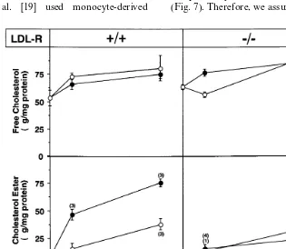
3.5. Cellular cholesterol mass in MPM stimulated by lipoproteins
Wild type and LDL-R(− / −) macrophages were
incubated with eitherb-VLDL-E(+) orb-VLDL-E(−
) and CE mass was measured (Fig. 7). While b
-VLDL-E(+) remarkably increased the CE mass in wild type
MPM, it had only a small effect in LDL-R(− / −)
MPM. b-VLDL-E(−) induced poor accumulation of
CE mass irrespective of the expression of the LDL-R by the macrophages. Free cholesterol mass was
in-creased by the addition of b-VLDL independently of
the MPM phenotype or the origin of the lipoprotein.
4. Discussion
It has been shown thatb-VLDL-E(+) strongly
stim-ulates CE accumulation in macrophages including MPM [16]. By using blocking antibody against the LDL-R, Koo et al. [17] and Ellsworth et al. [18] have
shown that the uptake of b-VLDL-E(+) by MPM is
largely mediated by the LDL-R. However, other recep-tors (i.e. LSR [28] and TGRLP receptor [29,30]) have been postulated to be involved in the uptake of triglyc-eride-rich lipoproteins, although their molecular iden-tity is yet to be determined. The use of MPM obtained from mice whose LDL-R is genetically ablated, is ideal to address the question of whether the LDL-R pathway
is involved in the uptake of b-VLDL-E(+).
Compari-son of the degradation of 125I-b-VLDL-E(+) between
wild type and LDL-R(− / −) MPM clearly indicates
that the LDL-R pathway accounts for as much as
80 – 90% of the degradation ofb-VLDL-E(+) (Fig. 4).
Previously, Whitman et al. [43] described that b
-VLDL prepared from apoE deficient mice stimulates Fig. 6. Stimulation of cholesterol ester formation byb-VLDL-E(+),
CE accumulation only slightly in J774A.1 cells, which do not express endogenous apoE. This confirmed the results of other studies using human lipoprotein in which apoE amount or ability to bind to receptors play
an important role in the CE accumulation in
macrophages [44 – 47]. In line with this, our current results showed that the presence of apoE on lipo-proteins is absolutely required for LDL-R-mediated
degradation of 125I-b-VLDL (Fig. 4). Furthermore, b
-VLDL-E(+), but not b-VLDL-E(−), completely
in-hibited the degradation of 125
I-b-VLDL-E(+), and
human LDL had intermediate effects (Fig. 5). These results implicate that the other receptor pathways, which recognize apoE, such as the LRP and VLDL receptors, play negligible roles in the uptake of apoE-rich lipoproteins by MPM.
Lipoprotein lipase is secreted by MPM [48] and potentially may mediate the direct uptake of lipo-proteins by bridging the lipoprotein to the receptor [49]. However our current results indicate that the contribu-tion of the bridge funccontribu-tion [50] of LPL is minimal in terms of overall uptake of the apoE-rich lipoproteins compared with the LDL-R-dependent pathway.
Similar studies addressing the role of the LDL-R have been performed with cells from other species such as humans and rabbits with natural mutations in the LDL-R. Koo et al. [19] used monocyte-derived
macrophages from homozygous FH individuals and concluded that the LDL-R accounts for the uptake of
b-VLDL-E(+) in these cells. Daugherty and Rateri
[51] used alveolar macrophages from WHHL rabbits, an animal model of FH, however the LDL-R in
WHHL rabbits retains binding affinity to b
-VLDL-E(+) [52] and this limits the interpretation of the
results.
Although the degradation ofb-VLDL-E(−) is lower
than that of b-VLDL-E(+) in wild type MPM, the
mechanism involved remains an interesting question. Zsigmond et al. [53] reported that serum VLDL in
apoE(− / −) mice fed a chow diet are resistant to
lipolysis by LPL, and this may affect their uptake by macrophages. However, the poor induction of
choles-terol ester formation by b-VLDL-E(−) compared to
b-VLDL-E(+) may not be explained by increased
triglyceride content since in our experimental condition where mice were fed a high fat diet, triglycerides were
not higher in b-VLDL-E(−). Another potential
expla-nation for the degradation of theb-VLDL-E(−) is that
radiolabeled apolipoprotein may be selectively taken up and therefore the degradation data do not reflect
lipo-protein particle endocytosis. However b-VLDL-E(−)
at 50 mg/ml induced a significant increase in CE mass
independently of the LDL-R expression by the MPM (Fig. 7). Therefore, we assume that degradation reflects,
at least partially, lipoprotein uptake. Since both b
-VLDL-E(+) and b-VLDL-E(−), but not LDL,
dis-placed the degradation of 125I-b-VLDL-E(
−) in a
similar manner, molecules common to these lipo-proteins mediate its degradation; apoB-48, apoA-I and apoA-IV are likely candidates (Fig. 1).
Further studies are needed to know whether the
receptor(s) responsible for the uptake ofb-VLDL-E(−
) are identical to certain known receptors such as the TGRLP receptors [29]. Recently, Hendriks et al. [54] have reported that VLDL from apoE-deficient mice were taken up by J774 macrophages, leading to an increase in CE mass, and they speculated that the triglyceride-rich lipoprotein (TGRLP) receptor pro-posed by Gianturco et al. accounted for the uptake. However we cannot rule out the possibility that slight
oxidation or aggregation of the b-VLDL-E(−)
lipo-proteins might occur during preparation and storage and may in part be responsible for the LDL-R indepen-dent uptake of the lipoproteins.
In agreement with the results of the lipoprotein degradation, the stimulation of CE formation as well as
the accumulation of CE byb-VLDL-E(+) was similar
to that ofb-VLDL-E(−) in the LDL-R(− / −) MPM
(Figs. 6 and 7). Because apoE is secreted by the LDL-R(− / −) MPM [36], these results indicate that the
secreted apoE did not further enrich theb-VLDL-E(+
) leading to uptake by the LRP that is expressed by these cells (data not shown). Furthermore, the CE
formation and accumulation stimulated by b
-VLDL-E(−) in wild type MPM were similar to those in
LDL-R(− / −) MPM (Figs. 6 and 7). This indicates
that macrophage-secreted apoE does not interact with
b-VLDL-E(−) nor does it directb-VLDL-E(−) to the
LDL-R pathway as postulated in the secretion-recap-ture hypothesis [49,55,56]. In contrast, Kowal et al. [23]
reported that b-VLDL enriched with apoE stimulates
CE formation in ldlA7 cells, CHO cells lacking the LDL-R. Potential explanations to reconcile the dis-crepancy between Kowal et al.’s and our experiments are as follows: (1) the amounts of apoE secreted by
MPM may not be sufficient to convertb-VLDL-E(−)
to be active in the stimulation of CE formation; and (2) MPM may express a lower level of LRP than CHO cells.
In conclusion, we demonstrate that the LDL-R is the
primary mechanism forb-VLDL uptake in mouse
peri-toneal macrophages and the presence of apoE at the
surface of the b-VLDL is critical for the uptake
pro-cess. Furthermore the slight uptake of b-VLDL by
LDL-R(− / −) MPM indicates that other potential
secondary mechanisms such as the LRP, b-VLDL
re-ceptor or the TGLRP rere-ceptor which may or may not
require apoE for b-VLDL uptake, play only a minor
role.
Since lipoprotein retention has been described as an important event in the initiation of the development of atherosclerotic lesions, the LDL-R may be an impor-tant factor to remove native lipoproteins inside the arterial wall prior to denaturation. The cellular choles-terol-related regulation of the LDL-R may suggest a beneficial role since the down-regulation of its expres-sion would allow cells to avoid becoming overwhelmed beyond their capacity of accommodating increased cholesterol. On the other hand, with LDL-R down-reg-ulation native lipoprotein would become trapped by the extracellular matrix and be more susceptible to oxida-tive modification, resulting in cellular uptake by scav-enger receptors, which are not subject to regulation by intracellular cholesterol.
Acknowledgements
This work was supported by Grant-in-Aid for Scien-tific Research from the Ministry of Education, Science and Culture, the Promotion of Fundamental Studies in Health Science of The Organization for Pharmaceutical Safety and Research (OPSR) and Health Sciences Re-search Grants (ReRe-search on Human Genome and Gene Therapy) from the Ministry of Health and Welfare. We thank Kimiko Saito, Emi Herai and Mihoko Kusubae for technical assistance and secretarial work, Masako Shimada and Hitoshi Shimano for helpful discussion and Alyssa M. Hasty for critical reading of the manuscript.
References
[1] Gerrity RG. The role of monocyte in atherogenesis. I. Transition of blood-borne monocytes into foam cells in fatty lesions. Am J Pathol 1981;103:181 – 90.
[2] Brown MS, Goldstein JL. Lipoprotein metabolism in the macrophages: implications for cholesterol deposition in atherosclerosis. Annu Rev Biochem 1983;52:223 – 61.
[3] Steinberg D. Oxidative modification of LDL and atherogenesis. Circulation 1997;95:1062 – 71.
[4] Goldstein JL, Hobbs HH, Brown MS. Familial hypercholes-terolemia. In: Scriver RS, Beaudet AL, Sly WS, Valli D, editors. The Metabolic and Molecular Bases of Inherited Disease. New York: McGraw-Hill, 1995:1981 – 2030.
[5] Yla¨-Herttuala S, Rosenfeld ME, Parthasarathy S, Sigal E, Sa¨rkioja T, Witztum JL, Steinberg D. Gene expression in macrophage-rich human atherosclerotic lesions. J Clin Invest 1991;87:1146 – 52.
[6] Luoma J, Hiltunen T, Sa¨rkioja T, Moestrup SK, Glieman J, Kodama T, Nikkari T, Yla¨-Herttuala S. Expression of a 2-macroglobulin receptor/low density lipoprotein receptor-related protein an scavenger receptor in human atherosclerotic lesions. J Clin Invest 1994;93:2014 – 21.
[8] Kodama T, Freeman M, Rohrer L, Zabrecky J, Matsudaira P, Krieger M. Type 1 macrophage scavenger receptor contains a-helical and collagen-like coiled coils. Nature 1990;343:531 – 5. [9] Endemann G, Stanton LW, Maden KS, Bryant CM, White RT,
Protter AA. CD36 is a receptor for oxidized low density lipo-protein. J Biol Chem 1993;268:11811 – 6.
[10] Stanton LW, White RT, Bryant CM, Protter AA, Endemann G. A macrophage Fc receptor for IgG is also a receptor for oxidized low density lipoprotein. J Biol Chem 1992;267:22446 – 51. [11] Ramprasad MP, Fischer W, Witztum JL, Sambrano GR,
Que-henberger O, Steinberg D. The 94- to 97kDa mouse macrophage membrane protein that recognized low density lipoprotein and phosphatidylserine-rich liposomes is identical to macrosialin, the mouse homologue of human CD68. Proc Natl Acad Sci USA 1995;92:9580 – 4.
[12] Elomaa O, Kangas M, Sahlberg C, Tuukkanen J, Sormunen R, Liakka A, Thesleff I, Kraaf G, Tryggvason K. Cloning of a novel bacteria-binding receptor structurally related to scavenger receptors and expressed in a subset of macrophages. Cell 1995;80:603 – 9.
[13] Haberland M, Fong D, Cheng L. Malondialdehyde-altered protein occurs in atheroma of Watanabe heritable hyperlipi-demic rabbits. Science 1988;241:215 – 8.
[14] Yla¨-Herttuala S, Palinski W, Rosenfeld ME, Parthasarathy S, Carew TE, Butler S, Witztum JL, Steinberg D. Evidence for the presence of oxidatively modified low density lipoprotein in atherosclerotic lesions of rabbit and man. J Clin Invest 1989;84:1086 – 95.
[15] Williams KJ, Tabas I. The response-to-retension hypothesis of early atherosclerosis. Arterioscler Thromb Vasc Biol 1995;15:551 – 61.
[16] Goldstein JL, Ho YK, Brown MS, Innerarity TL, Mahley RW. Cholesteryl ester accumulation in macrophages resulting from receptor-mediated uptake and degradation of hypercholes-terolemic canine b-very low density lipoproteins. J Biol Chem 1980;255:1839 – 48.
[17] Koo C, Wernette-Hammond ME, Innerarity TL. Uptake of canine b-very low density lipoproteins by mouse peritoneal macrophages is mediated by a low density lipoprotein receptor. J Biol Chem 1986;261:11194 – 201.
[18] Ellsworth JL, Kraemer FB, Cooper AD. Transport ofb-VLDL and chylomicron remnants by macrophages is mediated by the LDL receptor pathway. J Biol Chem 1987;262:2316 – 25. [19] Koo C, Wernette-Hammond ME, Garcia Z, Mallo MJ, Uauy R,
East C, Bilheimer DW, Mahley RW, Innerarity TL. Uptake of cholesterol-rich remnant lipoproteins by human monocyte-derived macrophages is mediated by low density lipoprotein receptors. J Clin Invest 1988;81:1332 – 40.
[20] Herz J, Hamann U, Rogne S, Myklebost O, Gausepohl H, Stanley KK. Surface location and high affinity for calcium of a 500 kDa liver membrane protein closely related to the LDL-re-ceptor suggest a physiological role as lipoprotein reLDL-re-ceptor. EMBO J 1988;7:4119 – 27.
[21] Moestrup S, Glieman J, Pallesen G. Distribution of the alpha2-microglobulin receptor/low density lipoprotein receptor-related protein (LRP) in human tissues. Cell Tissue Res 1992;269:375 – 82.
[22] Watanabe Y, Inaba T, Shimano H, Gotoda T, Yamamoto K, Mokuno H, Sato H, Yazaki Y, Yamada N. Induction of LDL receptor-related protein during the differentiation of monocyte-macrophages. Arterioscler Thromb Vasc Biol 1994;14:1000 – 6. [23] Kowal R, Herz J, Goldstein JL, Esser V, Brown MS. Low
density lipoprotein receptor-related protein mediates uptake of cholesteryl ester derived from apolipoprotein E-enriched lipo-proteins. Proc Natl Acad Sci USA 1989;86:5810 – 4.
[24] Beisiegel U, Weber W, Bengtsson-Olivecrona G. Lipoprotein lipase enhances binding of chylomicrons to low density
lipo-protein receptor related lipo-protein. Proc Natl Acad Sci USA 1991;88:8342 – 6.
[25] Nykjaer A, Bengtsson-Olivecrona G, Lookene A, Moestrup SK, Petersen CM, Weber W, Beisiegel U, Gliemann J. The alpha 2-macroglobulin receptor/low density lipoprotein receptor-re-lated protein binds lipoprotein lipase and beta-migrating very low density lipoprotein associated with the lipase. J Biol Chem 1993;268:15048 – 55.
[26] Takahashi S, Kawarabayashi Y, Nakai T, Sakai J, Yamamoto T. Rabbit very low density lipoprotein receptor: a low density lipoprotein receptor-like protein with distinct ligand specificity. Proc Natl Acad Sci USA 1992;89:9252 – 9.
[27] Suzuki J, Takahashi S, Oida K, Shimada A, Kohno M, Tamai T, Miyabo S, Yamamoto T, Nakai T. Lipid accumulation and foam cell formation in Chinese hamster ovary cells overexpress-ing very low density lipoprotein receptor. Biochem Biophys Res Commun 1995;206:835 – 42.
[28] Yen F, Mann CJ, Guermani LM, Hanouche NF, Hubert N, Hornick CA, Bordeau VN, Agnani G, Bihain B. Identification of a lipolysis-stimulated receptor that is distinct from the LDL receptor and the LDL receptor-related protein. Biochemistry 1994;33:1172 – 80.
[29] Gianturco SH, Lin AH-Y, Hwang S-LC, Young J, Brown SA, Via DP, Bradley WA. Distinct murine macrophage receptor pathway for human triglyceride-rich lipoproteins. J Clin Invest 1988;82:1633 – 43.
[30] Ramprasad M, Li R, Gianturco SH, Bradley WA. Purification of the human THP-1 monocyte-macrophage triglyceride-rich lipoprotein receptor. Biochem Biophys Res Commun 1995;210:491 – 7.
[31] Lupu F, Heim D, Bachmann F, Kruithof EKO. Expression of LDL receptor-related protein/a2-macroglobulin receptor in hu-man normal and atherosclerotic arteries. Arterioscler Thromb Vasc Biol 1994;14:1438 – 44.
[32] Multhaupt H, Gafvels ME, Kariko K, Jin H, Arenas-Elliot C, Goldman BI, Strauss JF, 3rd, Angelin B, Warhol MJ, McCrae KR. Expression of very low density lipoprotein receptor in the vascular wall. Analysis of human tissues by in situ hybridization and immunohistochemistry. Am J Pathol 1996;148:1985 – 97. [33] Ishibashi S, Brown MS, Goldstein JL, Gerard RD, Hammer RE,
Herz J. Hypercholesterolemia in low density lipoprotein receptor knockout mice and its reversal by adenovirus-mediated gene delivery. J Clin Invest 1993;92:883 – 93.
[34] Zhang S, Reddick RL, Piedrahita JA, Maeda N. Spontaneous hypercholesterolemia and arterial lesions in mice lacking apolipoprotein E. Science 1992;258:468 – 71.
[35] Plump AS, Smith JD, Hayek T, Aalto-Setala K, Walsh A, Verstuyft JG, Rubin EM, Breslow JL. Severe hypercholes-terolemia and atherosclerosis in apolipoprotein E deficient mice created by homologous recombination in ES cells. Cell 1992;71:343 – 53.
[36] Basu S, Brown MS, Ho YK, Havel RJ, Goldstein JL. Mouse macrophages synthesize and secrete a protein resembling apolipoprotein E. Proc Natl Acad Sci USA 1981;78:7545 – 9. [37] Ishibashi S, Herz J, Maeda N, Goldstein JL, Brown MS. The
two-receptor model of lipoprotein clearance: tests of the hypoth-esis in ‘knockout’ mice lacking the low density lipoprotein receptor, apolipoprotein E, or both proteins. Proc Natl Acad Sci USA 1994;91:4431 – 5.
[38] Havel RJ, Eder HA, Bragdon JH. The distribution and chemical composition of ultracentrifugally separated lipoproteins in hu-man serum. J Clin Invest 1955;34:1345 – 53.
[39] Goldstein JL, Basu SK, Brown MS. Receptor-mediated endocy-tosis of low density lipoprotein in cultured cells. Methods Enzy-mol 1983;98:241 – 60.
cholesterol metabolism in homozygous familial hypercholes-terolemia fibroblasts. Proc Natl Acad Sci USA 1976;73:3178 – 83. [41] Lowry O, Rosenbrough NJ, Farr AL, Randall RJ. Protein measurement with Folin reagent. J Biol Chem 1951;193:265 – 75. [42] Heider JG, Boyett RL. The picomole determination of free and total cholesterol in cells in culture. J Lipid Res 1978;19:514 – 8. [43] Whitman SC, Hazen SL, Miller DB, Hegel RA, Heinecke JW,
Huff MW. Modification of type III VLDL, their remnants, and VLDL from apoE-knockout mice by p-hydroxyphenylacetlde-hyde, a product of myeloperoxidase activity, causes marked cholesteryl ester accumulation in macrophages. Arterioscler Thromb Vasc Biol 1999;19:1238 – 49.
[44] Evans AJ, Sawyez CG, Wolfe BM, Connelly PW, Maguire GF, Huff MW. Evidence that cholesteryl ester and triglyceride accu-mulation in J774 macrophages induced by very low density lipoprotein subfractions occurs by different mechanisms. J Lipid Res 1993;34:703 – 17.
[45] Whitman SC, Sawyez CG, Miller DB, Wolfe BM, Huff MW. Oxidized type IV hypertriglyceridemic VLDL-remnants cause greater macrophage cholesteryl ester accumulation than oxidized LDL. J Lipid Res 1998;39:1008 – 20.
[46] Whitman SC, Miller DB, Wolfe BM, Hegele RA, Huff MW. Uptake of type III hypertriglyceridemic VLDL by macrophages is enhanced by oxidation, especially after remnant formation. Arterioscler Thromb Vasc Biol 1997;17:1707 – 15.
[47] Innerarity TL, Arnold KS, Weisgraber KH, Mahley RW. Apolipoprotein E is the determinant that mediates the receptor uptake of beta-very low density lipoproteins by mouse macrophages. Arterosclerosis 1986;6:114 – 22.
[48] Khoo J, Mahoney E, Witztum JL. Secretion of lipoprotein lipase by macrophages in culture. J Biol Chem 1981;256:7105 – 8. [49] Rumsey S, Obunike JC, Arad Y, Deckelbaum RJ, Goldberg IJ.
Lipoprotein lipase-mediated uptake and degradation of low
den-sity lipoproteins by fibroblasts and macrophages. J Clin Invest 1992;90:1504 – 12.
[50] Salinelli S, Lo JY, Mims MP, Zsigmond E, Smith LC, Chan L. Structure-function relationship of lipoprotein lipase-mediated en-hancement of very low density lipoprotein binding and catabolism by the low density lipoprotein receptor. J Biol Chem 1996;271:21906 – 13.
[51] Daugherty A, Rateri D. Failure of the intracellular itinerary ofb very low density lipoproteins to augment cholesterol esterifica-tion in macrophages from Watanabe Heritable Hyperlipidemic rabbits. J Biol Chem 1991;266:17269 – 75.
[52] Yamamoto T, Bishop RW, Brown MS, Goldstein JL, Russell DW. Deletion in cysteine-rich region of LDL receptor impedes transport to cell surface in WHHL rabbit. Science 1986;232:1230 – 7.
[53] Zsigmond E, Fuke Y, Kobayashi K, Chan L. Resistance of chylomicron and VLDL remnants to post-heparin lipolysis in apoE-deficient mice: the role of apoE in lipoprotein lipase-medi-ated lipolysis in vivo and in vitro. J Lipid Res 1998;39:1852 – 61. [54] Hendriks W L, van der Sman-de Beer F, van Vlijmen BJM, van Vark LC, Hofker MH, Havekes LM. Uptake by J774 macrophages of very-low-density lipoproteins isolated from apoE-deficient mice is mediated by a distinct receptor and stimu-lated by lipoprotein lipase. Arterioscler Thromb Vasc Biol 1997;17:498 – 504.
[55] Mahley RW. Apolipoprotein E: cholesterol transport protein with expanding role in cell biology. Science (Wash DC) 1988;240:622 – 30.
[56] Ishibashi S, Yamada N, Shimano H, Mori N, Mokuno K, Gotoda T, Kawamaki M, Murase T, Takaku F. Apolipoprotein E and lipoprotein lipase secreted from human monocyte-derived macrophages modulate very low density lipoprotein uptake. J Biol Chem 1990;265:3040 – 7.