Journal of Pain Research
Dove
press
open access to scientific and medical research
Open Access Full Text Article O R i g i n A L R e s e A R c h
high TnF-alpha plasma levels and macrophages
inOs and TnF-alpha expression as risk factors
for painful diabetic neuropathy
Thomas eko Purwata
neurology Department, Udayana University/sanglah general hospital, Denpasar, Bali, indonesia
correspondence: Thomas eko Purwata neurology Department, Udayana University, Jalan Diponegoro
Denpasar, Bali, indonesia Tel/Fax +62 361 246082 email [email protected]
submit your manuscript | www.dovepress.com
Dovepress
http://dx.doi.org/10.2147/JPR.S21751
Abstract: Painful diabetic neuropathy (PDN) is one of the most common complications of diabetes mellitus. Recently it has become clear that nitric oxide (NO) and proinflammatory cytokines play an important role in the pathogenesis of PDN. We investigated whether the cytokine tumor necrosis factor alpha (TNF- ) and NO play a role in PDN pathogenesis by performing a crosssectional and a case–control study in 110 type 2 diabetic patients. Of 110 subjects, 59 patients suffered from PDN (cases) and the remaining were painless DN (controls). Cross-sectionally, plasma TNF- levels and immunoreactivity for inducible NO synthase (iNOS) and TNF- were higher in patients with more severe pain on the visual analog scale. There were statistically significant differences between mild and severe pain for TNF- levels, iNOS immunoreactivity, and TNF- immunoreactivity. There were statistically significant differences between mild and severe pain for TNF- levels (mean 15.24 pg/mL ± 5.42 vs 20.44 ± 10.34),
iNOS immunoreactivity (9.76% ± 8.60% vs 15.48% ± 11.56%), and TNF- immunoreactivity
(13.0% ± 9.48% vs 20.44% ± 11.75%). The case–control study showed that TNF- had an odds
ratio of 5.053 (P , 0.001), TNF- immunoreactivity of 4.125 (P , 0.001), and iNOS immunoreactivity of 3.546 (P = 0.002). DN patients with high TNF- levels, and high iNOS and TNF- expression in macrophages are at risk of suffering from pain. The higher the TNF- level, and iNOS and TNF- immunoreactivity, the more severe the pain. These findings could form the basis of further research into better management of PDN.
Keywords: diabetic neuropathic pain, tumor necrosis factor-alpha, iNOS
Background
Uncontrolled diabetes can lead to acute and chronic complications, disability, and death. Diabetic neuropathy (DN) is a common complication of diabetes that can cause significant morbidity and mortality. It can be painful or painless. The reason for this difference in clinical presentation is as yet unknown.1 The prevalence of painful diabetic neuropathy (PDN) in a population-based sample is 26.4%.2 It can have a serious impact on quality of life, leading to severe depression and even suicide. The pathogenesis of PDN is complex and still poorly understood. In the past, research on neuropathic pain was focused on neurons and neurotransmitters. Recently it has become clear that nitric oxide (NO) and proinflammatory cytokines play an important role in the pathogenesis and maintenance of neuropathic pain. Neuroinflammation and neuroimmune activation occur following injury to peripheral nerves and nerve roots as well as in peripheral inflammatory models of persistent pain.3,4
Purwata
170 Journal of Pain Research 2011:4 Journal of Pain Research 2011:4 169–
175
© 2011 Purwata, publisher and licensee Dove Medical Press Ltd. This is an Open Access article which permits unrestricted noncommercial use, provided the original work is properly cited.
of cytokines in painful and painless neuropathies.5 Peripheral nerve injury induces activation of resident immune cells as well as recruitment of inflammatory cells to the site of lesion. There is increasing knowledge of the intracellular cascades that are activated in nociceptors by some mediators such as tumor necrosis factor alpha (TNF- ), interleukin (IL)-1 , IL-6, NO, prostaglandins (PG), nerve growth factor (NGF), and cyclo-oxygenase 2 (COX-2), which ultimately either activate or sensitize these neurons. Nerve injury also initiates Schwann cell de-differentiation and the release of several algesic mediators such as pro-inflammatory cytokines, NGF, prostaglandin E2 (PGE2), and ATP. This cocktail of mediators serves as a mechanism of enhanced inflammatory response in the injured nerve and contributes to neuropathic pain.6,7
TNF- has been shown to be directly involved in the production of pain in several models of nerve injury. The effect of TNF- on neurons seems to be mediated, directly and/or
indirectly, by the
phosphorylation of
extracellularregulated kinase and p38 mitogen-activated protein kinase,8 translocation of nuclear factor kappa B (NFκB) to the nucleus, and activation of COX-2-dependent prostanoid release.9 The phosphorylation of p38
mitogen-activated protein kinase may mediate mechanical allodynia via a modulation of tetrodotoxin-resistant Na channels.10 TNF- also activates NFκB for initiation of NOS (NO synthase), and NO production whereas NO is a pain neurotransmitter.11 Inhibition of serum TNF- and NO levels with insulin and its combinations with resveratrol and curcumin contribute in attenuating DN pain, since TNF- plays an important role in the development of DN.12
The role of NO in pathogenesis of PDN seems to be via upregulation of inducible NO synthase (iNOS) as a result of chronic inflammation. Lowenstein et al showed that there were reciprocal interactions between NO and PG, high levels of NO increased PDN, and iNOS inhibitor attenuated PDN.11 With this background, the present study was designed to explore the plasma levels of TNF- and iNOS and TNF- expression in macrophages as risk factors for PDN.
Methods
subjects
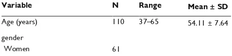
One hundred and ten type 2 diabetic patients who fulfilled inclusion criteria were enrolled in this study (61 women and 59 men, mean age 54.1, range 37–65 years). Patients were recruited consecutively at the outpatient Clinic of Neurology and Internal Medicine Sanglah Hospital Denpasar, Bali from 1 October 2008 until 31 July 2009. The patients had a mean duration of diabetes of 5.30 years (2–10 years). Of 110 subjects, 59 patients suffered from PDN (cases) and the remaining 51 patients had painless DN (controls). The clinical characteristic of subjects and comparison of case and control group are given in Tables 1 and 2.
Inclusion criteria for the cross-sectional study were type 2 diabetes patients, age 20–65 years, duration of diabetes 2–10 years, and signed written informed consent. For the case–control study, the criteria were the same as for the cross-sectional study plus painless DN for control and PDN for case.
Exclusion criteria included: ongoing local and systemic infection (documented with a blood cell count and erythrocyte sedimen rate), active ulcer in the leg, patients under treatment with glyceryl trinitrate or sildenafil, herbal or synthetic medications containing antioxidant or anti- inflammatory substances, and patients suffering from other types of neuropathic pain. To minimize confounding factors for blood cytokine levels, the study conditions were standardized by applying the following exclusion criteria: heavy physical activity in the last 3 days, food intake within 60 minutes before blood sampling, alcohol consumption up to 24 hours before the examination, and any current infectious disease or fever.
Procedure
The study was carried out in two steps. The first step was cross-sectional study and the second step was a case– control study. In the cross-sectional study, type 2 diabetic patients who visited the outpatient Neurology and Internal Medicine Clinic Sanglah Hospital underwent anamnesis,
Table 1 characteristics of diabetic neuropathy patients
Variable N Range Mean ± SD
Age (years) 110 37–65 54.11 ± 7.64 gender
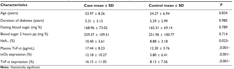
Dovepress Risk factors for painful diabetic neuropathy hbA1c, glycosylated hemoglobin; inOs, inducible nitric oxide synthase; pp, post prandial; sD, standard deviation; TnF- , tumor necrosis factor alpha.
necrosis factor alpha.
neurological, laboratory, and electromyoneurography (ENMG) examinations. Anamnesis included: sensory, motor, and autonomic symptoms of DN. The neurologic examination included: sensory, motor, autonomic functions, and reflexes. The diagnosis of a DN was based on a typical history and thorough neurologic and ENMG examinations. Laboratory studies included: erythrocyte sedimentation rate, whole blood, blood sugar, glycosylated hemoglobin
(HbA1C), urea, creatinine, and liver function test. A complete electrophysiologic assessment with standard nerve conduction studies in motor and sensory nerves at upper and lower extremities was performed in all diabetic patients. Nerve conduction studies revealed low compound muscle action potentials and sensory nerve action potentials or low conduction velocities.
Patients who had DN and fulfilled inclusion criteria were recruited in the case– control study and were asked to assess their pain intensity on a visual analog scale (VAS), with 0 representing no pain and 10 representing the worst pain imaginable. Patients were assigned to two groups according to their VAS: 1–5 mild pain and 6– 10 severe pain. Patients were examined by blood tests for plasma TNF- levels by ELISA and for iNOS and TNF- expression in macrophages immunohistochemically.
Blood for TNF- analysis was drawn in the morning, stored at 4°C, centrifuged at 1500 rpm for 10 minutes, and the plasma was stored at −20°C until assayed. The presence of TNF- in plasma was detected by a commercially available ELISA Kit
(R&D system, Wiesbaden, Germany).
Detection of TNF- and iNOS expression in macrophages was performed immunohistochemically.
Approximately 3 mL blood was collected from DN patients. The blood was centrifuged at 1500 rpm for 10 minutes and the plasma collected into small aliquots. Buffy coat layer at the interface was collected into a clean centrifuge tube. Red blood cells present in the buffy coat were lyzed using
0.8% NH4Cl for 1 minute and leukocytes were pelleted by centrifugation as above. Leukocytes were washed twice by repeated centrifugation and a thin smear of leukocytes at a density of 1 million per 1 mL phosphate-buffered saline (PBS) was prepared on poly-L-lysine microscope slides. After air drying, the leukocytes were fixed with cold acetone for 20 minutes. The endogenous peroxidase of the leukocytes was then inactivated by 3% H2O2 in PBS for 30 minutes at room temperature. After blocking with 2% skim milk in PBS, monoclonal antibodies (Mab) against TNF- (BIODESIGN International®, Saco, ME) and against iNOS (Santa Cruz Biotechnology, Inc, Santa Cruz, CA) was added into leukocytes smear on microscope slides. After three washes with PBS (pH. 7.4) and incubation for 1 hour at room temperature, the bound Mabs were detected by biotinylated goat anti-mouse IgG (BIODESIGN International) and avidin-horse radish peroxidase (Sigma-Aldrich, St Louis, MO). The expression of TNF- and
Table 2 comparison of clinical data of painful Dn (cases) and painless Dn (controls)
Characteristics Case mean ± SD Control mean ± SD P
Purwata
172 Journal of Pain Research 2011:4
iNOS was then visualized by adding diazinobenzidine substrates (Sigma-Aldrich; 50 mg/50 mL PBS containing 0.07% H2O2). The mononuclear cells expressing iNOS and TNF- were counted under 450× magnification by observation of 100 cells.
Data obtained from this study comprised descriptive analysis,
Student’s t-test, bivariate
correlation test, and cross tab (chi-square test and odds ratio). The data were analyzed using SPSS software, version 15, with significance set at P , 0.05.
Results
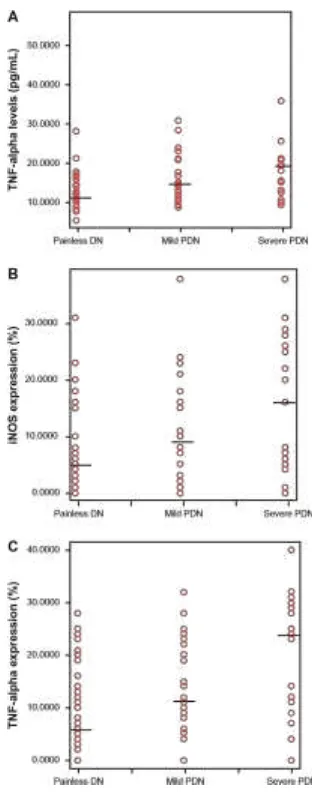
cross-sectional study
Of 110 subjects, 59 patients suffered from PDN which consisted of mild pain in 34 and severe pain in 25. Plasma TNF- levels, and iNOS and TNF- expression were higher in patients with more severe pain in the VAS (P , 0.05) as shown in Figure 1.Figure 1 scatter plots illustrating plasma TnF- levels, inOs and TnF- expression in patients with Painless Dn (n = 51 ), mild PDn (n = 34) and severe PDn (n = 25). The short horizontal line marks median values. (A) Patients with severe PDn had higher plasma TnF- levels than patients with mild PDn (P, 0.001) and Painless Dn (P, 0.001) (B) Patients with severe PDn had higher inOs expression than patients with mild PDn (P, 0.001) and Painless Dn (P, 0.001). (C) Patients with severe PDn had higher plasma TnF- levels than patients with mild PDn (P, 0.001) and Painless Dn (P, 0.001). in patients with PDn plasma TnF- levels were 1.4 (P, 0.05), inOs expression was 2.1 (P, 0.05), and TnF- expression were 1.98 (P, 0.05) fold higher than in Painless Dn. Abbreviations: PDn, painful diabetic neuropathy; TnF- , tumor necrosis factor alpha; hbA1c, glycosylated hemoglobin; inOs, inducible nitric oxide synthase; pp, post prandial.
Correlation between pain intensity and risk factors for PDN (age, duration of diabetes, blood sugar level, HbA1C, plasma TNF- level, TNF- , and iNOS expression) are given in Table 3.
Correlation between pain intensity and age, duration of diabetes, fasting blood sugar, blood sugar 2 hours postprandial, and HbA1C were not statistically significant (P . 0.05) whereas correlation between pain intensity and plasma TNF- level, TNF- , and iNOS expression were statistically significant (r = 0.330, 0.284, and 0.275, respectively; P , 0.05). To investigate the correlation
Table 3 correlation between pain intensity and some risk factors of painful diabetic neuropathy
Dovepress Risk factors for painful diabetic neuropathy
173
R P
Age −0.144 0.275 Duration of diabetes −0.101 0.448 Fasting blood sugar 0.196 0.136 Blood sugar 2 hours post prandial 0.177 0.180 hbA1c 0.138 0.298
Plasma TnF- 0.330 0.011a
inOs expression 0.284 0.029a
TnF- expression 0.275 0.035a Note:aStatistically significant.
Abbreviations: TnF- , tumor necrosis factor alpha; hbA1c, glycosylated hemoglobin; inOs, inducible nitric oxide synthase.
between plasma TNF- , and iNOS and TNF- expression in macrophages as risk factors of PDN, a bivariate correlation test was conducted. There were positive correlations between 3 risk factors above that statistically significant (P , 0.05). Correlation between plasma TNF- and iNOS expression was moderate (r = 0.351), correlation between plasma TNF- and TNF- expression was also moderate (r = 0.389), whereas correlation between TNF- and iNOS expression was macrophages, the more severe the pain. There were correlations between the three risk factors above.
case-control study
DN patients with high plasma TNF- levels had a higher risk of getting PDN than those with low plasma TNF- levels (odds ratio [OR] 5.053, 95% confidence interval [CI] 2.241–11.392; P , 0.001).
DN patients with high TNF- expression had a higher risk of getting PDN than those with low TNF- expression (OR 4.125; 95% CI, 1.805–9.425; P , 0.001).
DN patients with high iNOS expression had a higher risk of getting PDN than those with low TNF- expression (OR 3.546; 95% CI, 1.613–7.795; P , 0.002).
High HbA1C levels did not increase the risk of getting PDN compared with low plasma HbA1C levels (OR 2.380; 95% CI, 0.996–5.688; P . 0.05).
In the case-control study, plasma TNF- was consistently the most important risk factor for PDN, both directly and indirectly, by increasing macrophage expression of TNF- and iNOS.
Discussion
In this study of 110 subjects who fulfilled cross-sectional inclusion criteria, all subjects also fulfilled inclusion criteria for the case–control study; 59 subjects suffered from PDN and 51 subjects suffered from painless DN. All patients who had had diabetes for 2 years or more suffered from DN, a finding supported by previous work that showed that DN as a complication of diabetes was already present in the early stage of hyperglycemia before clinical diagnosis.13
Plasma TNF- and expression of iNOS and TNF- in macrophages according to pain intensity are compared in Figure 1. The higher the plasma TNF- levels, and the iNOS and TNF- expression in macrophages, the more severe the pain. These differences were statistically significant. These results are in agreement with those of a previous study of cytokine expression in nervus suralis biopsy specimen of neuropathy patients. Patients with painful neuropathies showed a stronger
immunoreactivity than patients with nonpainful neuropathy.14 Another study reported that patients with a painful neuropathy had about 2-fold higher TNF- mRNA and protein levels than healthy control subjects, and about 2-fold higher IL-2 and TNF mRNA and protein levels than patients with painless neuropathy (all differences significant).15
Purwata
174 Journal of Pain Research 2011:4
In our study, the highest levels of plasma TNF- , and
iNOS and TNF-
expression, were also found in patients with PDN. Pro-inflammatory cytokines (IL-1 , IL-2, IL-6, IFN- , TNF-) in the plasma were correlated with increasing pain intensity. Chronic pain patients show a significant increase in plasma levels of NO in comparison with healthy controls.4 Another study also showed a correlation between pro- inflammatory cytokine and pain intensity or severity of the disease in juvenile rheumatoid arthritis18 and fibromyalgia.19
There were positive correlations between pain intensity and plasma TNF- level, and iNOS and TNF- expression, and also a moderate to strong correlation between these three risk factors. Correlation between pain intensity and age, duration of diabetes, fasting blood sugar, blood sugar 2 hours postprandial and HbA1C were not statistically significant, indicating that above factors were not proven to increase PDN.
According to these results and those of some other
investigations, a
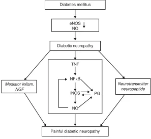
pathophysiological relationship can be shown between TNF- , iNOS, and PDN, as depicted in Figure 2.
Prolonged hyperglycemia in diabetic patients can cause diabetic pain through many mechanisms, including
metabolic, oxidative stress, immunologic, advanced glycosylation of end products, and vascular. All of these mechanisms are closely related to decreased endothelial NOS (eNOS), which is followed by a decrease in NO. NO derived from the eNOS could inhibit platelet aggregation and proliferation of smooth muscle. NO also a potent vasodilator.20 A decrease in NO will result in vessel endothelium disruption followed with nerve fiber disruption. Under chronic hyperglycemia, endogenous TNF- production is accelerated in microvascular and neural tissues, which may undergo increased microvascular permeability, hypercoagulability, and nerve damage, thus initiating and promoting the development of DN.21
Dysfunction or lesion in peripheral nerve fibers leads to membrane remodeling and hyperexcitability which in turn cause diabetic pain. Lesion in primary afferent nerve fibers will induce activation of immune resident cells and recruitment of inflammatory cells to the inflamed cell. Then inflammation cascade takes place as activated mast cells release TNF- which sensitizes nociceptors and contribute to neutrophils and macrophages recruitment. In nerve cells, neutrophils and macrophages produce and secrete neurotransmitter, neuropeptide and inflammatory mediators such as TNF- , PGE2, bradykinin, serotonin, histamine, and NGF. These mixed mediators activate nociceptors directly, cause sensitization, and contribute to neuropathic pain, which result in pain, spontaneous or primary hyperalgesia.6
Dovepress Risk factors for painful diabetic neuropathy
175
tumor necrosis factor alpha.
a positive feedback loop in which NO might activate transcription factors, which in turn could increase NO synthesis by causing additional iNOS expression.
The role of NO is thought to be through increased iNOS following chronic inflammation in diabetic pain. Strong evidence supports a reciprocal relationship
between NO and PGs
biosynthetic pathways.23 The interaction between NO and PGs biosynthesis occurs in some levels. NO directly influences COX expression and PGs biosynthesis. Conversely, arachidonic acid and its metabolites, which are produced by COX isoform, also have an effect on NO biosynthesis. The ability of NO to directly activate COX-2 is supported by evidence that NO increases purified
COX-2 recombinant enzymes. NO also influences COX-COX-2 enzyme activities by post-transcription and translation processes which make macrophages increase their PGs production. There is evidence that NO also activates COX-2.24 NO release by constitutive NOS in basal conditions deactivates iNOS by inhibition of NFκB signal. NFκB signal is one of the signaling pathways for iNOS expression by means of extracellular mediators, including TNF- (proinflammatory cytokine) and endotoxin.16 At low NO concentration, NFκB remains inactive,23 while at high NO concentration, NFκB is not suppressed by NO, so that it activates iNOS by producing combination anion superoxide from peroxynitrite. NFκB is a strong COX-2 inducer and the inhibitory role of NO in COX-2 is mediated by NFκB inhibition. NO is produced by many types of cells in peripheral tissues after inflammation has occurred. Once expressed, iNOS produces NO in large amounts for a long period.25
High NO is related to PDN while iNOS inhibitor can decrease PDN symptoms.12,26,27 Evidence that TNF- and iNOS have important roles in the pathogenesis of PDN can lead to a new strategy of therapy – that of modulating the NO pathway and neuroimmune system. Modulation can be achieved by specifically inhibiting iNOS, antibody cytokine receptors, and TNF- .
Conclusion
DN patients with high TNF- levels, and high iNOS and TNF- immunoreactivity of macrophages, are at risk of suffering from pain. The higher the TNF- level, and iNOS and TNF- immunoreactivity, the more severe the pain. This supports the hypothesis that TNF- and iNOS have a role in PDN pathogenesis. The results of Figure 2 Pathophysiological relationship between TnF- , inOs, and painful diabetic neuropathy.
Abbreviations: enOs, endothelial nitric oxide synthase; nFκB, nuclear factor kappa beta; inOs, inducible nitric oxide synthase; nO, nitric oxide; Pg, prostaglandins; TnF- ,
Diabetes mellitus
eNOS NO
Diabetic neuropathy
Painful diabetic neuropathy TNF
NFκB
iNOS
Mediator inflam. NGF
Neurotransmitter neuropeptide
PG
Purwata
176 Journal of Pain Research 2011:4
this research could be used as a basis for further research in pursuit of better management of PDN.
Acknowledgments
The author would like to acknowledge Claudia Sommer University Bali for his support in
this research.
This research was funded by Indonesian Department of Health as part of dissertation from doctoral study of the neuropathies. Current Pain and Headache Report. 2008;12:159–164. 2. Davies M, William R, Brophy S, inflammatory mechanisms in neuropathic pain. Brain Res Rev. 2006;51:240–264.
4. Koch A, Zacharowski K, Boehm O, et al. Nitric oxide and pro-inflammatory cytokines correlate with pain intensity in chronic pain patients. Inflamm Res. 2007;56:32–37.
5. Sommer C, Schäfers M. Mechanisms of neuropathic pain: the role of cytokines. Drug Discov Today. 2004;1:441–446.
6. Moalem G, Tracey DJ. Immune and inflammatory mechanisms in neuropathic pain. Brain Res Rev. 2006;51:240–264.
7. Thacker MA, Clark AK, Marchand F, McMahon SB. Pathophysiology of peripheral neuropathic pain: immune cells and molecules. Anesth Analg. 2007;105:838–847.
8. Schäfers M, Svensson CI, Sommer C, Sorkin LS. Tumor necrosis factor-alpha induces mechanical allodynia after spinal nerve ligation by activation of p38 MAPK in primary sensory neurons. J Neurosci. 2003;23:2517–2521.
9. Dinarello CA. 1999. Overview of inflamatory cytokines and their role in pain. In : Watkins LR, Maier SF, editors. Cytokines and Pain. Basel: Birkhauser Verlag; 2003:1–39.
10. Jin X, Gerean R. Acute p38-mediated modulation of tetrodotoxinresistent sodium channels in mouse sensory neuron by tumor necrosis factor- . J Neurosci. 2006;26:246–255.
11. Lowenstein CJ, Dinerman JL, Snyder SH. Nitric oxide: a physiologic messenger. Ann Intern Med. 1994;120:227–237.
12. Sharma S, Chopra K, Kulkarni SK. Effect of insulin and its combination with resveratrol or curcumin in attention of diabetic neuropathic pain: participation of nitric oxide and TNF-alpha. Phytother Res. 2007;21:278–283.
13. Quan D. Diabetic neuropathy. http://emedicine.medscape.com/ article/1170337-overview. Accessed Sep 20, 2008.
14. Emp M, Renaud S, Erne B, et al. TNF-alpha expression in painful and nonpainful neuropathies.
Neurology. 2001;56:1371–1377.
15. Üçeyler N, Rogausch JP, Toyka KV, Sommer C. Differential expression of cytokines in painful and painless neuropathies. Neurology. 2007;69:42–49.
16. Dogonay S, Evereklioglu C, Turkoz Y, Servinc A, Mehmet N, Savli H. Comparison of serum NO,
TNF-, sIL-2RTNF-, IL-6 and IL-8 levels with grades of retinopathy in patients with diabetes mellitus. Eye. 2002;16:163–170.
17. Üçeyler N, Kafke W, Riediger N, et al. Elevated proinflammatory cytokine expression in affected skin in small fiber neuropathy.
Neurology. 2010;74:1806–1813
18. Chen DY, Lan JL, Lin FJ, Hsieh TY. Proinflammatory cytokine profiles in sera and pathological tissues of patients with active untreated adult onset Still’s disease. J Rheumatol. 2004;31:2189–2198.
19. Gur A, Karakoc K, Cevik R, Denli A, Sarac J. Cytokine and depression in cases with fibromyalgia. J Rheumatol. 2002;29:358–361.
20. De Caterina R, Libby P, Peng HB, et al. Nitric oxide decreases cytokine-induced endothelial activation: nitric oxide selectively reduces endothelial expression of adhesion molecules and proinflammatory cytokines. J Clin Invest. 1995;96:60–68.
21. Satoh J, Yagihashi S, Toyota T. The possible role of tumor necrosis factor-alpha in diabetic polyneuropathy. Exp Diabesity Res. 2003;4: 65–71.
22. Tegeder L, Niederberger E, Schmidt R, et al. Specific inhibition of IkappaB kinase reduces hyperalgesia in inflammatory and neuropathic pain models in rats. J Neurosci. 2004;24:1637–1645.
23. Mollace, V, Muscoli C, Rotiroti D, Nisticom G. Spontaneous induction of nitric oxide- and prostaglandin E2-release by hypoxic astroglial cells is modulated by interleukin 1 beta. Biochem Biophys Res Commun. 1997;238:916–919.
24. Togashi H, Sasaki M, Frohman E, et al. Neuronal (type I) nitric oxide synthase regulates nuclear factor kappaB activity and immunologic (type II) nitric oxide synthase expression. Proc Natl Acad Sci U S A. 1997;94:2676–2680.
25. Nathan C. NO as a secretory product of mammalian cells. FASEB J. 1992;6:3051–3064
26. De Alba JD, Clayton NM, Collins SD, Colthup P, Chessell IK. GW 274150, a novel and highly selective of the iNOS, shows analgesic effects in rat models of inflammatory and neuropathic pain. Pain. 2006;120:170–181.
Journal of Pain Research
Publish your work in this journal
The Journal of Pain Research is an international, peer-reviewed, open access, online journal that welcomes laboratory and clinical findings in the fields of pain research and the prevention and management of pain. Original research, reviews, symposium reports, hypothesis formation and commentaries are all considered for publication.
Submit your manuscript here: http://www.dovepress.com/journal-of-pain-research-journal
Dove
press
Dovepress Risk factors for painful diabetic neuropathy
177