. Bali Medical Journal (BMJ) 2013, Volume 2, Number 2: 51-54 P-ISSN.2089-1180, E-ISSN.2302-2914
Open access:
www.balimedicaljournal.com
or
www.ojs.unud.ac.id
51
OPERATION TECHNIQUE OF
ANTERIOR MENINGOENCEPHALOCELE:
Transcranial Combined with Anterior Resection (Case Report)
Muhammad-Zafrullah, A., Yudoyono, F., Mirna-Sobana, A., and Faried, A.
Department of Neurosurgery, Faculty of Medicine Padjadjaran University
Dr Hasan Sadikin General Hospital, Bandung-Indonesia
Meningoencephaloceles are a frequent condition characterized by protrusion of meningeal and brain
tissue through a skull-base defect. Here, we report three cases of anterior meningoencephalocele
operated on one-stage procedure, a combined of transcranial and anterior approach, for invagination
removal of non-functional extracranial cerebral tissue with watertight closure of the dural defect;
written consent was taken. All cases were nasoethmoid, the congenital anomalies between the nasal
bones and the nasal cartilage.
Keywords: Menigoencephalocele, Nasoethmoid, Transcranial and Anterior Resection
INTRODUCTION
Encephaloceles is a serious congenital anomaly characterized by protrusion of meningeal and brain tissue along the midline of the cranial vault or through a skull-base defect. The case is far less common compare to spinal dysraphism. It may content meninges (meningocele) or brain tissue and meninges (meningoencephalocele).1 Encephaloce- les occur in 1 : 5.000 to 10.000 births worldwide.2 The primary abnormality in the development of an encephalocele is a mesodermal defect resulting in a defect in the calvarium and dura associated with herniation of cerebrospinal fluid (CSF), brain tissues and meninges through defect. According to the location, encephaloceles are classified in: cranial vault, frontoethmoid, basal, occipital and posterior fossae.3 For nasal encephalocele devided into sincipital and basal variety. Sincipital form consists of nasofrontal, nasoethmoid and nasoorbital, whereas basal form consists of: transethmoidal, sphenoethmoidal, trans sphenoidal, and sphenoorbita. The commonest site of encephalocele is occipital (75-80%), followed by frontoethmoidal (13-15%), parietal (10-12%) or sphenoidal. Occipital and posterior encephalocele are more common in Western population (European and North America), whereas anterior encephaloceles are higher in Southeast Asia and Southern Asia.2,4 It is not easy to manage the congenital case of encephalocele, since there were many perioperative problem and we try to manage each cases carefully. We present here three cases with nasoethmoid meningoencephalocele that
Correspondence: Faried Yudoyono.
Address: Department of Neurosurgery, Faculty of Medicine Padjadjaran University/Dr Hasan Sadikin General Hospital, Bandung-Indonesia Email: [email protected]
was operated on one-stage procedure which combine the transcranial and anterior resection.
Cases Report Case 1
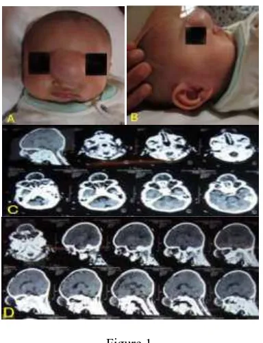
A 3 month-old baby boy was complaint by his parents, that there is a lump on his nose since he was born, the lump has gradually increased in size, impulse on crying and coughing was felt over the
swelling, the lump’s size as big as a pingpong ball (Figure 1).
Figure 1
A) and B) The features of a midline nasoethmoidal encephalocele. Note the interorbital hypertelorism, the inferior canthal dystopia; C) and D) There was bone defect at nasoethmoid bone, with cyst like
. Bali Medical Journal (BMJ) 2013, Volume 2, Number 2: 51-54 P-ISSN.2089-1180, E-ISSN.2302-2914
Open access:
www.balimedicaljournal.com
or
www.ojs.unud.ac.id
52
He was born from P2A1 mother, aterm helped by traditional midwife, spontaneous delivery, immediately crying, baby birth weight (BBW) 2800 grams, head circumference 40 cm (with normal size are 39-43 cm), painless swelling, transilumination test positive and anterior fontanelle still open but not tense. Associated hypertelorism was noted.
Case 2
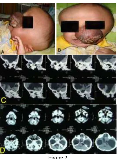
A 8 month-old baby girl had a lump on her nose since she was born, the lump getting bigger during time especially when she was crying, until the size as big as a tenis ball (Figure 2). She was born from P4A0 mother, aterm and helped by traditional midwife, spontaneous delivery, immediately crying, BBW 4000 grams, head circumference 54 cm (47-52 cm), painless swelling, transilumination test positive and anterior fontanelle still open but not tense. Associated hypertelorism was noted.
Figure 2
A) and B) This baby girl demonstrates the features of a nasoethmoidal encephalocele. Note the interorbital hypertelorism, the inferior canthal
dystopia. C) and D) There was bone defect at nasoethmoid bone with cyst like CSF content
Case 3
One-year old baby girl has had a lump on her nose since she was born, the lump as big as a bird egg, and not getting bigger during time or when patient crying or coughing (Figure 3). She was born from P3A0 mother, 36 weeks, spontaneously and helped by midwife, immediately crying, BBW 2700 gram. Fontanela anterior was closed, head circumference 42cm (41-46 cm), there was a mass
size 4x3 cm at nasion, soft and tender, translumination negative. Associated hypertelorism was noted.
Figure 3
A) and B) This baby girl demonstrates the features of a nasoethmoidal encephalocele. Note the interorbital hypertelorism. C) and D) There was
bone defect at nasoethmoid bone
Operation Technique
Inform concern for all patients were signed by their parents. This study approved with ethical clearance obtained from local authority ethical committee medic.
Imaging by computed tomography (CT) scan was carried out for all patients, herniation of meninges and brain tissue of frontal lobus through a defect in the skull base presented as a lump on the nose and suggestive at nasoethmoid (Figure 1, 2, and 3 C and D).
. Bali Medical Journal (BMJ) 2013, Volume 2, Number 2: 51-54 P-ISSN.2089-1180, E-ISSN.2302-2914
Open access:
www.balimedicaljournal.com
or
www.ojs.unud.ac.id
53
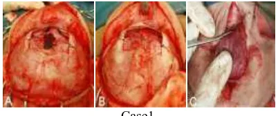
elevator. Bifrontal craniotomy was made using the Smith craniotomes and dura spatulas. The encephalocele was approached via the extradural route without injuring the dural protective layer. The nasal bone defect was identified and the herniated portion of abnormality was opened and neck of the sac was identified, after the occlution of
the sac’s pedicle, the whole sac that associated with dysplastic brain tissue was cut. The cut ends of sac closed waterthight and supported with galea tissues and 1 ml of fibrinogen product was injected onto was made in Y shape manner at the fore head-nasal junction and dysplastic brain tissue in remaining sac was removed, along with closure of skin defect made by the lump (Figure 4). All patients were in a very good condition after the surgery.
Case1
A) we exposed the invagination of the duramater, B) Duramater close with duroplasty watertight,
C) Non functional extracranial brain tissue.
Case 2
D) We cut the invagination of the meningen. E) Duramater close with duroplasty watertight.
F) Non functional extracranial brain tissue
Case 3
G) We exposed the invagination of the meningen. H) Duramater close with duroplasty watertight. I) Non functional extracranial brain tissue from
anterior.
Figure 4
Operation Technique For all Cases
DISCUSSION
Encephalocele is a severe malformation with the involvement of many facial anatomical areas, treatment of which requires a multidisciplinary neuro-craniofacial team. Fronto ethmoidal encephaloceles present with a facial mass. Nasofrontal encephaloceles appear at the root of the nose above the level of nasal bone. Nasoethmoidal encephaloceles are situated inferior to the nasal bones and naso-orbital encephalcels cause proptosis and displacement of eye ball. The incidence of sincipital encephalocele compared with occipital encephalocele is substantially greater in the tropical latitudes, particularly in parts of Asia and Africa, and its also more common in parts of Russia. However, the occipital-to-sincipital encephalomeningocele ratio is reversed in North America, Australia, Europe, and parts of South America, varies from 2.5:1 to 15:1. The occipital encephaloceles occur in 1 to 3 per 10.000 live births. The etiology of sincipital encephalocele is unknown but its includes ethnic, genetic, and environmental factors and paternal age.2,6
Anterior encephalocele are associated with hypertelorism, midline craniofacial dysraphism, agenesis of corpus callosum, lipoma and subependymal heterotopias.7 In the growing-child patient, it is necessary to reconstruct at an early age to avoid further distortion of the craniofacial anatomy. Many authors have described the various operation techniques in repairing the fronto-nasoethmoidal encephalocele. In the past, operations for meningoencephaloceles were performed in two stage procedures; as the first stage of transcranial resectione done, the second stage of anterior resection will perform six months up to 1 year after the fist surgery, waiting until the fibrotic scars occurs. Since the development of advanced neurosurgical technique, an intracranial approach combined with extracranial resection as one stage procedure is one of the option.
Details of the surgical techniques are described above. In authors opinion, the two step procedures has disadvantages which requires a longer period for perfom the second surgery and has additional psychological effect to the family and the patient; for not removed the nose-mass skin or lump skin of their children. The advantages of our transcranial combined with aterior resection are: it will profide a wide exposure of the dural defect that make us easier to close the dural in water tight manner, decrease the possibility of CSF leakage, decrease the possibility of infection even and easier to performed duroplasty compare with two stage procedure.
. Bali Medical Journal (BMJ) 2013, Volume 2, Number 2: 51-54 P-ISSN.2089-1180, E-ISSN.2302-2914
Open access:
www.balimedicaljournal.com
or
www.ojs.unud.ac.id
54
(anterior encephalocele) is usually cosmetic. The principal is to return the cerebral components in to the cranial cavity along with amputation of dysplastic tissue, closured of bony defect and reconstruct the skin. The presence of microcephaly with a large occipital encephalocele containing significant brain tissue also a predictor of poor neurological outcome.6-8
CONCLUSION
The difficulties of management of anterior meningoencephalocele can reduced with a good perioperative management and right decision to performed surgery that give more beneficial. We believe this combine technique, one stage procedure, has many advantages then the coventional two stage procedure. Thus rarity of occurrence of nasoethmoidal meningoencepha locele, highlights the importance of research in the field diagnosis and surgical management of these cases.
REFFERENCES
1. McLone DG. Congenital malformations of the central nervous system. Clin Neurosurg. 2000;47:346-77.
2. Mahapatra AK, Agrawal D. Anterior
encephaloceles: A series of 103 cases over 32 years. J Clin Neurosci. 2006;13:536-9.
3. Suwanwela C, Suwanwela N. A morphological classification on sincipital encephalo meningoceles. J Neurosurg. 1972;36:202-11. 4. Hoving EW. Nasal encephaloceles. Child’s
Nerv Syst. 2000;16:702-6.
5. Raja RA, Qureshi AA, Memon AR, Ali H, Dev V. Pattern of encephaloceles: A case series. J Ayub Med Coll Abbottabad. 2008;20:125-8. 6. Holmes AD, Meara JG, Kolker AR, Rosenfeld
JV, Klug GL. Frontoethmoidal encephaloceles: Reconstruction and refinements. J Craniofacial Surg. 2001;12:6-18.
7. Dubey D, Pande S, Dubey P, Sawhney A. A case of naso-ethmoidal meningo encephalocele. Int J Collaborative Res Internal Med Public Health 2011;3:666-7.
8. Bozinov O, Tirakotai W, Sure U, Bertalanffy H. Surgical closure and reconstruction of a large occipital encephalocele without parenchymal excision. Childs Nerv Syst 2005;21:144–7.
This work is licensed under