1
Impacted Button Battery in The Nasal Cavity
Bestari J. Budiman, Dedy Rusdi
Otorhinolaryngology Head and Neck Surgery Department Medical Faculty of Andalas University/Dr. M. Djamil Hospital
Abstract
A button battery in the nasal cavity of the children is an unnusual foreign body, which can cause liquefaction necrosis with subsequent severe local tissue destruction. All button batteries as foreign bodies in the nasal cavity should be removed immediately to prevent severe local tissue damage, resulting in late sequelae, such as septal perforation or stenosis of the nasal cavity. Sometimes their removal were relatively easy, but sometimes could be very challenging.
One case of button battery in nasal cavity in a 3 years old boy was reported, which had been performed an extraction under general anesthesia.
Key word: Button battery, nasal foreign body, septal perforation
Abstrak
Baterai kancing di rongga hidung anak-anak merupakan benda asing yang tidak biasa, yang dapat menyebabkan nekrosis disertai kerusakan jaringan yang luas. Baterai kancing sebagai benda asing di rongga hidung harus dikeluarkan sedini mungkin untuk mencegah sekuele seperti perforasi septum atau stenosis rongga hidung. Usaha untuk mengeluarkannya relatif mudah tapi kadang-kadang juga merupakan tantangan.
Dilaporkan satu kasus baterai kancing di hidung pada anak laki-laki usia 3 tahun yang telah dilakukan ekstraksi dalam narkose umum.
Kata kunci: Baterai kancing, benda asing di hidung, perforasi septum
Korespondensi: dr. Dedy Rusdi : [email protected]
INTRODUCTION
Nasal foreign bodies in children often present to both general practice and the emergency physicians. There are usually innocuos with the majority being plastic objects (particularly beads), foam, paper or cotton.1 Foreign bodies in the nasal cavity are common,
especially in children and are relatively easily removed in outpatient department, but if the foreign body is a battery, special attention must be thought, and can be very challenging. These kind of foreign bodies have the potential to cause extensive damage.2
The use of small button batteries can be attributed to the advent as well as the reduction in size of many technological devices such as hearing aids, electronic games, watches, digital planner and new electronic gadget. In addition, their smooth and shinny appearance makes them quite attractive and noticeable to children.3
Despite improvement in the safety designs of the products, children are still able to remove these batteries from the devices. Being small, they can be easily inserted into various orrifices such as the nose, ears and mouth.2,4
Inside the body cavity, surrounding moisture result in corrosion of the battery casing, thereby liberating its alkaline contents. More important, the batteries can generate localcurrents resulting in thermal
burns and production of more alkaline materials through electrolysis, resulting in extensive damage to surrounding mucosa.4
Fortunately, the majority of these batteries can be removed in outpatient clinics. In cases in which the batteries have been left in the nasal cavities for a longer duration, serosanguinous nasal discharge and crusting may obscure these foreign bodies, making their removal difficult. Removal of the batteries under general anesthesia will then become necessary.4
CASE REPORT
A 3 years old boy presented to the emergency department on March, 29th 2011 at 00.15 am with a chief
complain foreign body (a button battery) in the right nostril since 10 hours before admission. Previously the patient was playing and suddenly he inserted a battery into his right nostril. His parent didn’t try to pull out the foreign body and brought him to district hospital, and referred to M. Djamil Hospital. No history of bleeding from the nose, no choking sensation after the incident, no difficulty in breathing and no nausea and vomitting.
On physical examination, general condition was moderately ill, composmentis not cooperative, pulse rate 88 x/mnt, respiratory rate 24 x/mnt and temperature 36.80C. There were no stridor, wheezing and retraction
2
On ENT examination revealed no abnormality was detected in the ear and throat. Right nasal cavity was narrowed, inferior turbinate was edema, middle turbinate was difficult to evaluated, there was serosanguineus discharge, no bleeding, no laceration and foreign body couldn’t be seen. Left nasal cavity was wide, inferior and middle turbinate was eutrophy, no bleeding, no discharge and no foreign body was seen.
Because the foreign body could not be seen on physical examination, then we decided to performed radiological examination. Anteroposterior and lateral x-ray views of the nose revealed the presence of metallic disc foreign body in the posterior part of right nasal cavity (Figure 1 and 2).
Figure 1: Anteroposterior x-ray views
Figure 2: Lateral x-ray views
Patient was diagnosed with foreign body (a button battery) at the right nasal cavity and planned for extraction of foreign body on general anasthesia, consult to pediatric and anestesiology departement.
Laboratory finding were haemoglobin 11.4 g/dl, leucocytes 15,700/mm3, thrombocytes 407,000/mm3,
haematocrytes 35% and PT/APTT 10.1’’/42.0’’. Patient was treated with ceftriaxon injection 400 mg.
Extraction of foreign body was performed at 07.45 am. Operating report:
- Patient laid down on operation table.
- Aseptic and antiseptic procedure was performed in the operating field.
- Applied oral packing.
- Evaluation with scope 00: right nasal cavity was
narrowed, inferior turbinate was edema, middle turbinate was difficult to evaluated, there was serosanguinus discharge and necrotic tissue.
- Discharge was suctioned → a button battery was adhere to septal. There were blackish crusting and necrotic tissue
- A battery was extracted with a hook → succeded - Evaluation of right nasal cavity →necrotic tissue (+)
in septal, approximately 0.5 cm in size, minimal bleeding (+)
- Irrigation with H2O2 3% + povidone iodine and then
washed out with NaCl 0,9%.
- Evaluation of right nasal cavity →middle turbinate was eutrophy, necrotic tissue (+), minimal bleeding (+)
- Performed nasal packing with sofratulle
- Evaluation of left nasal cavity →was wide, inferior and middle turbinate was eutrophy, there was septal perforation approximately 2 mm in size, no discharge and bleeding.
- Oral packing was released and operation had been finished
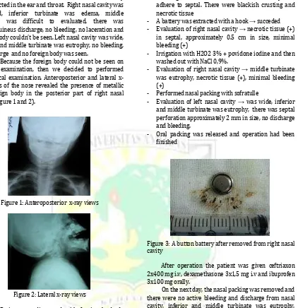
Figure 3: A button battery after removed from right nasal cavity
After operation the patient was given ceftriaxon 2x400 mg i.v, dexamethasone 3x1,5 mg i.v and ibuprofen 3x100 mg orally.
On the next day, the nasal packing was removed and there were no active bleeding and discharge from nasal cavity, inferior and middle turbinate was eutrophy, necrotic tissue and septal perforation (+). Patient was discharge with therapy amoxicillin clavulanic acid 3x125 mg and suggest patient to control on the next 7 days.
Patient controlled to ENT outpatient after 7 days ( April 5th 2011). From anamnesa there was rhinorrhoea
but there were no fever, epistaxis and noisy breathing. From examination with nasoendoscopy, right nasal cavity was wide, inferior and middle turbinate was eutrophy, there was serous discharge, necrotic tissue, nasal septal perforation 1-2 mm in size and adhesion in 1/3 middle of nasal septum. On left nasal cavity , inferior and middle turbinate was eutrophy, no discharge and bleeding, there was a nasal septal perforation. Then we released adhesion/sinechia with respatoriu and set up a handscoen nasal pack moistured with povidone iodine
and chloramfenicol cream to prevent the
3
amoxicillin-clavulanic acid 3x125 mg and suggest patient to control on the next 3 days to released nasal pack.
On April 8th 2011, patient controlled to ENT
outpatient. After released nasal pack, examination was performed with nasoendoscopy. On right nasal cavity, inferior and middle turbinate was eutrophy, no bleeding, there was serous discharge, necrotic tissue, and the nasal septal perforation still the same size with previous examination. On left nasal cavity we found nasal septal perforation in the same size with previous examination. Patient was given therapy amoxicillin-clavulanic acid 3x125 mg, ambroxol syrup 3x15 mg and suggest to control on the next 7 days.
On April 15th 2011, patient was controlled to
ENT outpatient. From anamnesa there were no fever, rhinorrhoea, epistaxis and noisy breathing. From examination on right nasal cavity, inferior and middle patient was suggest to control for a next 14 days.
On April 29th 2011, patient was controlled.
From anamnesa there were no fever, rhinorrhoea, epistaxis and noisy breathing. From examination on right nasal cavity, inferior and middle turbinate was eutrophy, no bleeding, no discharge and no necrotic tissue, but the nasal septal perforation was decrease in size from previous examination and there was a thickened on nasal septal. On left nasal cavity we found septal perforation was decrease in size from previous examination. Patient was suggest to control to ENT outpatient if there is a complaining.
DISCUSSION
The removal of foreign bodies in children is very common in the otolaryngologists daily routine. Nasal foreign bodies are commonly encountered in emergency departments. Although more frequently seen in the pediatric setting, they can also affects adults, especially those with mental retardation or psychiatric illness. Children’s interests in exploring their bodies make them more prone to lodging foreign bodies in their nasal cavities. As benign as nasal foreign body may seen, it harbors the potential for morbidity and even mortality if the object is dislodged into the airway.5,6
Despite the high frequency of foreign body insertion into the nose, there are very few studies on this problem in literature. Success in removing a foreign body nose depends on a number of factors including the size, shape and texture of the foreign body, time duration of foreign body, the cooperation of patient at the time of removal, the ability to visualize the foreign body and surrounding structure, trauma to the nasal cavity due to insertion or attempted removals of the foreign, the equipment available for removal and skill of the doctor attempting the removal.5,7
Foreign bodies can be classified as either inorganic or organic. Inorganic materials are typically plastic or a metal. Common examples include beads, button, stones, paper and small parts from toys. These materials are often asymptomatic and may be discovered incidentally. Organic foreign bodies, including food, rubber, wood, sponge, and metallic batteries, tend to be more irritating to the nasal mucosa and thus may produce earlier symptoms.5,6
Button batteries are widely used in daily life and can be easily found in electronic games, toys, calculators, watches, cameras, hearing aids, laser pointers and other electronic instruments. These batteries contain various heavy metals, including mercury, zinc, silver, nickel, cadmium, manganese, or lithium, and a concentrated alkaline electrolyte solution of 26% to 45% potassium or sodium hydroxide.8
Button batteries are found as foreign bodies in the nasal cavity, external auditory canal, esophagus and gastrointestinal tract where its battery substances can cause liquefaction necrosis by contact with the human moist tissue in a fast period of time resulting in tissue necrosis followed by perforation.9
The most common locations for nasal foreign bodies to lodge are just anterior to the middle turbinate or below the inferior turbinate. Unilateral foreign bodies affect the right side about twice as often compared to the left. This may be due to preference of right handed individuals to insert objects in their right naries.5,6,7 In this patient the foreign body was found in the right nasal cavity in front of middle turbinate.
Various reports describe the mechanism for damage to the nasal mucosa and surrounding tissue that can occur if a button battery lodges in the nose. One mechanism of injury is leakage of battery contents. Most miniature batteries contain sodium or potassium hydroxyde in the anoda mixture. A plastic seal separates the anode and catode mixture. The seal can be corroded easily in a moist environment, resulting in the leakage of the corrosive contents with secondary alkaline burns. This events could explain the observation that some patient suffer maximum tissue damage at the anode side of the battery.1,24,8,10,11 In this case, the necrotic tissue was
found in nasal septal which the anode side of battery was adhered.
Another mechanism of injury is production of local currents. When the battery is in contact with the nasal tissue, it can discharge current through the surrounding tissue. Electrons generated from the anode will combine with hydrogen ions in the tissue to form hydrogen gas. The remaining hydroxyl ion will combine with sodium or potassium ions to produce alkaline by-products, causing more damage to the surrounding tissue.1,2,4,8,10,11
4
resulting from direct currents following through the tissue. Similar thermal injury may occur in the nasal cavity.1,2,4,8,10,11
Battery lodged in a tight nasal cavity can cause pressure ischemic necrosis of the surrounding tissue. However this seems to play a minor role in tissue injury, as the batteries are usually small in comparison to the size of the nasal cavity.1,2,4,8,10,11
Liberation of the contents of the battery causes various types of lesions depending on the localization, with an intense local tissue reaction and liquefaction necrosis. The nasal injuries noted in the literatures included localized nasal mucosal necrosis, septal perforation, facial cellulitis and lateral nasal wall necrosi.1,2,4
In 2004, Loh et al4 reviewed the history of 6
children hospitalized for removal of nasal button batteries; 4 of the 6 children suffered septal perforations. The duration of button battery impaction ranged from 4 to 72 hours. The shortest duration of battery impaction for septal perforation to occur was 7 hours. In 2001, Lee
et al9 reviewed the history of 2 children age 4 and 6 years
old hospitalized for removal of nasal button batteries, both of them suffered septal perforation after impacted of button battery for 8 and 48 hours. In this patient, we found septal perforation and nasal mucosal necrosis after total duration of battery lodgement in the nasal cavity for 17 hours.
Frequently the foreign body can be seen on anterior rhinoscopy (and sometimes on posterior rhinoscopy), but on several occasions the mucosal oedema or granulation will hide it. If the foreign body is easily seen, and the patient is a cooperative child, it is usually possible to remove the object through the anterior nares, either without anesthesia, or after spraying local anesthetic solution such as tetracaine or lidocaine. In this case, the battery was not seen on anterior rhinoscopy, the patient was not cooperative and needed x-ray examinations to localize the battery.2
In the literature, most authors agree with the need for urgent removal of batteries lodged in the nasal cavity. Some authors emphasized that unskilled attempts to remove the foreign body in the emergency department, by personnel without appropriate training, may result in disaster; the foreign body may be displaced backwards and may even reach the nasopharynx with risk of inhalation; epistaxis may occur; and a docile child may become terrified and require a general anesthesia and admission to hospital which might have been avoided.2,4
However, few recommended immediate
removal in the operating room if one cannot retrieve them in the outpatient setting. It is important to wait for ideal facilities, especially an experienced anesthetist. Unskilled manipulation in adverse conditions can lead to inhalation of the foreign body or of blood. At present, there is no guideline as to its removal to prevent occurrence of irreversible complications.2,4
Ransom cited by Herawati2 state that a general
anesthetic will be required in the following circumstances : 1. if the patient is uncooperative or very apprehensive ; 2. if there is likely to be troublesome bleeding, for instance if the foreign body is firmly embedded in granulation tissue ; 3. if the foreign body is posteriorly placed with a risk of pushing it back into the nasopharynx ; 4. If a foreign body is strongly suspected but cannot be found, and more extensive examination of the nose is required, with the opportunity to deal with whatever is found.2 Meanwhile Loh et al4 state that in cases in which
the batteries have been left in the nasal cavities for a longer duration, serosanguinous nasal discharge and crusting may obscure these foreign bodies, making their removal difficult, then removal under general anesthesia will become necessary. In this case we decided to performed extraction of button batteries under general anesthesia because the patient did not cooperative and we found a serosanguinous discharge but anterior we couldn’t see the foreign body on anterior rhinoscopy.
Nasal septal perforation is one of complication due to impacted button battery in nasal cavity. Nasal septal perforation vary in symptomatology depending on both the size and location.12,13 Small perforations refer to
those with a diameter of ≤ 0.5 cm; medium perforations with a diameter ranging between 0.5-2 cm; large perforation with a diameter > 2 cm.13
An estimated two-third of perforations are either asymptomatic or cause minimal symptoms. In general, the larger an the more anteriorly located a perforation is, the more likely it is to cause symptoms, such as crusting, nasal obstruction, rhinorrhoea, epistaxis, and sometimes headache. Smaller perforations may lead to noisy breathing or whistling. Treatment is only necessary for symptomatic perforations and may be conservative, prosthetic or surgical.14,15 Repeated
application of moistening, and when indicated,
antibacterial oinments and nasal douching with saline, is sometimes all that is needed to reduce or cure symptoms of crusting and bleeding.14 Surgery aimed at correcting
nasal septal perforation is based on two main principle:
repairing the perforation using mucosal
mucoperichondrial and/or mucoperiosteal flaps from the internal nasal cavity, and connective tissue autograft interposed between the mucosal flaps.13
In this patient, the nasal perforation was decreased in size after several examination and there were no symptom according to perforation, therefore we didn’t give any special treatment for the perforation and suggest patient to control if there were any sign and symptoms.
5
REFERENCES
1. Guidera AK, Stegehuis HR. Button batteries: the worst case scenario in nasal foreign bodies. NZMJ 2010; 123(1313): 68-73.
2. Herawati S. Impacted button battery in the nasal cavity. Folia Medica Indonesiana 2004; 40(3): 139-42. 3. Lin VYW, Daniel SJ, Papsin BC. Button batteries in the ear, nose and upper aerodigestive tract. Int J Pediatric Otol Rhino Laryngol 2004; 68: 473-9.
4. Loh WS, Leong JL, Tan HKK. Hazardous foreign bodies: complications and management of button batteries in nose. Ann Otol Rhinol Laryngol 2003; 112: 379-83.
5. Hafeez M, Zakirullah, Inayatullah. Foreign body in children: a common problem with social roots. Abasyn University Journal of Social Sciences 2009; 2(1): 22-5.
6. Mackle T, Conlon B. Foreign bodies of nose and ears in children, should these be managed in accident and emergency setting?. Internal J Pediatric Otolaryngol 2006; 70: 425-8.
7. Hafeez M, Zakirullah, Inayatullah. Foreign body in children presenting at tertiary care teaching hospital in pakistan. Pak J Med Sci 2009; 27(1): 124-7. 8. Chan YL, et al. Button battery ingestion: an analysis of
25 cases. Chang Gung Med J 2002; 25(3): 169-73. 9. Lee HM, et al. Nasal septal perforation due to button
battery. J Rhinol 2001; 8(1): 69-72.
10. Fannon MP, Hernandez EM, Baxter A. Button battery nasal foreign body: a case report. The American Journal for Nurse Practitioners 2009; 13(7): 43-8. 11. Chung S, Forte V, Campisi P. A review of pediatric
foreign body ingestion and management. Elsevier 2010; 11(3): 225-30.
12. Price DL, Sherris DA, Kern EB. Computed tomography
for constructing custom nasal septal buttons. Arch Otolaryngol Head Neck Surg 2003; 129: 1236-39 13. Paolucci L, Romeo R, Mallardi V. Surgical treatment of
nasal septal perforations, our experience. Acta Otorhinolaryngol Ital 2006; 26 (102): 102-9.
14. Andre RF, Lohuis PJFM, Vuyk HD. Nasal septum perforation repair using differently designed, bilateral intranasal flaps, with nonoposing suture lines. Journal of Plastic Reconstructive & Aesthetic Surgery 2006; 59: 829-34.