VOL. 5, NO. 3, pp. 128-132, September, 2015
Cocoa Extract Indicated Has Activity on Selectively Killing Breast Cancer Cells
Ariza Budi Tunjung Sari1*, Mahriani2, Gusti Agung Perias Tiningrum2, Teguh Wahyudi1, Misnawi1
1Indonesian Coffee and Cocoa Research Institute (ICCRI), Jl. PB Sudirman 90, Jember 68118, East Java,
Indonesia, www.iccri.net, [email protected]
2Department of Biology, Faculty of Mathematics and Sciences, Jember University, Jl. Kalimantan 37, Jember 68121,
East Java, Indonesia
ABSTRACT
Effect of the cocoa crude extract on mortality of breast cancer cell lines i.e. MCF-7, T47D and normal cell (Vero), was observed. Crude cocoa extract prepared from a freshly dried cocoa bean that was containing 14% catechin and 0.6% caffeine. Catechin and caffeine content were modulated to 2folds (28% catechin or 1.2% caf -feine) and 3-folds (42% catechin or 1.8% caf-feine) by adding pure compounds. Extracts were dissolved in dimethylsulfoxide (DMSO) at concentrations ranging from 200 to 1600 μg/ml. The positive control was
doxoru-bicin (0.5-16 μg/ml in DMSO). Cell lines (MCF-7, T47D, and Vero) were incubated in test sample for 24h at 37°,
prior to 3-(4,4-dimetylthiazole-2-yl)-2,5-diphenyltetrazolium bromide (MTT) assay. The absorbance of each well was measured at 550 nm, and lethal concentration (LC50) was calculated. The cocoa extract induced mortality of breast cancer cell lines but not in Vero cells. The effect on MCF-7 was greater than on T47D, given the LC50 was 1236 μg/ml (MCF-7) and 1893 μg/ml (T47D). Cytotoxic potential of cocoa extract was much lower than
doxoru-bicin whose LC50 was 0,777 μg/ml (MCF-7) and 0,082 μg/ml (T47D). Increasing catechin content to 2-folds did
not significantly affect LC50 value, but 3-folds catechin content reduced LC50 to 1021 μg/ml. Meanwhile
increas-ing caffeine content to 2-folds significantly reduced LC50 to 750 μg/ml, however, 3-fold content resulted in
slightly higher LC50 at 780 μg/ml. This indicates that cocoa extract have anti-cancer potential, and purification
may improve this property.
Keywords: breast cancer, cocoa, cytotoxicity, MCF-7, T47D
Statistics revealed 32.6 million people ever lived with cancer, and 8.2 million died due to cancer in 2012. The numbers are increasing from 2008 whereas 7.6 million cancer-related deaths occurred. It is esti-mated that 14.1 million cases arose in 2012, compared with 12.7 million cases in 2008. Breast cancers account for 11.9% cases among diagnosed cancers, in which data from 2008-2012 showed increasing 20% incidence and 14% mortality [1]. Smoking, alcohol consumption, and imbalance diet are among main factors [2]. Op-tions for treatment fall into three categories i.e. surgical treatment (breast-conserving surgery and mastectomy), radiation and chemotherapy. Those therapies indeed carry side effects such as lymphedema, chest discom-fort, impaired fertility, premature menopause and a
higher risk of osteoporosis [3].
To improve the chance of combating cancers, and to reduce the feeling of sickness following therapy, can-cer patients seek for complementary and alternative medicines, in which most of them incorporate herbal extract [4]. Green tea, phytoestrogens, and mistletoe are among frequently used herbs [5] while breast can-cer survivors also supplement with flaxseed, vitamin C and vitamin E [6]. The anti-cancer potency of plants extract is due to secondary metabolites, particularly ter-penoids and phenolic compounds, that promote sensi-tivity and prevent the resistance of cancerous cells to-wards conventional therapies [7].
Cocoa is cultivated in southern hemisphere coun-tries, to be consumed mostly by northern hemisphere population. The production is continually enhanced to overcome escalating consumption [8]. Cocoa provides a significant amount of phenolic and methylxanthine compounds that have been employed as an anti-inflam-matory and stimulatory agent [9,10]. This study aims INTRODUCTION
*Corresponding author: Ariza Budi Tunjung Sari
to observe the activity of cocoa extract in inducing can-cer cells death, and to compare with doxorubicin as a chemotherapy agent. The composition of two sec-ondary metabolites (catechin and caffeine) were modu-lated to determine the putative anti-cancer compound.
Cocoa extract (CE) powder (Theobroma cacao L.) was obtained from Indonesian Coffee and Cocoa Re-search Institute. MCF-7, T47D and Vero cell lines cul-tures were obtained from Gajah Mada University, Yo-gyakarta. Reagents and solutions were Trypsin-EDTA 1x (Gibco), RPMI 1640 medium (Gibco), FBS (Fetal Bovine Serum) (Gibco), 0.5% fungizone (Gibco), 2% penicillin-streptomycin (Gibco), MTT (3-(4,5 dimetiltiazol-2-yl) -2,5 diphenyl tetrazolium bromide) (Sigma), DMSO (Dimethyl Sulfoxide) (E-Merck), SDS (Sodium Dodecyl Sulphate), HCl (E-Merck), PBS (Phosphate Buffer Saline). Other chemicals are doxoru-bicin (Kalbe), catechin (Sigma) and caffeine (Merck).
Cocoa Extract Preparation
CE was prepared by soaking cocoa powder (fat re-moved, unroasted, unfermented) in ethanol for overnight. The liquid was filtered and was concentrated by using vacuum evaporator to result in CEE powder. CEE containing 14% catechin (assayed by Folin-ciocal-teu’s reagent [11]) and 0,6% caffeine (assayed using acid-base chromatographic column [12]).
Cell Culture and Sample Preparation
Cells were grown in RPMI 1640 medium contain-ing 2% Penicillin-streptomycin, 0.5% fungizone, and 10% FBS. Cells were distributed in 96-well microtiter plate at density 2x10⁴ cells/ml and allowed to attach during incubation at 37○C for 24 h under the presence
of CO2. The test samples were prepared by dissolving 10 mg CE in 50 ml DMSO and 950 ml culture medium. The solution was gradually diluted in medium, to reach concentration 600, 800, 1000, 1200, 1400, and 1600 µg/ml. Cocoa extract was modified by adding pure catechin, so the final concentration is 28% (CE-cat2) and 42% (CE-cat3). Caffeine was also added to crude extract to concentration 1.2% (CE-caf2) and 1.8% (CE-caf3). The modified extract was prepared in a similar manner with crude extract with concentra-tions at 600, 1000, 1400, and 1800 µg/ml. Doxorubicin as the positive control was dissolved in DMSO and cul-ture medium with concentration ranges from 0.25-8 µg/ml.
Cell Viability Assay
Cytotoxic activity were evaluated through MTT as-say described by Hamedeyazdan et al. [13]. Briefly, cells were separately incubated in the presence of CE, modified extracts, or doxorubicin at 37○C for 24 h.
Af-ter removing the medium, cells were washed with PBS solution prior receiving 3-(4,5 dimetiltiazol-2-yl) -2,5 diphenyl tetrazolium bromide (MTT) reagent and in-cubated for another four hours. MTT reaction was stopped by addition of 10% SDS in 0.01 N HCl. The absorbance value was read at 550 nm wavelength using ELISA reader.
Tests were done triplicate. A concentration that re-sult cells death by 50% population (IC50) was esti-mated from probit analysis by using SPSS 18 statistical software.
Cytotoxicity of ethanolic cocoa extract
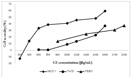
CE induced cytotoxicity in MCF-7 cells while demonstrated weak toxicity on T47D cells. The initial concentration of 600 μg/ml has already resulted in mortality 33±5,2% of MCF-7 cells, while, at the same concentration, T47D cell death was 1.15±5.13%. In-creasing CE concentration significantly enhanced MCF-7 cells mortality (p<0,01, α=0,05). The concentra-tion of 1600 μg/ml resulted in cell death more than
50% population of MCF-7 cells, but for T47D cells the mortality was only 37±2.2% [Figure 1].
Vero cells representing normal cell lines were mildly affected by the presence of CE. At concentration 900 μg/ml, CE induced 14±6,5% Vero cells death. The
RESULTS AND DISCUSSION MATERIALS AND METHODS
viability of Vero cells exceeded 50% even after incuba-tion in CE at very high concentraincuba-tion (2100 μg/ml). This data indicates the selectivity of CE towards breast cancer cells, particularly MCF-7, over normal cells.
Anticancer Potential of Cocoa Extract
The concentration that inhibits 50% viable cells (IC50) of CE was 1236 μg/ml against MCF-7. The value could not be calculated for T47D and Vero cells since CE was unable to induce 50% mortality even at high concentration. The positive control, doxorubicin, exhibited IC50 value 0,07 μg/ml, which is 1600-fold
greater than CE.
Modulation of Catechin and Caffeine Concentration Affected Cytotoxic Potential
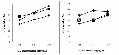
Higher catechin and caffeine content in CE increased cytotoxic potential. At concentration 600
μg/ml, MCF-7 cells mortality was increased from
33±5,2% to 40±2,2% when catechin content was 2-fold, and to 48±1,7% at 3-fold. The same manner observed when caffeine content was increased from the initial concentration (33±5,21%) to 2-fold (47±1,8%) and 3-fold (40±5,7%). Catechin augmentation may improve IC50 value from 1236 μg/ml to 1208 μg/ml and 1021
μg/ml. On the other hand, increasing caffeine content to 2-fold may enhance IC50 to 750 μg/ml, however,
3-fold caffeine resulted in a slightly higher IC50 780
μg/ml [Figure 2]. This research suggests caffeine plays a major role in the cytotoxic activity of CE towards MCF-7 cells.
Cytotoxic potential of CE is lower than other plant extracts such as Mangifera indica kernel (15 μg/ml), Elephantopus scaber (14,69 μg/ml) and Dillenia
suffru-ticosa (76 μg/ml) [14-16]. However CE is stronger
than Argyreia nervosa leaf (>2500 μg/ml),
Dypterocar-pus turbinatus leaf (>2500 μl/ml), and Saraca asoca leaf
(>2500 μg/ml) [17]. There are reports from other stud-ies that MCF-7 cells is more sensitive than T47D cells. Both cancer cells are attributed to estrogen-sensitive proliferation; cancer treatments target thus estrogen re-ceptor. Resistant cells alters estrogen receptor expres-sions [18] while the expresexpres-sions were increased in re-sistant MCF-7 cells, it is found to be negative or un-changed in resistant T47D cells. Estrogen receptor ex-pression affects the sensitivity of cells towards tamox-ifen or fulvestrant, whereas MCF-7 was more sensitive than T47D cells [18]. Further investigation by Brandie et al. [19] indicates that T47D cells tolerate cellular stress better than MCF-7 cells.
Cancer therapies attempt to induce programmed death in cancer cells. It is approached by either intrin-sic or extrinintrin-sic apoptotic pathway. As an important drug for breast cancer therapy, doxorubicin mode of action is promoting cellular stress that leads to death through several mechanisms i.e. inhibiting topoiso-merase II enzyme, forming doxorubicin-DNA adduct, stimulating oxidative stress, and increasing ceramide production that sensitizes cancer cells [20-22]. Further-more, Yang et al. [23] suggested that doxorubicin is more likely altering DNA topography, by enhancing nucleosome turnover around promoter gene and triggering DNA instability.
Anti-cancer properties of catechin are associated with inhibition of proliferation and induction of apop-tosis. Studies showed catechin regulates apoptosis through downregulation of anti-apoptotic protein Bcl-2 and survivin, while upregulating pro-apoptotic protein Bax [24]. In the other hand, catechin suppresses cell proliferation by inhibiting nuclear factor κB (NF-κB) activation, vascular endothelial growth factor (VEGF) expression, and protecting from reactive oxygen species (ROS) stimulation [25-27]. Schlachterman et al. [28] reported that catechin along with resveratrol and quercetin, could be incorporated in a diet, to reduce breast tumor growth.
Caffeine has long history as an agent to sensitize cancer cells against ionizing radiation [29,30]. It has been investigated for cell cycle modulation and DNA damage signals over-riding [31,32]. Caffeine was also reported to stimulate apoptotic cells either through p53-dependent or p53-independent pathways since it Figure 2. MCF-7 cells mortality (%) after incubation in CE for
enhanced UV-induced cell death in wild-type mice as well as in p53-knockout mice [33,34]. Besides, caffeine also induces autophagy, when it is found to increase autophagic vacuoles in SH-SY5Y cells at 10 or 25 mM. The level of microtubule-associated protein 1-light chain-3 (LC3) II as the autophagosomal marker was elevated, and this was associated with PI3K/Akt /mTOR/p70S6K pathway inhibition [35].
Greater cytotoxic potential exhibited in CE contain-ing a higher concentration of phytoalexin compound suggests that purification may improve its anti-cancer properties. Lower IC50 value resulted from higher caf-feine content instead of catechin indicates the purifica-tion method should consider preserving methylxan-thine as a polar constituent, rather than semi-polar flavonoid. Even though IC50 of CE is much lower than doxorubicin and other plant extract, CE might be uti-lized as an anti-cancer agent by using it as a preventive or co-treatment agent. To validate anticancer property of CE, an investigation of immunomodulatory proper-ties, cell sensitization and co-administration with dox-orubicin are required.
This research confirmed the cytotoxic potential of the cocoa extract against breast cancer cells. The effect is more pronounced in MCF-7 cells instead of T47D cells. High concentration of CE induced very low Vero cells mortality, indicating the selectivity of CE on can-cerous cells rather than normal cells. Augmentation of phytoalexin constituent lowered IC50 value of CE, whereas caffeine gave greater effect than catechin.
1. IARC (2013) Latest world cancer statistics, global cancer burden rises to 14.1 million new cases in 2012, marked in-crease in breast cancers must be addressed. Press Release. pp. 1-3.
2. Danaei G, Vander Hoorn S, Lopez AD, Murray CJ, Ezzati M (2005) Causes of cancer in the world: comparative risk assessment of nine behavioural and environmental risk factors. The Lancet. 366: 1784-1793.
3. Siegel R, DeSantis C, Virgo K, Stein K, Mariotto A, et al. (2012) Cancer treatment and survivorship statistics, 2012. CA A Cancer Journal for Clinicians. 62: 220-241. 4. Molassiotis A, Fernadez-Ortega P, Pud D, Ozden G, Scott
JA, et al. (2005) Use of complementary and alternative medicine in cancer patients: a European survey. Annals of Oncology. 16: 655-663.
5. Olaku O, White JD (2011) Herbal therapy use by cancer patients: a literature review on case reports. European
Journal of Cancer. 47: 508-514.
6. 6. Boon HS, Olatunde F, Zick SM (2007) Trends in com-plementary/alternative medicine use by breast cancer sur-vivors: comparing survey data from 1998 and 2005. BMC Women's Health.7: 4.
7. Wink M, Ashour ML, El-Readi MZ (2012) Secondary me-tabolites from plants inhibiting ABC transporters and re-versing resistance of cancer cells and microbes to cytotoxic and antimicrobial agents. Frontiers in Microbiology. 3: 1-15.
8. ICCO (2012) The world cocoa economy: past and present. Executive committee 146th meeting: International Cocoa Organization. pp. 43.
9. Murphy KJ, Chronopoulos AK, Singh I, Francis MA, Mo-riarty H, et al. (2003) Dietary flavanols and procyanidin oligomers from cocoa (Theobroma cacao) inhibit platelet function. The American journal of clinical nutrition. 77: 1466-1473.
10. Radin D, Hayssen G, Walsh J (2007) Effects of intention-ally enhanced chocolate on mood. Explore: The Journal of Science and Healing. 3: 485-492.
11. Anesini C, Ferraro GE, Filip R (2008) Total polyphenol content and antioxidant capacity of commercially available tea (Camellia sinensis) in Argentina. Journal of Agricul-tural and Food Chemistry. 56: 9225-9229.
12. EC (1979) Laying down community methods for analysis for coffee extracts and chicory extracts. Official Journal of European Communities. 327: 18-28.
13. Hamedeyazdan S, Fathiazad F, Sharifi S, Nazemiyeh H (2012) Antiproliferative activity of Marrubium persicum extract in the MCF-7 human breast cancer cell line. Asian Pacific Journal of Cancer Prevention. 13: 5843-5848. 14. Abdullah A-SH, Mohammed AS, Abdullah R, Mirghani
ME, Al-Qubaisi M (2014) Cytotoxic effects of Mangifera indica L. kernel extract on human breast cancer (MCF-7 and MDA-MB-231 cell lines) and bioactive constituents in the crude extract. BMC Complementary and Alternative Medicine. 14: 199.
15. Wan YH, Swee KY, Chai LH, Abdul RR, Abdul AS, et al. (2011) Elephantopus scaber induces cytotoxicity in MCF-7 human breast cancer cells via p53-induced apoptosis. Jour-nal of MediciJour-nal Plants Research. 5: 5741-5749.
16. Armania N, Yazan LS, Ismail IS, Foo JB, Tor YS, et al. (2013) Dillenia suffruticosa extract inhibits proliferation of human breast cancer cell lines (MCF-7 and MDA-MB-231) via induction of G2/M arrest and apoptosis. Mole-cules. 18: 13320-13339.
17. Akter R, Uddin SJ, Grice ID, Tiralongo E (2014) Cyto-toxic activity screening of Bangladeshi medicinal plant ex-tracts. Journal of natural medicines. 68: 246-252.
18. Lykkesfeldt A, Yde C, Thrane S, Larsen S, Pedersen A, et REFERENCES
al. (2013) Abstract P5-09-02: Cell culture models to study mechanisms for endocrine resistant breast cancer, new treatment options and new biomarkers. Cancer Research 73: P5-09-02-P05-09-02.
19. Brandie NR, Margarita MI, Huy XM, Joshua KS, Brad-ford GH, et al. (2015) Bioenergetic differences between MCF-7 and T47D breast cancer cells and their regulation by oestradiol and tamoxifen. Biochemical Journal 465: 49-61.
20. Pritchard JE, Dillon PM, Conaway MR, Silva CM, Par-sons SJ (2012) A mechanistic study of the effect of dox-orubicin/adriamycin on the estrogen response in a breast cancer model. Oncology 83: 305-320.
21. Yang F, Teves SS, Kemp CJ, Henikoff S (2014) Doxoru-bicin, DNA torsion, and chromatin dynamics. Biochimica et Biophysica Acta (BBA)-Biomembranes 1845: 84-89. 22. Swift LP, Rephaeli A, Nudelman A, Phillips DR, Cutts
SM (2006) Doxorubicin-DNA adducts induce a non-topoisomerase II–mediated form of cell death. Cancer Re-search 66: 4863-4871.
23. Yang F, Kemp CJ, Henikoff S (2013) Doxorubicin en-hances nucleosome turnover around promoters. Current Biology 23: 782-787.
24. Papademetrio DL, Trabucchi A, Cavaliere V, Ricco R, Costantino S, et al. (2013) The catechin flavonoid reduces proliferation and induces apoptosis of murine lymphoma cells LB02 through modulation of antiapoptotic proteins. Revista Brasileira de Farmacognosia 23: 455-463.
25. Rodríguez ML, Estrela JM, Ortega Á (2013) Natural Polyphenols and Apoptosis Induction in Cancer Therapy. Journal of Carcinogenesis and Mutagenesis 6: 1-10. 26. Gu J-W, Makey KL, Tucker KB, Chinchar E, Mao X, et
al. (2013) EGCG, a major green tea catechin suppresses breast tumor angiogenesis and growth via inhibiting the
activation of HIF-1 and NF B, and VEGF expression.α κ
Vascular Cell 5: 9.
27. Rathore K, Choudhary S, Wang H-CR (2012) Green tea catechin intervention of reactive oxygen species-mediated ERK pathway activation and chronically induced breast cell carcinogenesis. Carcinogenesis 33: 174-183.
28. Schlachterman A, Valle F, Wall KM, Azios NG, Castillo L, et al. (2008) Combined resveratrol, quercetin, and cate-chin treatment reduces breast tumor growth in a nude mouse model. Translational Oncology 1: 19-27.
29. Sarkaria JN, Busby EC, Tibbetts RS, Roos P, Taya Y, et al. (1999) Inhibition of ATM and ATR kinase activities by the radiosensitizing agent, caffeine. Cancer Research 59: 4375-4382.
30. DeFrank JS, Tang W, Powell SN (1996) p53-null cells are more sensitive to ultraviolet light only in the presence of caffeine. Cancer Research 56: 5365-5368.
31. Bode AM, Dong Z (2007) The enigmatic effects of caf-feine in cell cycle and cancer. Cancer Letters 247: 26-39. 32. Kawabe T (2004) G2 checkpoint abrogators as anticancer
drugs. Molecular Cancer Therapeutics 3: 513-519. 33. Lu Y-P, Lou Y-R, Li XH, Xie JG, Brash D, et al. (2000)
Stimulatory effect of oral administration of green tea or caffeine on ultraviolet light-induced increases in epidermal wild-type p53, p21 (WAF1/CIP1), and apoptotic sunburn cells in SKH-1 mice. Cancer Research 60: 4785-4791. 34. Lu Y-P, Lou Y-R, Peng Q-Y, Xie J-G, Conney AH (2004)
Stimulatory effect of topical application of caffeine on UVB-induced apoptosis in the epidermis of p53 and Bax knockout mice. Cancer Research 64: 5020-5027.