DOI:http://dx.doi.org/10.7314/APJCP.2016.17.5.2683 Potassium Pentagamavunon and Doxorubicin Combination Inhibitory Effects in Breast Cancer Cells
Asian Pac J Cancer Prev,17 (5), 2683-2688
Introduction
Long-term use of chemotherapeutic agent, doxorubicin for the treatment of cancer cause several side effects including toxicity and drug resistance (Tacar et al., 2013). Therefore, there is development of chemotherapy to reduce its side effects, called co-chemotherapy. Combination of chemotherapeutic agent with chemorepentive agent (co-chemotherapy) is an alternative to overcome the drug resistance, improve efficacy, and reduce its toxicity (Parhi et al., 2012).
Curcumin, a natural compound has been extensively explored and has the potential to be developed as co-chemotherapeutic agent. Single treatment of curcumin inhibited cancer cell growth through various mechanisms (Yoysungnoen et al., 2005; Lin et al., 2009; Prasad et al., 2009). However, based on previous study, curcumin was found to be poorly soluble in water, thus curcumuin has low bioavailability (Tønnesen et al., 2002). To improve the bioavailability of curcumin in the body, curcumin has been developed to a salt compound, namely sodium
1Cancer Chemoprevention Research Center, Faculty of Pharmacy, Universitas Gadjah Mada, 2Research Center for Chemistry,
Indonesian Institute of Science, Yogyakarta, Indonesia *For correspondence: [email protected]
Abstract
A salt compound of a curcumin analogue, potassium pentagamavunon-0 (K PGV-0) has been synthesized to improve solubility of pentagamavunon-0 which has been proven to have anti-proliferative effects on several cancer cells. The purpose of this study was to investigate cytotoxic activity and metastasis inhibition by K PGV-0 alone and in combination with achemotherapeutic agent, doxorubicin (dox), in breast cancer cells. Based on
MTT assay analysis, K PGV-0 showed cytotoxic activity in T47D and 4T1 cell lines with IC50 values of 94.9 µM
and 49.0±0.2 µM, respectively. In general, K PGV-0+dox demonstrated synergistic effects and decreased cell viability up to 84.7% in T47D cells and 62.6% in 4T1 cells. Cell cycle modulation and apoptosis induction were
examined by flow cytometry. K PGV-0 and K PGV-0+dox caused cell accumulation in G2/M phase and apoptosis
induction. Regarding cancer metastasis, while K PGV-0 alone did not show any inhibition of 4T1 cell migration, K PGV-0+dox exerted inhibition. K PGV-0 and its combination with dox inhibited the activity of MMP-9 which has a pivotal role in extracellular matrix degradation. These results show that a combination of K PGV-0 and doxorubicin inhibits cancer cell growth through cell cycling, apoptosis induction, and inhibition of cell migration and MMP-9 activity. Therefore, K PGV-0 may have potential for development as a co-chemotherapeutic agent.
Keywords:K PGV-0 - doxorubicin - breast cancer - cell lines - apoptosis - cell cycling - metastasis
RESEARCH ARTICLE
Combination of Potassium Pentagamavunon-0 and Doxorubicin
Induces Apoptosis and Cell Cycle Arrest and Inhibits Metastasis
in Breast Cancer Cells
Herwandhani Putri
1, Riris Istighfari Jenie
1, Sri Handayani
1,2, Ria Fajarwati
Kastian
1, Edy Meiyanto
1*
curcuminate (Mukhopadhyay et al., 1982). Sodium curcuminate has been shown to have anti-inflammatory activity better than curcumin (Ghatak and Basu, 1972).
Faculty of Pharmacy Universitas Gadjah Mada developed a curcumin analogue as chemopreventive agent, namely [2,5-bis (4’-hydroxy-3’-methoxyibenzylidene) cyclopentanone] or pentagamavunon-0 (PGV-0) (Meiyanto et al., 2006). PGV-0 has been proven to have several pharmacological activities, especially to inhibit cancer cell growth (Meiyanto et al., 2006; Hermawan et al., 2011; Meiyanto et al., 2014). The salt compound of PGV-0, potassium pentagamavunon-0 (K PGV-0) had been developed to increase its bioavailability. This compound has a good solubility in water and is expected to have same or better activity than PGV-0 (Margono and Verawati, 2005)
inhibition of the activity of MMP-9. Our results suggest that K PGV-0 is potential to be developed as an effective co-chemotheraphy to inhibit the breast cancer cells metastasis.
Materials and Methods
Cell culture
T47D and 4T1 breast cancer cells were obtained from Prof. Masashi Kawaichi (NAIST, Japan). Cells were grown in Dulbecco’s Modified Eagles Medium (DMEM) high glucose (Sigma) supplemented with 10% FBS (Sigma), HEPES, sodium bicarbonate, 1.5% Penicillin-Streptomycin and Fungizone (Gibco). Cells were incubated in 37°C with 5% CO2.
MTT Cytotoxicity assay
MTT assay was performed according to (Mosmann, 1983), with slight modification. Cells were treated with various concentration of K PGV-0 (obtained from Curcumin Research Center, Faculty of Pharmacy, Universitas Gadjah Mada), doxorubicin (dox) (Sigma, Germany) or the combination of K PGV-0 and dox. After 24 h of treatment, cells were treated with 0.5 mg/mL of 3-(4,5-Dimethylthiazol-2-yl)-2,5-diphenyltetrazolium bromide (MTT) (Biovision) and incubated further for 4 h. Cells were lysed using SDS stopper containing 0.01 N HCl and incubated in the dark condition for overnight. After incubation, cells absorbance was measured by ELISA reader plate at λ 595 nm. Cells absorbance was converted to % cell viability. Linear regression between concentration and % cell viability giving the equation y = Bx + A were used to calculate IC50 value, that is the
concentration inhibiting 50% cell proliferation.
Apoptosis assay
Cells were treated with K PGV-0, doxorubicin and the combination for 24 h. After the treatment, cells were harvested by trypsinisation and stained with Annexin-V-FLUOS staining kit (Roche) consisting of 100 mL of binding buffer, 2 mL of 2 mL Anexin V and propidium iodide (PI) and incubated for 10 minutes in the dark room, according to manufacturer’s instruction, and analyzed by flow cytometer (BD FACS Calibur, BD Bioscience)
Cell cycle analysis
Cells were seeded, treated, harvested and stained with the staining solution contains propidium iodide (PI) 1 mg/mL protease inhibitor, 10 mg/mL RNase and 0,1% (v/v) Triton X-100 (Merck). Cells were incubated for 5 minutes in the dark room, transferred into a flow cytometric tube and analyzed by BD FACS Calibur (BD Bioscience, USA). Cell cycle distribution was acquired by using Flowing software.
Wound healing assay
The 4T1 cells (7.5 x 10⁴ cells) were seeded in 24-well
plate and incubated for 24 hours. Cells starvation was done by incubating cells in culture medium supplemented with 0,5% FBS for 24 hours. After starvation, cells were scratched with sterile tip, and treated with various
concentration of samples. The cells were documented at 0, 18, 24, and 42 h after treatment. The results were analyzed by ImageJ software and presented as % of wound closure.
Gelatin zymography
The 8% of SDS-PAGE supplemented with 0.1% of gelatin was used to determined the activity of MMP-9 in the culture medium. After electrophoresis, gels were washed and incubated with distilled water containing 2% of Triton-X 100 (Merck) for 30 min at room temperature. The solution was removed from gels. 100 mL of reaction buffer (40 mM Tris-HCl pH 8, 10 mM CaCl2, 0.02% NaN3) was added and incubated for 24 h at 37°C. After removal of reaction buffer, gels were stained by Coomassie Brilliant Blue R-250 solution and destained by destaining solution (20% methanol, 10% acetic acid and
Figure 1. Cytotoxic Effect of K PGV-0 on T47D and
4T1 cells. T47D cells (8x10³ cells/well) and 4T1 cells (5x10³
cells/well) were seeded in 96 wellplate and incubated for 24h, then treated with K PGV-0. Cells viability was determined by using MTT assay as described in methods. (A) T47D and 4T1
cells morphology after treatment for 24 h. Arrows indicate cells
morphological changes. (B) Cell viability profile after single treatment of K PGV-0 for 24 h on T47D and 4T1 cells. Profiles of
cell viability were means ± SE from 3 independent experiments Cells Control K PGV-0 10 µM K PGV-0 50 µM
Cell viability (%)
K PGV-0 (µM) 4T1
T47D
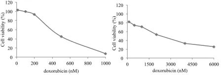
Figure 2. Cytotoxic effect of Doxorubicin on T47D and
4T1 cells. T47D cells (8x10³ cells/well) and 4T1 cells (5x10³
cells/well) were seeded in 96 well-plate and incubated for 24h,
then treated with doxorubicin. Cells viability was determined by using MTT assay as described in the methods. (A) Cell
viability profile after single treatment of Doxorubicin for 24 h on T47D cells. (B) Cell viability profile after single treatment of Doxorubicin for 24 h on 4T1 cells. Profiles of cell viability
were means ± SE from 3 independent experiments 0
Cell viability (%)
doxorubicin (nM)
Cell viability (%)
DOI:http://dx.doi.org/10.7314/APJCP.2016.17.5.2683 Potassium Pentagamavunon and Doxorubicin Combination Inhibitory Effects in Breast Cancer Cells 70% water) until clear bands with dark blue background
appear. The results were documented and analyzed by ImageJ software.
Statistical analysis
Data were expressed as mean ± SE. One-way analysis of variance (ANOVA) followed by the least significant difference (LSD) test were used for statistical analyses. P-values less than 0.05 were considered significant.
Results
Single Treatment of K PGV-0 and doxorubicin Revealed Cytotoxic Effect on T47D and 4T1 cells
Cytototoxic assay was conducted to analyse cytotoxicity of K PGV-0 and Dox on T47D and 4T1 breast cancer cells with IC50 parameter. Cytotoxic effect of K PGV-0 was observed after 24 h of treatment. Single treatment of K PGV-0 for 24 h induced morphological changes. K PGV-0 10 μM induced cell shrinkage. At
higher concentrations, K PGV-0 50 μM there were more number of cell shrinkage and fragmentation. In addition, single treatment of K PGV-0 for 24 h revealed cytotoxic effect on T47D and 4T1 cells with IC50 value of 94.9 µM
dan 48.97±0.2 µM, respectively (Figure 1).
We also examined the cytotoxic activity of doxorubicin on T47D and 4T1 cells. Doxorubicin performed cytotoxic effect with IC50 value of 383.67±83.70 nM and 2.65±0.27 µM, respectively. Subsequently, doxorubicin was combined with K PGV-0 to enhance the effectiveness and decrease side effects of chemotherapeutic agents for cancer therapy (Figure 2).
Combination K PGV-0 and Doxorubicin Decreased Cells Viability on T47D cells
Combination of doxorubicin and K PGV-0 reduced cell viability of T47D cells. The results showed that the combination of cytotoxic K PGV-0 in combination with doxorubicin for 24 hours decreased cell viability up to 84.69% (Figure 3), in combination of K PGV-0 48 μM (1/2 IC50 K PGV-0) with 200 nM doxorubicin (1/2
Figure 3. Cytotoxic Effect of Combination of K PGV-0
and Doxorubicin on T47D Cells. T47D cells (8x10³ cells/
well) were seeded in 96 wellplate and incubated for 24h, then
treated with doxorubicin and K PGV-0 as indicated in the figure.
Cells viability was determined by using MTT assay as described in the methods
Cells viability (%)
Dox (nM)
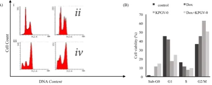
Figure 4. Effects of Combination of Doxorubicin and
KPGV-0 on Cell Cycle Modulation of T47D Cells. Cells
were harvested after 24 h of treatment, stained with propidium
iodide and DNA content were analyzed by using flowcytometry. (A) The profiles of cells in the phases of sub-G1, G1, S and G2-M (i) control cells, (ii) doxorubicin 200 nM, (iii) K PGV-0 48 μM, (iv) a combination of doxorubicin 200 nM - K PGV-0 48 µM. X-axis showed the relative content of DNA and the Y axis showed the relative cell numbers. (B) Quantification
of cells distribution in each phase of the various treatments.
The percentage of sub-G1 phase cells showed apoptotic cell
population
Cell viability (%)
Kontrol sel Dox KPGV-0 Dox+KPGV-0
DNA Content
Figure 5. Effect of Combination of K PGV-0 and doxorubicin on Apoptosis Induction on T47 D Cells.
Cells were harvested after treatment of the sample (200 nM
doxorubicin, 48 μM K PGV-0, or a combination of both) for 24 hours, stained with reagents annexin V and propidium iodide (PI) and analyzed using flowcytometer. (A) The distribution profiles
of living cells (bottom-left), early apoptotic (bottom-right), late apoptosis (upper-right), necrosis (upper-left) in various indicated
treatments. (B) Quantification of cells undergoing early and
late apoptosis
apoptosis awal apoptosis akhir
(A) (B)
Figure 6. Cytotoxic Effect of Combination of K PGV-0
and Doxorubicin on 4T1 Cells. 4T1 cells (8x10³ cells/well)
were seeded in 96 wellplate and incubated for 24h, then treated
with doxorubicin and K PGV-0 as indicated in the figure. Cells
IC50 doxorubicin). Several combination K PGV-0 with doxorubicin showed synergistic effect with value of CI
<1 (Table 1).
Modulation of Cell Cycle of Single Treatment of K PGV-0 and Its Combination with Doxorubicin
Based on the analysis of the cell cycle, single treatment of K PGV-0 increased T47D cells accumulation in G2/M phase. Combination of K PGV-0 and doxorubicin also increased cells accumulation in G2/M, however, with less percentage compared to single treatment of K PGV-0 (Figure 4). This phenomenon probably due to the differences of the target of K PGV-0 and doxorubicin in modulating cell cycle. Furthermore, combination of K PGV-0 and doxorubicin increased the accumulation of cells in sub-G1 phase. Cells in sub-G1 phase were hipodiploid cell with low DNA content due to the DNA fragmentation. Hipodipoid cells were the sign of cell death. Thus, the combination of K PGV-0 with doxorubicin increased the cell death of T47D.
K PGV-0 and its combination with Doxorubicin Induced Apoptosis on T47D cells
Results showed that treatment of K PGV-0 combination with doxorubicin increased the number of apoptotic cells up to 66.92% (Figure 5). We suggested that the combination of K PGV-0 with doxorubicin induced cell death through apoptosis mechanism.
Combination K PGV-0 and Doxorubicin Revealed Synergistic Effect on 4T1 cells
Combination of Doxorubicin with K PGV-0 reduced cell viability greater than single treatment of K PGV-0 and doxorubicin on 4T1 cells. Treatment of K PGV-0 in combination with doxorubicin for 24 hours reduced the viability of 4T1 cells up to 62.46% (Figure 6). In addition, combination of doxorubicin and K PGV-0 showed synergistic effect. Synergistic effect that caused cell viability less than 50% were performed by combination of K PGV-0 12.5 μM (1/4 IC50 K PGV-0) with doxorubicin
1350 nM (1/2 IC50 doxorubicin) and combination of K
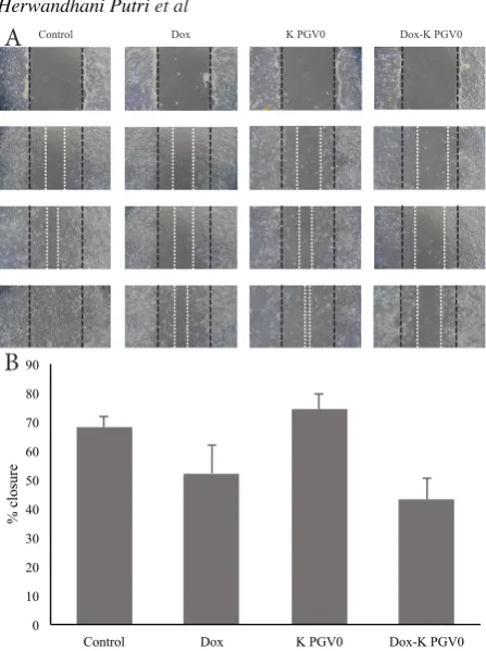
Figure 7. Effect of Single Treatment of K PGV-0 and its Combination with Doxorubicin on 4T1 Cells
Migration. (A) The morphology of the cells after scratch
and treated with 675 nM doxorubicin, K PGV-0 12.5 μM and their combination. Observations were made after 0, 18, 24
and 42 hour of treatment under an inverted microscope with
magnification of 100x. (B) The percentage of 4T1 cells closure after 24 h of treatment. Values were means of % closure ± SD
from 3 independent experiments. The area of the scratch were
analyzed using ImageJ software then % closure was calculated in
accordance with the procedures of the analysis. The asterisk (*)
indicates p < 0.05; n = 3. Black dashed line showed the boundary
area of scratches (scratch) h-0 and white dotted line indicated scratching area at the time of observation
ime Control Dox K PGV0 Dox-K PGV0
0 10 20 30 40 50 60 70 80 90
Control Dox K PGV0 Dox-K PGV0
% closure
A
B
Figure 8. Effect of Single Treatment of K PGV-0 and its Combination with Doxorubicin in MMP-9 Activity on
4T1 Cells. Cells were treated with 675 nM doxorubicin, K
PGV-0 12.5 μM and their combination. MMP-9 activity assay was
conducted using gelatin zymography according to the method.
Analysis of the results were done by using ImageJ software to measure the intensity of gelatin degradation by MMP-9 in the gel
0 20 40 60 80 100 120
KS+E Dox
KPGV -0
Dox+KPGV -0
Relative MMP-9 activity (%)
control
MMP-9
Table 1. CI Values of Combination of K PGV-0 and Doxorubicin on T47D Cells
CI
Doxorubicin (nM)
25 50 100 200
K PGV
-0
(µM)
6 3.23 17.45 0.69 0.52
12 -0.73 1.45 0.8 0.65
24 3.28 0.8 1.32 0.7
48 0.71 0.59 0.66 0.58
Table 2. CI Values of Combination of K PGV-0 and Doxorubicin on 4T1 Cells
CI
Dox (nM)
270 337.5 675 1350
PGV
-0
(µM)
5 0.45 0.53 0.65 0.79
6.25 0.45 0.49 0.58 0.74 12.5 0.62 0.67 0.76 0.7
DOI:http://dx.doi.org/10.7314/APJCP.2016.17.5.2683 Potassium Pentagamavunon and Doxorubicin Combination Inhibitory Effects in Breast Cancer Cells PGV-0 25 μM (1/2 IC50 K PGV-0) with doxorubicin 1350
nM (1/2 IC50 doxorubicin) with CI values of 0.70 and 0.78,
respectively (Table 2).
Combination of K PGV-0 with Doxorubicin Inhibited 4T1 Cell Migration
Results of scratch wound healing assay up to 42 h showed that single treatment of K PGV-0 did not inhibit 4T1 cell migration. In addition, combination of K PGV-0 with doxorubicin up to 24 h inhibited the migration of 4T1 breast cancer cells with % closure up to 43.21% (Figure 7).
Combination of K PGV-0 with Doxorubicin Decreased MMP-9 Activity on 4T1 Cells
Results showed that single treatment of 1.5 µM K PGV-0 and 675 nM doxorubicin decreased gelatinase activity of MMP-9 (Figure 8). Combination treatment of doxorubicin and K PGV-0 decreased activity of MMP-9 greater than single treatment of K PGV-0 and doxorubicin. We suggested that combination of K PGV-0 and doxorubicin plays a role in breast cancer invasion process.
Discussion
Single treatment of K PGV-0 showed cytotoxic effect on T47D and 4T1 breast cancer cells with IC50 value
94.94 µM and 48.97 µM, respectively. Single treatment of doxorubicin also performed cytotoxic effect on T47D and 4T1 cells wit IC50 value 400 ± 71.76 nM and 2.7 ± 0.16 µM, respectively. The IC50 value of K PGV-0 in T47D was about 10-fold greater than IC50 value of curcumin analogue. PGV-0. The IC50 values of PGV-0 on T47D cells
was 11 µM (Meiyanto et al., 2006). This phenomenon occurs due to the polarity difference between PGV-0 and K PGV-0. The polarity difference will cause a difference in the process of drug diffusion into the cell. The process drug diffusion into the cell is important since the protein targets of both curcumin and its analogues are in the cell.
Combination of K PGV-0 with doxorubicin reduced cell viability greater than single treatment of K PGV-0 and doxorubicin on T47D cells and 4T1 cells. In general, the combination of various concentrations of K PGV-0 and doxorubicin showed synergistic effect and reduced cell viability up to 84.69% on T47D cells and 62.6% on 4T1 cells. Synergistic effect of combination PGV-0 and doxorubicin in breast cancer cells has been proven in the previous studies, PGV-0 increased the effect of doxorubicin on MCF-7 cells (Hermawan et al., 2011). Based on the result of sinergistic effect of combination K PGV-0 ad doxorubicin. this present study was continued to investigate possibility pathway in growth cells inhibition through cell cycle modulation and apoptosis induction.
This study showed that K PGV-0 and doxorubicin inhibited T47 breast cancer cell growth through cell cycle modulation. Single treatment of K PGV-0 and doxorubicin caused cell accumulation in G2/M phase, while combination treatment of K PGV-0 and doxorubicin caused cell accumulation at G2/M and subG1 phase. This findings indicated that the cell cycle modulation mechanism of K PGV-0 on T47D cells is similar with
curcumin analogue. PGV-0 which also cause cell accumulation in G2/M phase. Probably, molecular mechanism occured after treatment of K PGV-0 and PGV-0 is similar with curcumin. Curcumin cause cell cycle modulation through down-regulation of cyclin A and up-regulation of cyclin-dependent kinase (CDK) inhibitor p21 (Park et al., 2006). Cell accumulation in subG1 phase is one of apoptosis property. Therefore, to prove it apoptosis induction. flow cytometric apoptosis assay was conducted.
Single treatment of K PGV-0 and doxorubicin inhibited T47D cell growth through apoptosis mechanism. Combination of K PGV-0 with doxorubicin increased apoptotic cells up to 64.58%. The molecular mechanisms causing apoptosis on T47D cells is through p53-independent pathway. Doxorubicin cleavage PARP and activate caspase-8 (Lee et al., 2002). In addition, doxorubicin induced caspase-independent cell death (Moreira et al., 2014), while curcumin analogue PGV-0 has been proven to cause apoptosis through PARP-cleavage mechanism (Da’i, 2008). Therefore, K PGV-0 alone or its combination with doxorubicin probably induced apoptosis in the same pathway with PGV-0.
In this present study. inhibition of cancer metastasis after treatment of combination K PGV-0 and doxorubicin was also observed. Cell migration is part of the process of metastasis. Single treatment of K PGV-0 did not inhibit 4T1 cells migration, while combination K PGV-0 and doxorubicin inhibited cell migration. Single treatment of K 0 did not inhibit cell migration because K PGV-0 did not inhibit FAK pathway. Therefore, lamelipodia formation and actomyosin that have a pivotal role in cell migration are still occur. Combination K PGV-0 and doxorubicin inhibited cell migration because the migration inhibitory activity of doxorubicin is greater than K PGV-0. This findings is consistent with previous research which perform that doxorubicin inhibit M5076 cell migration (Sugiyama and Sadzuka, 1999) and hepatocellular cancer HepG2 cells (Yang et al., 2014)
Although single treatment of K PGV-0 did not inhibit cell migration. this compound inhibited cancer cell invasion process by reducing the activity of matrix metalloproteinase-9 (MMP-9). The possible molecular mechanism which cause this finding is inhibition of NF-κB activation. MMP-9 transcription factor. Previous research states that curcumin and its analogue inhibit the activation of transcription factor NF-κB (Sandur et al., 2007).
Taken together, combination of K PGV-0 and doxorubicin inhibit cancer cell growth through cell cycle inhibition, apoptosis induction, and inhibition of cell migration and MMP-9 activity. Therefore, K PGV-0 performs potency to be developed as co-chemotherapeutic agent..
References
Da’i M (2008). Mekanisme molekuler aktivitas analog kurkumin pentagamavunon terhadap sel kanker payudara (T74D).
dissertation.
Ghatak N, Basu N (1972). Sodium curcuminate as an effective
235-6.
Lin SS, Lai KC, Hsu SC, et al (2009). Curcumin inhibits the migration and invasion of human A549 lung cancer cells through the inhibition of matrix metalloproteinase-2 and-9
and Vascular Endothelial Growth Factor (VEGF). Cancer
Letters, 285, 127-33.
Margono SA, Verawati EY (2005). Sintesis kalium
pentagamavunonat-0 meng-gunakan kalium hidroksida dengan pelarut tetrahidrofuran-etanol. Majalah Farmasi Indonesia, 16, 239-45.
Meiyanto E, Putri D, Susidarti RA, et al (2014). Curcumin and its analogues (PGV-0 and PGV-1) enhance sensitivity of resistant MCF-7 cells to doxorubicin through inhibition
of HER2 and NF-kB activation. Asian Pac J Cancer Prev,
15, 179-84.
Moreira AC, Branco AF, Sampaio SF, et al (2014). Mitochondrial
apoptosis-inducing factor is involved in doxorubicin-induced toxicity on H9c2 cardiomyoblasts. Biochimica et Biophysica Acta (BBA)-Molecular Basis of Disease, 1842, 2468-78. Mosmann T (1983). Rapid colorimetric assay for cellular growth
and survival: application to proliferation and cytotoxicity assays. J Immunological Methods, 65, 55-63.
Mukhopadhyay A, Basu N, Ghatak N, et al (1982). Anti-inflammatory and irritant activities of curcumin analogues
in rats. Agents and actions, 12, 508-15.
Parhi P, Mohanty C, Sahoo SK (2012). Nanotechnology-based
combinational drug delivery: an emerging approach for cancer therapy. Drug Discovery Today, 17, 1044-52.
Park C, Kim GY, Kim GD, et al (2006). Induction of G2/M arrest
and inhibition of cyclooxygenase-2 activity by curcumin in human bladder cancer T24 cells. Oncology Reports, 15, 1225-31.
Prasad CP, Rath G, Mathur S, et al (2009). Potent growth
suppressive activity of curcumin in human breast cancer
cells: Modulation of Wnt/β-catenin signaling.
Chemico-Biological Interactions, 181, 263-71.
Sandur SK, Ichikawa H, Pandey MK, et al (2007). Role of pro-oxidants and antioxidants in the anti-inflammatory and
apoptotic effects of curcumin (diferuloylmethane). Free Radical Biol Med, 43, 568-80.
Sugiyama T, Sadzuka Y (1999). Combination of theanine with
doxorubicin inhibits hepatic metastasis of M5076 ovarian
sarcoma. Clinical Cancer Res, 5, 413-6.
Tacar O, Sriamornsak P, Dass CR (2013). Doxorubicin: an
update on anticancer molecular action, toxicity and novel drug delivery systems. J Pharmacy Pharmacol, 65, 157-70. Tønnesen HH, Másson M, Loftsson T (2002). Studies of
curcumin and curcuminoids. XXVII. Cyclodextrin
complexation: solubility, chemical and photochemical stability. Int J Pharmaceutics, 244, 127-35.
Yang K, Ma L, Cheng X, et al (2014). Effect of
low-molecular-weight heparin combined with doxorubicin on hepatocellular cancer cell migration in vitro. J Southern Medical University, 34, 1048-52.
Yoysungnoen P, Wirachwong P, Bhattarakosol P, et al (2005).
Effects of curcumin on tumor angiogenesis and biomarkers,
COX-2 and VEGF, in hepatocellular carcinoma