Screening of several Indonesian medicinal plants for their
inhibitory effect on histamine release from RBL-2H3 cells
Zullies Ikawati
a, Subagus Wahyuono
b, Kazutaka Maeyama
a,*
aDepartment of Pharmacology Ehime Uni
6ersity School of Medicine,Shigenobu-cho,Onsen-gun,Ehime791-0295,Japan bFaculty of Pharmacy,Gadjah Mada Uni
6ersity,Yogyakarta, Indonesia
Received 17 July 1999; received in revised form 25 December 2000; accepted 21 January 2001
Abstract
Twelve alcoholic extracts and 12 hexane extracts of plant materials selected on the basis of medicinal folklore for asthma treatment in Indonesia were studied for their activity in inhibiting histamine release from RBL-2H3 cells (rat basophilic leukemia cell line), a tumor analog of mast cells. The results of screening indicated that five alcoholic extracts (Plantago major leaves,
Eucalyptus globulus leaves and fruit,Cinnamomum massoiae cortex, Vitex trifolia leaves) and two hexane extracts (Eucalyptus globulusleaves,Vitex trifolialeaves) inhibited IgE-dependent histamine release from RBL-2H3 cells. The inhibitory effects were found to be more than 80% for extract concentrations of 0.5 mg/ml. The results indicate that the extracts contain active compounds that inhibit mast-cell degranulation, and provide insight into the development of new drugs for treating asthma and/or allergic disease. © 2001 Elsevier Science Ireland Ltd. All rights reserved.
Keywords:Histamine; Indonesian medicinal plants; Jamu; RBL-2H3 cells
www.elsevier.com/locate/jethpharm
1. Introduction
The mechanism of the inflammatory response result-ing in asthma is complex and involves numerous cell types, including mast cells (Meltzer, 1998). The mast cell has long been associated with asthma, since it releases a variety of preformed and newly synthesized mediators that could account for several features of asthma (Barnes, 1993). Among the mediators released from mast cells, histamine is a well-characterized and the most potent vasoactive mediator in acute bron-choconstriction provoked by allergen, exercise, hyper-tonic stimuli and inhaled adenosin. Histamine may also contribute to the allergen-induced late asthmatic re-sponse probably following the recruitment and activa-tion of basophils (Holgate, 1999).
Rat basophilic leukemia (RBL-2H3) cells, a tumor analog of mast cells, display properties of mucosal-type mast cells. The 2H3 cells contain several hundred thou-sand IgE receptors on the membrane surface, and after
sensitization with mouse monoclonal IgE, the cells re-spond to antigen and release histamine. Although the mucosal mast-cell response to secretagogues and their sensitivity to inhibitors are different from those of cutaneous-type mast cells, the mechanism of histamine secretion is supposed to be general (Maeyama et al., 1992). Their advantage over peritoneal mast cells is that a great number of homogenous cells are obtained at once and can be used for experiments. They thus provide a good tool for studying the effect of unknown compounds on histamine release activity.
The use of herbal medicines has increased in recent years. Many medicinal plants provide relief of symp-toms comparable to that obtained from allopathic medicines. Specific chemical derivatives have been iso-lated from many plant products that act on the mecha-nisms and mediators that cause asthma and allergies (Bielory and Lupoli, 1999). In Indonesia, several medic-inal plants have been traditionally used in the treatment of respiratory disorders. These plants are: Amomum cardamomum Willd, Cinnamomum burmanii Nees ex BL, Cinnamomum massoiaeSchewc, Curcuma xanthor
-rhiza Roxb, Eucalyptus globulus Labill, Justicia gen
-darusa Linn, Orthosiphon stamineus Benth, Plantago * Corresponding author. Tel.: +81-89-9605258; fax: +
81-89-9605263.
E-mail address:[email protected] (K. Maeyama).
major L., Piper cubeba L.f., Thymus 6ulgaris L., and Vitex trifolia L. Some of these plants have been re-ported to have tracheospasmolytic activity on guinea-pig trachea contraction induced by histamine and other spasmogens, whereas for others, no scientific data con-cerning their activities for asthma therapy have been reported.
Broucke and Lemli (1983) and Meister et al. (1999)reported the spasmolytic activity of Thymus6ul -garis on guinea-pig isolated trachea. Such an effect is also found in Piper cubeba, Vitex trifolia, Curcuma xanthorrhiza, and Eucalyptus globulus (Wahyuono et al., 1998, 1999). The spasmolytic or bronchodilator effect is only one action of an anti-asthma drug due to the complexity of the disease. Other actions involve protection of the airways from precipitants that lead to bronchospasm or inflammation and contribute towards resolving airway inflammation (Szefler, 1993). Inhibi-tion of mast-cell degranulaInhibi-tion, which in turn inhibits release of mast-cell mediators, is also considered one means of treating asthma, as shown by the cromones drugs, such as sodium cromoglycate and nedocromil sodium, the most specific anti-allergic drugs discovered so far (Barnes, 1999).
Investigation of how traditionally used medicinal plants work is one way of discovering bioactive com-pounds (Verpoorte, 1999). In this study, we screened medicinal plants regarding their effects on histamine release from mast-cell models, believed to be one of the triggering events of allergic asthma disease. We dis-cussed whether the plants have a significant effect on asthma therapy and whether there is synergism between the bronchospasmolytic activity and the inhibitory ef-fect of histamine release of the medicinal plants. The results of this screening may provide useful information
for further discovering pharmacologically active com-pounds for treatment of asthma.
2. Materials and methods
2.1. Plant collection
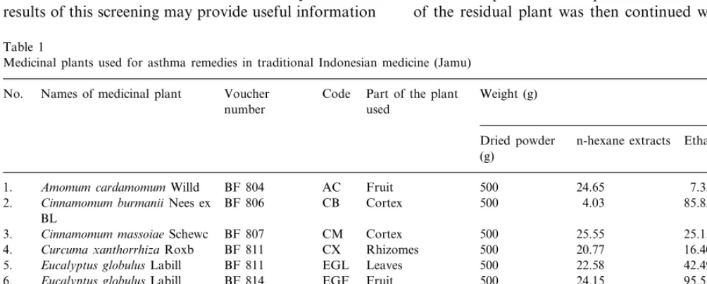
Eleven types of plants that are commonly used in ‘‘jamu’’ (traditional Indonesian medicine) as remedies for asthma or respiratory disorders were selected. These plant materials were collected from the plantation in the Medicinal Plants Research Office (BPTO), Tawang-mangu, Surakarta, Indonesia. These plant materials were identified by a botanist at the BPTO, Tawang-mangu, and their voucher specimens were deposited in the Department of Pharmacognosy, Faculty of Phar-macy, Gadjah Mada University, Yogyakarta, Indone-sia. The names of these medicinal plants, voucher numbers, and parts of each plant used for the experi-ments are listed in Table 1.
2.2. Preparation of medicinal plant extracts
The plant material was cut into small pieces and dried in an oven set at 50°C. The dried material was ground into powder, which was then extracted with n-hexane followed by ethanol to give n-hexane and ethanol extracts, respectively.
In brief, the powder obtained from each plant (500 g each) was macerated with 250 ml of n-hexane overnight. After filtration and evaporation of the filtrate, dried n-hexane extracts were obtained. This maceration procedure was performed twice. Maceration of the residual plant was then continued with ethanol,
Table 1
Medicinal plants used for asthma remedies in traditional Indonesian medicine (Jamu) Names of medicinal plant
No. Voucher Code Part of the plant Weight (g)
used number
Dried powder n-hexane extracts Ethanol extracts (g)
1. Amomum cardamomumWilld BF 804 AC Fruit 500 24.65 7.35
Cortex CB
BF 806 Cinnamomum burmaniiNees ex
2. 500 4.03 85.83
BL
Cinnamomum massoiaeSchewc BF 807 CM
3. Cortex 500 25.55 25.15
4. Curcuma xanthorrhizaRoxb BF 811 CX Rhizomes 500 20.77 16.40
BF 811 EGL Leaves 500 22.58 42.49
5. Eucalyptus globulusLabill
BF 814 EGF Fruit 500 24.15 95.55
6. Eucalyptus globulusLabill
23.92 14.26
500 Leaves
7. Justicia gendarusaLinn BF 816 JG
Leaves 500 4.92
8. Orthosiphon stamineusBenth BF 817 OS 25.92
Piper cubebaL.f. BF 819 PC
9. Fruit 500 82.97 30.57
Plantago majorL. 30.70
10. BF 820 PM Leaves 500 6.72
51.49 19.80
500 Leaves
11. Thymus6ulgarisL. BF 823 TV Vitex trifolia L.
and ethanol extracts were obtained by the procedure as described above.
2.3. Materials
Dinitrophenylated bovine serum albumin (DNP24
-BSA, which consists of 24 mol of dinitrophenol bound per 1 mol of BSA) was a gift from Dr. H. Metzger, NIH (Bethesda, MD). Monoclonal IgE against DNP-BSA was purified from the supernatant in IgE produc-ing hybridoma, which was obtained in our laboratory. Eagle’s minimum essential medium (MEM) and antibi-otics were obtained from Gibco (Grand Island, NY), fetal calf serum was purchased from JRH Biosciences (A SCL company), and PIPES [piperazine-1,4-bis(2-ethanesulfonic acid)] was purchased from Dosindo (Ku-mamoto, Japan). Other chemicals were of the highest grade available.
2.4. Preparation of RBL-2H3 cells line
RBL-2H3 cells were cultured in MEM containing 15% fetal calf serum in a flask in a humidified atmo-sphere of 5% of CO2 in air at 37°C according to
Barsumian et al. (1981). For the histamine release as-say, RBL-2H3 cells were seeded into 24-well culture plates (2×10⁵
cells/well) in 0.4 ml medium for each well. Cells were incubated overnight at 37°C and sensi-tized with 0.5mg/ml of monoclonal IgE against DNP24
-BSA.
2.5. Assay of histamine release
After sensitizing the cells with IgE, the medium was removed, and the cells were washed twice with 0.5 ml of PIPES buffer (25 mM PIPES, 119 mM NaCl, 5 mM KCl, 5.6 mM glucose, 0.4 mM MgCl2, 1 mM CaCl2, 40
mM NaOH, 0.1% BSA, pH 7.2) and preincubated with either 200ml of PIPES buffer (as control) or extracts of
medicinal plants (concentration 0.5 mg/ml) at 37°C for 10 min. For comparison, quercetin (50mM) and
thapsi-gargin (0.5mM) were used as a standard for inhibitory
effect and stimulatory effect, respectively. RBL-2H3 cells were stimulated with 20 ng/ml of DNP24-BSA as
antigen for 30 min, and histamine released into the medium was measured by HPLC-fluorometry accord-ing to Yamatodani et al. (1985).
Briefly, 100ml of the cell medium were collected and
centrifuged at 3000 rpm for 5 min. Fifty microlitres of supernatant were then collected and diluted with 250ml
of 3% perchloric acid in 5 mM Na2EDTA and added
by 30 ml 2 M KOH/1 M KH2PO4. This mixture was
then centrifuged at 10 000×g for 15 min at 4°C, and 50 ml of the supernatant were injected directly onto a
column packed with TSKgel SP-2SW Cation Ex-changer (Tosoh, Tokyo). Histamine was eluted with
0.25 M potassium phosphate at a flow rate of 0.6 ml/min. The histamine was post-labeled with o -phtha-laldehyde in alkaline conditions, and detected fluoro-metrically in an F1080 Fluorometer (Hitachi, Tokyo), using excitation and emission wavelengths of 360 and 450 nm, respectively.
The percentage of net histamine release was calcu-lated as follows:
Net histamine release (%)
=challenged release (pmol)−spontaneous release (pmol)
total histamine (pmol)−spontaneous release (pmol)
×100.
Spontaneous histamine release is the release of his-tamine in the absence of DNP-BSA antigen, and it was calculated with the following equation:
Spontaneous histamine release (%)
= histamine release (pmol)
total histamine content (pmol)×100.
The inhibition percentage of histamine release was cal-culated using the following equation:
Percentage inhibition
=histamine release without drugs−histamine release with drugs histamine release without drugs
×100.
3. Results
3.1. Extraction of medicinal plants
Each of the medicinal plant materials was extracted using the procedure described in Fig. 1, and the yields of each extract are reported in Table 1.
3.2. Effect of the extracts on histamine release
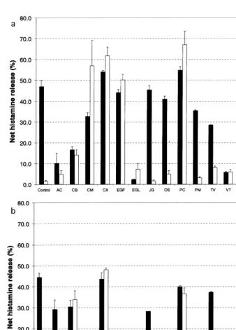
A total of 24 extracts derived from 11 different plant species commonly used in traditional Indonesian medicine to treat respiratory disorders were screened for their inhibitory effect on histamine release in 2H3 cells. Histamine release from IgE-sensitized RBL-2H3 cells was induced by DNP-BSA as antigen stimulation. The net histamine release from RBL-2H3 effected by medicinal plant extracts is shown in Fig. 2a and b, for hexane and ethanol extracts, respectively. The inhibition of histamine release by the extracts is shown in Table 2.
Fig. 1. Scheme of procedures for obtaining n-hexane and ethanol extracts from the medicinal plants.
4. Discussion
Asthma is defined as a lung disease with the follow-ing characteristics: (1) airway obstruction that is re-versible, but not completely so in some patients; (2) airway inflammation; and (3) increased airway respon-siveness to a variety of stimuli (Busse and Reed, 1993). Therapeutic strategies for asthma include bronchodila-tors, corticosteroids, mediator antagonists (antihis-tamines, antileukotrienes, etc.), anti-inflammatory drugs, and specific inhibitors drugs, such as cromones that act by inhibiting mast-cell degranulation, etc. (Barnes, 1999).
Recently, the use of herbal medicine as an alternative medicine for the treatment of various diseases (Bielory and Lupoli, 1999), including asthma, has increased dramatically. The use of herbal medicines is based on traditional healing, and is also influenced by culture. In Indonesia, the use of medicinal plants and herbal ther-apy has been practised long before recorded history. However, scientific knowledge concerning the mecha-nism of action of medicinal plants in asthmatic disease is very limited. It is likely that some of the plants used have no significant effect on respiratory disorders.
In this study, we investigated several medicinal plant extracts traditionally used in Indonesia for asthma ther-apy, and focused our attention on their inhibitory action against histamine release from mast cells. We used quercetin as a standard drug for an inhibitory effect, since the 2H3 cells are insensitive to commonly used mast-cell stabilizers like sodium cromoglycate or nedocromil (Maeyama et al., 1992). Quercetin, a natu-rally occurring flavonoid found in many plants, has been reported to potently inhibit histamine release from rat peritoneal mast cells (Fewtrell and Gomperts, 1977) and 2H3 cells (Cheong et al., 1998) by inhibiting protein kinase (Hagiwara et al., 1988). For the stimula-tory effect, we used thapsigargin, a sesquiterpen lacton isolated from the roots of Thapsia garganica, L., which is known to release histamine from mast cells (Patkar et al., 1979) by inhibiting the sarcoplasmic/endoplasmic reticulum (ER) calcium-dependent ATPase (Thastrup et al., 1994) that in turn elevates cytosolic Ca2+
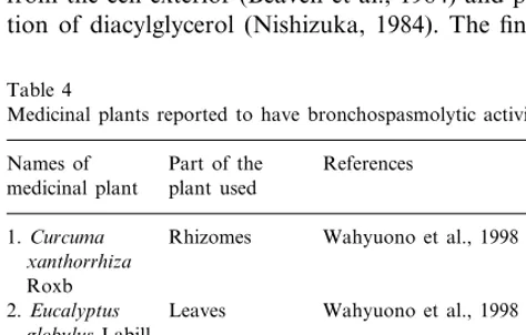
re-quired to induce histamine release from mast cells. The results of screening were also then compared to the known effects of the medicinal plants on bronchospas-molytic activity (Table 4) for further discussion.
Histamine and mast cells are of our interest, since they are closely related to asthmatic disease, and drugs acting on mast cells may be effective as a prophylactic agent in the treatment of mild to moderate asthma, although the precise mechanism of action is not com-pletely understood (Meltzer, 1998). It is best to begin treatment prophylactically and prevent the onset of significant symptomatology rather than attempting to lessen the ongoing symptoms (Meltzer, 1998).
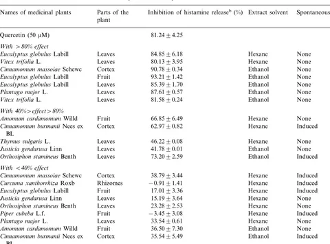
n-hexane extracts and five ethanol extracts were found to have high inhibitory effects on histamine release from 2H3 cells showing more than 80% inhibition of histamine release compared to controls, whereas quercetin (50mM) as a standard drug showed an
inhibi-tion of about 80% (Table 2). The extracts with a high inhibitory activity (in alphabetical order) are as follows: n-hexane extracts of Eucalyptus globulus (leaves) and
Vitex trifolia(leaves), and ethanol extracts ofCinnamo
-mum massoiae(cortex),Eucalyptus globulus(leaves and fruit), Plantago major (leaves), and Vitex trifolia
(leaves). Other plant extracts showed either a medium or low inhibitory activity.
We also found evidence for varying levels of induc-tion of histamine release by some medicinal plant ex-tracts, even though no antigen was added. The effect was considered significant if the extracts caused sponta-neous histamine release of more than 10%. Thapsi-gargin, as a standard drug, showed stimulation of histamine release of about 65%. Extracts having such an effect are: both n-hexane and ethanol extracts of
Cinnamomum burmanii cortex, Curcuma xanthorriza
rhizome, and Piper cubebafruits; hexane extracts only of Eucalyptus globulus fruits and Cinnamomum mas
Some of the medicinal plants were found to suppress histamine release from 2H3 cells to various extents (Table 2). Interestingly, we found that some extracts also had an opposite effect, i.e. they both inhibited histamine release and induced spontaneous histamine release, as found in hexane and ethanol extracts of
Cinnamomum bumanii and ethanol extracts of Or
-thosiphon stamineus. In such cases, it is difficult to derive any exact conclusions concerning the inhibitory effect of these plants, since the opposite effects were
mixed. This phenomenon is likely to happen since there are still various compounds and these require more separation.
We also noted that some plants did not have any considerable inhibitory effect and even induced sponta-neous histamine release, as in the case of hexane and ethanol extracts of Curcuma xanthorrhiza and Piper cubeba. The mechanism of induction of histamine re-lease from 2H3 cells by these extracts has not yet been investigated. One possibility is that the plant caused cell
Fig. 2. Effect of n-hexane extracts (a) and ethanol extracts (b) of medicinal plants on histamine release activity from 2H3 cells either in the presence (solid bar) or absence (open bar) of DNP-BSA as antigen to stimulate histamine release. Each of the data represents mean9S.E.M of three experiments performed in duplicate (n=6). Note that several medicinal plants induced significant spontaneous histamine release in the absence of antigen. The names of the medicinal plant were coded as shown in Table 1. QU is quercetin 50mM, and TH is thapsigargin 0.5mM
Table 2
Inhibition of histamine release from 2H3 cells by the medicinal plantsa Parts of the
Names of medicinal plants Inhibition of histamine releaseb(%) Extract solvent Spontaneous histamine release plant
81.2494.25 Quercetin (50mM)
With\80%effect
84.8596.18 Hexane
Leaves None
Eucalyptus globulusLabill
Leaves
Vitex trifoliaL. 80.1393.95 Hexane None
Cortex
Cinnamomum massoiaeSchewc 90.7890.34 Ethanol None
93.2191.42 Ethanol
Fruit None
Eucalyptus globulusLabill
85.3991.70 Ethanol None
Eucalyptus globulusLabill Leaves
87.6190.57 Ethanol
Leaves None
Plantago majorL.
Vitex trifoliaL. Leaves 81.5890.24 Ethanol None
With40%\effect\80%
Amomum cardamomumWilld Fruit 66.8596.49 Hexane None
Cortex 62.9790.82 Hexane
Cinnamomum burmaniiNees ex Induced
BL
Thymus6ulgarisL. Leaves 46.2290.08 Hexane None
41.7890.01 Ethanol
Leaves None
Justicia gendarusaLinn
73.2092.59 Ethanol Induced
Orthosiphon stamineusBenth Leaves WithB40%effect
Cinnamomum massoiaeSchewc Cortex 38.7993.44 Hexane Induced Curcuma xanthorrhizaRoxb Rhizomes −0.9191.41 Hexane Induced
17.0193.36 Hexane
Fruit Induced
Eucalyptus globulusLabill
Leaves
Justicia gendarusaLinn 15.1993.64 Hexane None
Leaves
Orthosiphon stamineusBenth 23.2892.53 Hexane None
−3.4593.08 Hexane
Fruit Induced
Piper cubebaL.f.
33.5490.61 Hexane
Plantago majorL. Leaves None
36.5097.30 Ethanol None
Amomum cardamomumWilld Fruit
Cortex 35.5495.49
Cinnamomum burmaniiNees ex Ethanol Induced
BL
Curcuma xanthorrhizaRoxb Rhizomes 8.3594.77 Ethanol Induced
15.8591.22 Ethanol
Fruit Induced
Piper cubebaL.f.
Leaves
Thymus6ulgarisL. 23.3090.92 Ethanol None
aData represent the mean9S.E.M of three experiments performed in duplicate (n=6), as a percentage of inhibition. bInhibition of histamine release by the medicinal plants was calculated using the equation mentioned in Section 2.5.
lysis, as found in hexane extracts of Piper cubeba. However, other mechanisms are possible and require further investigation.
In this study, we rather prefer to discuss the possible correlation between the inhibitory effect on histamine release and bronchospasmolytic activity of the medici-nal plants. We found that inhibition of histamine re-lease was not correlated with bronchospasmolytic activity, and some extracts even demonstrated the op-posite effect. We classified our findings into three
cate-gories. Plants in the first category have both
bronchospasmolytic activity and an inhibitory effect on histamine release. In this category areEucalyptus glob
-ulus leaves (ethanol extract) and Vitex trifolia leaves (both n-hexane and ethanol extracts). This dual action may be due to the presence of various compounds in those plants that may have a significant effect on asthma therapy.
Plants in the second category have bronchospas-molytic activity, but only a mild or no effect on his-tamine release from 2H3 cells. This category includes
Thymus 6ulgaris leaves. This plant has long been used
for the treatment of coughs, and it has been reported to have bronchospasmolytic activity (Broucke and Lemli, 1983; Meister et al., 1999). Both n-hexane and ethanol extracts of this plant showed inhibition of histamine release of less than 40%.
Table 3
Spontaneous histamine release induced by several medicinal plantsa
Part of the plant used
Names of medicinal plant Extract solvent
n-hexane Ethanol
Thapsigargin (0.5mM) 66.4094.31
14.0592.51
Cortex 33.8394.23
Cinnamomum burmaniiNees ex BL
Cortex
Cinnamomum massoiaeSchewc 56.86912.17 None
Rhizomes
Curcuma xanthorrhizaRoxb 61.7194.25 47.9990.86
50.1792.59
Fruit None
Eucalyptus globulusLabill
Orthosiphon stamineusBenth Leaves None 13.9290.18
67.0496.41 36.4792.89
Piper cubebaL.f. Fruit
aData represent the mean9S.E.M of three experiments performed in duplicate (n=6), as a percentage. The percentage of spontaneous histamine release was calculated using the equation mentioned in Section 2.5.
anti-inflammatory effect would inhibit histamine release from mast cells. However, we did not find any evidence that this plant inhibited histamine release from mast cells, and it is likely that the site of action on the target cells is different. In fact, we found that the extract from
Piper cubeba caused lysis of 2H3 cells, and this may account for the high histamine release.
There is no other available literature concerning the bronchospasmolytic activity of the other medicinal plants, apart from that shown in Table 4. To our knowledge, our report is the first concerning the effects of Indonesian medicinal plants on the activity of his-tamine release from 2H3 cells.
The mechanism of histamine release from 2H3 cells involves several pathways. The cascades responsible for release have been considered to involve aggregation of IgE receptors (Maeyama et al., 1986), tyrosine phos-phorylation of phospoliphase C (Park et al., 1991) via
src-related tyrosine kinase (Jouvin et al., 1994), hydrol-ysis of inositol phospholipids (Maeyama et al., 1988), and mobilization of calcium by generated inositol triphospate (Streb et al., 1983), enhanced calcium influx from the cell exterior (Beaven et al., 1984) and produc-tion of diacylglycerol (Nishizuka, 1984). The final two
messengers, calcium signal and activation of protein kinase C, then elicit the release of histamine synergisti-cally (Beaven et al., 1987).
The inhibitory action of the medicinal plants against histamine release from 2H3 cells has not yet been investigated and might influence one or more of these proposed mechanisms. Further study is required to investigate the mechanism by which the medicinal plants inhibit histamine release from mast cells. Isola-tion and purificaIsola-tion of some potent extracts are being carried out by our group for such purposes. If this mechanism is also applied in an in-vivo experiment, it would provide useful information to further explain drug action.
We conclude that some of the Indonesian medicinal plants traditionally used for asthma treatment might have significant effects on mast-cell degranulation and provide insight for the discovery of new drugs for treating respiratory disorders involving mast cells.
References
Barnes, P.J., 1993. Pathophysiology of allergic inflammation. In: Middleton, E., Jr., Reed, C.E., Ellis, E.F., Adkinson, N.F., Jr., Yunginger, J.W., Busse, W.W. (Eds.), Allergy, Principles and Practice, vol. I, fourth ed. Mosby, MO, p. 243.
Barnes, P.J., 1999. Therapeutic strategies for allergic diseases. Nature 402 (supplement), B31 – B38.
Barsumian, E.L., Isersky, C., Petrino, M.G., Siraganian, R.P., 1981. IgE-induced histamine release from rat basophilic leukemia cell lines: isolation of releasing and nonreleasing clones. European Journal of Immunology 11, 317 – 323.
Beaven, M.A., Guthrie, D.F., Moore, J.P., Smith, G.A., Hesketh, T.R., Metcalfe, J.C., 1987. Synergistic signals in the mechanism of antigen-induced exocytosis in 2H3 cells: Evidence for an uniden-tified signal required for histamine release. Journal of Cellular Biology 105, 1129 – 1136.
Beaven, M.A., Rogers, J., Moore, J.P., Hesketh, T.R., Smith, G.A, Metcalfe, J.C., 1984. The mechanism of the calcium signal and correlation with histamine release in 2H3 cells. Journal of Biolog-ical Chemistry 259, 7129 – 7136.
Bielory, L., Lupoli, K., 1999. Herbal intervention in asthma and allergy. Journal of Asthma 36, 1 – 65.
Table 4
Medicinal plants reported to have bronchospasmolytic activity Part of the References
Names of
plant used medicinal plant
Rhizomes
1.Curcuma Wahyuono et al., 1998 xanthorrhiza
Roxb
2.Eucalyptus Leaves Wahyuono et al., 1998 globulusLabill
3.Piper cubeba Fruit Wahyuono et al., 1999 L.f.
Broucke and Lemli, 1983; 4.Thymus Leaves
Meister et al., 1999 6ulgarisL.
Leaves Wahyuono et al., 1998 5.Vitex trifolia
Broucke, V.D., Lemli, J.A., 1983. Spasmolytic activity of the flavonoids fromThymus6ulgaris. Pharmaceutical Weekblad Sci-entific Edition 5, 9 – 14.
Busse, W.W., Reed, C.F., 1993. Asthma, definition and pathogenesis. In: Middleton, E., Jr., Reed, C.E., Ellis, E.F., Adkinson, N.F., Jr., Yunginger, J.W., Busse, W.W. (Eds.), Allergy, Principles and Practice, vol. II, fourth ed. MO, Mosby, pp. 1173 – 1201. Cheong, H., Ryu, S.Y., Oak, M.H., Cheon, S.H., Yoo, G.S., Kim,
K.M., 1998. Studies of structure activity relationship of flavonoids for the anti-allergic actions. Archive of Pharmaceutical Research 21, 478 – 480.
Claeson, P., Pongprauoon, U., Sematong, T., Tuchinada, P., Reutrakul, V., Soontornsaratune, P., Taylor, W.C., 1996. Non-phenolic linear diarylheptanoids from Curcuma xanthorrhiza: a novel type of topical anti-inflammatory agents: structure – activity relationship. Planta Medika 62, 236 – 240.
Fewtrell, C.M., Gomperts, B.D., 1977. Quercetin: a novel inhibitor of Ca2+ influx and exocytosis in rat peritoneal mast cells. Biochim
Biophys Acta 469, 52 – 60.
Hagiwara, M., Inoue, S., Tanaka, T., Nunoki, K., Ito, M., Hidaka, H., 1988. Differential effects of flavonoids as inhibitors of tyrosine protein kinase and serine/threonine protein kinases. Biochemical Pharmacology 37, 2987 – 2992.
Holgate, S.T., 1999. Experimental models in asthma. Clinical Experi-mental Allergy 29 (supplement 3), 82 – 86.
Jouvin, M.H.E., Adamczewski, M., Numerof, R., Letoutrneur, O., Valle, A., Kinet, J.P., 1994. Differential control of the tyrosine kinases Lyn and Syk by two signaling chains of the high affinity immunoglobulin E receptor. Journal of Biological Chemistry 269, 5918 – 5925.
Maeyama, K., Hohman, R.J., Ali, H., Cunha-melo, J.R., Beaven, M.A., 1988. Assesment of hydrolysis of membrane inositol phos-pholipids. The Journal of Immunology 140, 3919 – 3927. Maeyama, K., Hohman, R.J., Metzger, H., Beaven, M.A., 1986.
Quantitative relationships between aggregation of IgE receptors in rat basophilic leukemia (2H3) cells. Journal of Biological Chem-istry 261, 2583 – 2592.
Maeyama, K., Yagi, S., Watanabe, T., 1992. Histamine release from rat basophilic leukemia cells (RBL-2H3). Annals of Psychiatry 3, 67 – 77.
Meister, A., Bernhardt, G., Chistoffel, V., Buschauer, A., 1999. Antispasmodic activity of Thymus 6ulgaris extract on isolated guinea-pig trachea: discrimination between drug and ethanol ef-fects. Planta Medica 65, 512 – 516.
Meltzer, E.O., 1998. Pharmacological treatment options for allergic rhinitis and asthma. Clinical Experimental Allergy 28 (supplement 2), 27 – 36.
Nishizuka, Y., 1984. The role of protein kinase C in cell surface transduction and tumorpromotion. Nature 308, 693 – 698. Ozaki, Y., 1990. Antiinflammatory effect of Curcuma xanthorrhiza
Rozb, and its active principles. Chemical Pharmaceutical Bulletin 38, 1045 – 1048.
Park, D.J., Min, H.K., Rhee, S.G., 1991. IgE-induced tyrosine phos-phorylation of phospholipase C-g1 in rat basophilic leukemia
cells. Journal of Biological Chemistry 266, 24237 – 24240. Patkar, S.A., Rasmussen, U., Diamant, B., 1979. On the mechanism
of histamine release induced by thapsigargin fromThapsia gar-ganicaL. Agents Actions 9, 53 – 57.
Streb, H., Irvine, R.F., Berridge, M.J., Schulz, I., 1983. Release of Ca2+ from a nonmitochondrial intracellular store in pancreatic
acinar cells by inositol-1,4,5-triphosphate. Nature 306, 67 – 69.
Szefler, S.J., 1993. Principles of therapeutics. In: Middleton, E., Reed, C.E., Ellis, E.F., Adkinson, N.F., Yunginger, J.W., Busse, W.W. (Eds.), Allergy, Principles and Practice, vol. I, fourth ed. Mosby, MO, pp. 761 – 777.
Thastrup, O., Dawson, A.P., Scharff, O., Foder, B., Cullen, P.J., Drobak, B.K., Bjerrum, P.J., Christensen, S.B., Hanley, M.R., 1994. Thapsigargin, a novel molecular probe for studying intracel-lular calcium release and storage. Agents Actions 43, 187. Wahyuono, S., Mulyono, Nurlaila, Astuti, P., Mursyidi, A., 1998.
Tracheospasmolytic screening of Curcuma xanthorrhiza Roxb. rhizomes, and the leaves ofVitex trifoliaL.,Eucalyptus globulus labill,Justica gendarusaBurm. F. Majalah Farmasi Indonesia 9 (3), 110 – 115.
Wahyuono, S., Mulyono, Wahyono, Mursyidi, A., 1999. Tra-cheospamolytic activity of Piper cubeba L.,f., fruits. Majalah Farmasi Indonesia 10 (1), 48 – 56.
Verpoorte, R., 1999. Exploration of nature’s chemodiversity: the role of secondary metabolites as leads in drug development. Drug Discovery Today 3, 232 – 238.
Yamatodani, A., Fukuda, H., Wada, H., Iwaeda, T., Watanabe, T., 1985. High-performance liquid chromatographic determination of plasma and brain histamine without previous purification of biological samples: cation-exchange chromatography coupled with post-column derivatization fluorometry. Journal of Chro-matography 344, 115 – 123.