Assessing Microleakage on Class V Composite Resin
Restorations after Er:YAG Laser Preparation Varying the
Adhesive Systems
REGINA GUENKA PALMA DIBB, D.D.S., Ph.D., SILMARA APARECIDA MILORI CORONA, D.D.S., Ph.D.,
MARIA CRISTINA BORSATTO, D.D.S., Ph.D., KAREN CRISTINA FERREIRA, D.D.S., RENATA PEREIRA RAMOS, D.D.S., and JESUS DJALMA PÉCORA, D.D.S., Ph.D.
ABSTRACT
Objective:The aim of this study was to assess the performance of three bonding agents in preventing mi-croleakage of class V cavities prepared and treated by Er:YAG laser associating with acid etching. Back-ground Data: There has been very little research comparing the efficiency of single-component and self-etching adhesive systems in preventing microleakage of cavities prepared and conditioned with Er:YAG laser. Materials and Methods: Thirty cavities—with occlusal margin in enamel and cervical in dentin/ cementum—were prepared in sound human third molars using a short pulsed Er:YAG laser (500 mJ/5 Hz) The enamel and dentin surfaces were conditioned for 30 sec using lower dosimetries (120 mJ/4 Hz), and the samples were randomly assigned into three groups, according to the adhesive system: (I) Bond-1; (II) Prime & Bond NT; and (III) Etch & Prime 3.0. Groups I and II were acid-etched for 15 sec, and group III did not re-ceive any acid treatment once a self-etching system was employed. Cavities were restored with a light-cured composite resin (JEK-Z250 Filtek-250), and after finishing, the samples were thermocycled, isolated with epoxy resin and nail varnish, immersed in a 0.2% Rhodamine B solution for 24 h, and sectioned longitudinally. The sections obtained were analyzed for leakage using an optical microscope connected to a computer and a video camera. We digitized the images using a special software program that allowed a quantitative evaluation of microleakage in millimeters. Results:Statistical analysis using the Kruskal-Wallis test showed statistically significant difference between both margins, and the occlusal region presented better marginal sealing. Com-paring the three resin bonding systems, Prime & Bond NT entirely sealed both margins, while Etch & Prime 3.0 provided the poorest overall results, showing a statistically significant difference (p< 0.01). Conclusion:It may be concluded that, for all the tested materials, microleakage values were higher in cervical (dentin/cemen-tum) margins. Additionally, Prime & Bond NT provided a complete elimination of marginal infiltration at both margins, after treating the dental surface with laser irradiation associated with a sequent acid-etching.
129 INTRODUCTION
R
ECENTLY, the applicability of newer methods for preparingdental hard tissue, such as Er:YAG laser irradiation, has been greatly widespread in an attempt to replace the high-speed air turbine and low-high-speed drills conventionally used, of-fering improved patient comfort, by reducing the pressure, heat, vibration, and noise associated with rotational methods.1
The Er:YAG laser was cleared by FDA in 19971for use on human teeth and has been reported as a reliable and safe tech-nology to cut or ablate tooth structure, for removal of carious lesion, to treat enamel/dentin surfaces, and for cavity prepara-tion with limited loss of sound dental tissue and minimum in-jury to the pulp and surrounding hard and soft tissue.1,2
Although it is a subject of controversy, a thermally induced mechanical process may explain the high ablation efficiency
Ribeirão Preto School of Dentistry, University of São Paulo, São Paulo, Brazil.
and absence of heat damage.1,2The emission wavelength of 2.94 mm coincides with the main absorption peak of water.1–3
As a result, the incident Er:YAG laser irradiation is absorbed in a thin surface layer, causing heating and evaporation of water. High steam pressure then leads to microexplosions with erupt-ing particles, and, once the substrate is disintegrated into frag-ments (but not vaporized) and no melting is observed, most of the incident energy is consumed by the ablation process.1–6 Consequently, these results reduced free energy and minimal thermal injury.1–3
However, in spite of the well-known efficacy of Er:YAG laser, it has been demonstrated that preparing and treating tooth surface exclusively by laser irradiation does not result in optimal sealing because the laser irradiation was not able to produce a high-quality, dye penetration–resistant interface. It was also observed that the laser treatment does not eliminate the need of a sequent acid-etching because the resin materials bonded to nonetching lased surfaces lack the seal obtained with acid conditioning.7–9 Several investigations7–13 have shown that, when the phosphoric acid etching was accom-plished following laser conditioning, marginal integrity of the restorations was considerably enhanced and microleakage val-ues comparable to conventional preparation (dental drill + acid etching) were possible. These findings were confirmed and re-composition, pretreatment required, and mechanism of adhe-sion may interfere with the marginal sealing of these cavities.16 Therefore, the aim of this study was to assess the perfor-mance of three bonding agents—a filled single-bottle acetone-based, an unfilled acetone-based single-bottle, and a self-etching system—in preventing marginal microleakage at class V cavities prepared and treated by Er:YAG laser associ-ated with a subsequent 37% phosphoric acid etching.
MATERIALS AND METHODS
Fifteen sound human third molars, extracted within a 6-month period and stored in saline solution, were selected and cleaned with water/pumice slurry in a dental prophylactic cup. Thirty class V cavities, with the occlusal margin in enamel and cervical margin located 1 mm below the amelocemental junction, were prepared on the buccal and lingual surfaces. Cavity dimensions were standardized utilizing a template to trace an outline onto both surfaces with a mesiodistal width
used to treat tooth surface. After cavity preparation, the enamel and dentin surfaces were conditioned for 30 sec using lower dosimetries (120 mJ/4 Hz), and the specimens were randomly assigned into three equal groups of 10 cavities each, according to the bonding systems.
For groups I (Bond-1—Jeneric/Pentron) and II (Prime & Bond NT—Dentsply), a subsequent 37% phosphoric acid-etching was accomplished for 15 sec following laser condition-ing. For group III (Etch & Prime 3.0—Degussa), the dental surface was not acid etched once a self-etching adhesive sys-tem was employed. Then, two uniform layers of the bonding systems, according to the experimental group, were applied and air-thinned, and each layer was light-cured for 30 sec with a visible-light curing unit with an output of 450 mW/cm2 (XL 3000; 3M Dental Products, St Paul, MN). A hybrid light-acti-vated composite resin (Z-250; 3M Dental Products) was in-serted with appropriate instruments according to an incremental placement technique, and each increment, of about 1 mm thick, was light-cured for 40 sec. The specimens were stored for 7 days in distilled water at 37°C, and then restora-tions were polished with Super-Snap disks (Shofu Inc., Kyoto, Japan). The specimens were subjected to a thermocycling regi-men of 500 cycles in 5–55°C waterbaths. Dwell time was 1 min, with a 3-sec transfer time between baths.
In preparation for the dye penetration test, the specimens were dried superficially, and entirely sealed (including apical region) with epoxy resin and two coats of nail varnish, leaving a 2-mm window around the cavity margins and immersed in a 0.2% Rhodhamine B solution for 24 h. Then, the surface-adhered dye was rinsed in tap water, and the epoxy resin and nail varnish were removed with a sharp instrument. The teeth were included in chemically activated acrylic resin (JET; Clás-sico, São Paulo) and bisected longitudinally in a mesiodistal direction with a water-cooled diamond saw in the sectioning machine Minitom (Struers A/S, Copenhagen, Denmark). Then, the separated buccal and lingual halves were embedded again in blocks of acrylic resin and sectioned in a buccolingual direc-tion, providing two to three cuts of 1.0-mm thick for each tooth. This approach was preferred because the third molar’s accentuated convexity did not enable the buccal and lingual preparations to be centralized and therefore would not allow both restorations to be appropriately sectioned in the same slice. Following sectioning, the cuts were initially thinned in a polishing machine (Politriz; Struers A/S) using no. 180–600 sandpapers and then manually smoothed with water no. 1000–1200 sandpapers to obtain a flattened surface and a final thickness of approximately 0.25 mm (Fig. 1a).
This methodology was followed because the third molar’s nat-ural convexity would not allow standardized 1.5 mm-depth restorations for all the sections, and as a consequence, the dif-ferences in depths would lead to distorted results. The data were submitted to statistical analysis using the Kruskal-Wallis test.
RESULTS
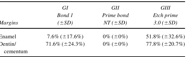
A statistically significant difference (p< 0.01) between the occlusal and cervical margins for all groups was observed, and, as a rule, the best marginal sealing was in the occlusal region, except for group II, which showed complete sealing at both margins. Table 1 shows the means of dye penetration and stan-dard deviation (percentage) for the experimental groups at both regions. Of the three adhesive systems, Etch & Prime 3.0 (group III) yielded the highest degree of microleakage (p<
0.01). Prime & Bond NT entirely sealed both margins and was statistically different (p < 0.01) from the other experimental
groups. In dentin/cementum margins, Bond 1 (group I) and Etch & Prime 3.0 (group II) no significant difference was observed.
DISCUSSION
Er:YAG laser irradiation has been widely indicated for differ-ent procedures in ddiffer-ental practice, and most recdiffer-ently, it has been suggested that this technology would be effective for condi-tioning tooth surface (with or without a subsequent phosphoric acid-etching) prior to an adhesive restorative system.17–19
Nevertheless, one of the concerns regarding this application of lasers has been whether the superficial alterations promoted by irradiation would result in a sealing and a higher degree of marginal infiltration. On the other hand, performing a subse-quent acid-etching would provide an enhanced surface condi-tioning and improve the adhesion of resin systems to cavity preparation. Therefore, supported by the review of the cited lit-erature7–13 and based on our experience,14,15 in the research conducted, the laser irradiation was accomplished coupled with a subsequent phosphoric acid-conditioning, except for group III, because the self etching systems did not indicate acid conditioning as a pretreatment. These observations would probably explain the Etch & Prime 3.0 undesirable perfor-mance, once this bonding system does not indicate acid-etching.
On the other hand, the findings of the present study dis-closed that Prime & Bond NT adhesive system promoted a complete sealing of both analyzed margins. A possible expla-nation for such performance would be that this system pro-vided a homogenous hybrid layer and uniform resin tags.25In a previous study, Latta et al.26observed that Prime & Bond 2.1 was able to promote hybrid layer and tag formation in cavities prepared and treated by Er:YAG laser.
Studies have yet to state some important features related to Er:YAG laser application, such as the specific type of etch pat-tern determined by laser irradiation, the aspects in which this conditioning differs from that obtained with acid-etching and how it may interfere in the integrity of adhesive interface. Likewise, supplementary research is required to investigate the performance of bonding systems when the laser is used for conditioning tooth surface. It is important to emphasize that, FIG. 1. Digitized image. (a) Buccal restoration. (b) Dye penetration (arrows) along cervical margin. (c) Quantitative assess-ment in millimeters: microleakage (x) and depth of cavity (y).
TABLE1. MICROLEAKAGE(%) OFDIFFERENTEXPERIMENTALGROUPS ATOCCLUSAL ANDCERVICALMARGINS
GI GII GIII
Bond 1 Prime bond Etch prime
Margins (6SD) NT (6SD) 3.0 (6SD)
Enamel 7.6% (617.6%) 0% (60%) 51.8% (632.6%)
Dentin/ 71.6% (624.3%) 0% (60%) 77.8% (620.7%)
the intrinsic aspects related to the adhesive systems, such as the pretreatment technique required, composition and micro-mechanically compromised adhesive interface and thus could interfere in the marginal sealing of restorations.13,20–23
Keller and Hibst12reported that treatment with Er:YAG laser would create surfaces that appear similar to acid-etched sur-faces. Other investigations19,24have shown that when bonding composite to tooth structure, the Er:YAG laser alone or com-bined with acid-etching produces a surface with bonding strength equal or better than that produced by acid-etching alone. however, Eduardo and others13 (1996) observed that composite resin shear bond strength to enamel was superior for acid-etched group compared to the group prepared by Er:YAG laser, because the morphological alterations created on enamel surface by laser irradiation were not sufficient to effectively bond composite to dental surface.
Tanji23analyzed the enamel and dentin morphological modi-fications in class V cavities prepared by the Er:YAG laser and the irradiation was considered efficient to ablate dental tissue, although preparations do not resemble conventional, precise, clearly identifiable outlines showing irregular walls and mar-gins. The dentin surface showed opened dentinal tubules and only few areas with melting and crystallization.
Although the laser mechanism has not been yet well cleared up, the morphological observation of lased surfaces by scan-ning electron microscope (SEM) reveals an irregular ablation pattern and an incomplete etching with the existence of non-conditioned dental surface areas,5possibly resulting from the difficulty to obtain a uniform pulse administration. The laser does not create the uniform microporosities characteristics of acid-conditioning and instead promotes a disorganized de-struction of enamel prisms, possibly due to its great ability to remove substance. The resultant microretentions clearly vary from acid-etching patterns and this irregular microstructure would result in poor mechanism of adhesion, may decisively influence their effectiveness in sealing cavity margins and pre-venting marginal microleakage.
Further investigation focusing on the long-term effect of ul-trastructural changes observed in Er:YAG laser irradiated den-tal substrates may provide restorations with increased marginal sealing and therefore lead to improved microleakage preven-tion as well as to a more widespread applicability of these new technologies in clinical practice.
CONCLUSION
Based on the findings of this study and, given the limitations of an in vitro investigation, it seems appropriate to conclude that higher microleakage values were observed in cervical mar-gins. Prime & Bond NT was the only bonding system to provide a complete elimination of marginal infiltration at both margins, after treating enamel and dentin/cementum surfaces with laser irradiation associated with a subsequent acid-etching.
REFERENCES
in dentistry: future possibilities with advanced technology. Quin-tessence Int. 23, 117–133.
5. Sekine, Y., Ebihara, A., Takeda, A., et al. (1995). Erbium:YAG laser application to cavity preparation: light microscopic investi-gation of tooth pulp. Lasers Dent. Int. Proc. 2128,167–172. by high-speed handpiece and Er:YAG laser—in vitroevaluation [Thesis]. São Paulo School of Dentistry.
9. Robles, F., Ramos, A., Zezell, D., et al. (2000). SEM and EDX evaluation of the microleakage in cavities prepared with Er:YAG laser. J. Dent. Res. 79, 1098. of class V composite restorations prepared conventionally with those prepared with an Er:YAG laser: a pilot study. Pediatr. Dent. 15, 425–426.
13. Eduardo, C.P., Myaki, S.I., Oliveira, Jr., W.T., et al. (1996). Micro-morphological evaluation of enamel surface and the shear bond strength of a composite resin after Er:YAG laser irradiation. An “in vitro” study. Presented at the 5th Congress of International Society for Laser in Dentistry, Jerusalem, Israel, May 5–9. Microleakage on class V composite restorations after bur, air-abra-sion and Er:YAG laser preparation. Oper. Dent. 26,491–499. 16. Roebuck, E.M., Whitters, C.J., and Saunders, W.P. (2001). The
in-fluence of three erbium:YAG laser energies on the in vitro mi-croleakage of class V compomer resin restorations. Int. J. Pediatr. Dent. 11, 49–56. by Er:YAG laser and phosphoric acid. Morphological analysis and bond strength [Thesis]. São Paulo School of Dentistry.
WA: The International Society of Optical Engineering, pp. 163–168.
23. Tanji, E.Y. (1998). Morphological alterations in enamel and dentin surface of class I cavities prepared by Er:YAG laser—in vitro
study [Thesis]. São Paulo School of Dentistry.
24. Hibst, R., and Keller, U. (1994). Sealing quality of composites after Er:YAG laser enamel conditioning, in: The International So-ciety for Optical Engineering (SPIE) Proceedings.Bellingham, WA: The International Society for Optical Engineering, pp. 260–266.
25. Ferrari, M., Mannocci, F., Kugel, G., et al. (1999). Standardized microscopic evaluation of the bonding mechanism of NRC/Prime & Bond NT. Am. J. Dent. 12, 77–83.
26. Latta, M.A., Blankenau, R.J., and Ellis, R.W. (1999). Hybrid zone microstructure dentin treatment with Er:YAG. J. Dent. Res. 78, 110.
Address reprint requests to:
Regina Guenka Palma Dibb, D.D.S., Ph.D. Faculdade de Odontologia de Ribeirão Preto Universidade de São Paulo Departamento de Odontologia Restauradora Av. do Café, S/N Monte Alegre CEP: 14040–904 Ribeirão Preto, SP, Brazil