Scientific Dental Journal
Level of Salivary Uric Acid in Gingivitis and Periodontitis Patients
Muhammad Ihsan Rizal1, Stiefani Vega1
1Department of Biochemistry and Molecular Biology, Faculty of Dentistry, Trisakti University–Indonesia
‘CorrespondingAuthor:Muhammad Ihsan Rizal, Faculty of Dentistry, Trisakti University–Indonesia. Email:[email protected] Received date:June 19, 2017.Accepted date:August 31, 2017.Published date:September 29, 2017.
Copyright:©2017 Rizal MI, Vega S. This is an open access article distributed under the terms of the Creative Commons Attribution License, which permits unrestricted use, distribution, and reproduction in any medium provided the original author and sources are credited.
Background: Periodontal disease is common chronic adult condition. Antioxidants are present in the body fluid as protection against free radical. Uric acid is one of antioxidants that can be found in saliva. Moreover, the relationship among the antioxidant enzymes activities and clinical periodontal status were investigated.
Objectives: The aim of the study was to observe uric acid level activities in the saliva of gingivitis and periodontitis patients. Methods: Six patients with gingivitis and six patients with periodontitis in Dental Hospital Trisakti University were included in the study. Clinical condition of each subject, the plaque index, and probing depth were determined. The salivary uric acid level was measured using the Folin-Wu method.
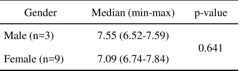
Result:Salivary uric acid levels in the periodontitis patients with a mean±SD 7.40±0.31 (p = 0.004) were found to be higher compared to the gingivitis patients (mean±SD = 6.84±0.19). In addition, there were no significant differences in salivary uric acid levels between gender (p = 0.641).Conclusion:Uric acid levels in periodontitis patients were found to be higher than in gingivitis patients. Moreover, uric acid has more role on periodontitis than in gingivitis as an antioxidant agent.
Keywords : gingivitis, periodontitis, saliva, uric acid
Periodontal diseases, e.g. gingivitis and
periodontitis, are among the most widespread chronic conditions affecting populations around the globe. Their prevalence has reached 50% of the total
adult population worldwide. Gingivitis is
characterized by an inflammation of the gum due to
the accumulation of plaque.1 Gingivitis and
periodontitis have the same clinical appereance of
inflamed gingiva.2 More severe destructive
periodontitis is associated with gum recession, loss of gingival tissue and deterioration of the underlying alveolar bone.1 Periodontitis is an oral inflammatory disorder that causes tissue damage and loss as a
result of the complex interaction between pathogenic bacteria and the host's immune response.3
Periodontal disease is initiated by bacterial
pathogens, such as Porphyromonas gingivalis,
Aggregatibacter actinomycetemcomitans and Bacteriodes forsythus, of which P. gingivalis is the most dominant.1 Phagocytosis is a cellular process for destruction of antigen particles and bacteria.4 Its system responses are divided into two pathways, oxidative and non-oxidative. The oxidative pathway is considered to have an important role in bacterial elimination, whereas the non-oxidative system produces enzymes for its work. Reactive oxygen
species (ROS) have been known to have a role in the pathogenesis of periodontitis. It has been suggested that Polymorphonuclear cells (PMN) produce and release significant quantities of ROS, culminating in increased oxidative damage to gingival tissue, periodontal ligaments and alveolar bone. The gingivitis response is dominated by an oxidative mechanism. In periodontitis, non-oxidative system responses are dominant due to anaerobic environment. Previous research on salivary antioxidant and periodontal disease has yielded unclear data about the comparison of specific uric acid level in gingivitis and periodontitis condition.5
Antioxidants are present in all body fluids and tissues and serve as a protection against endogenously formed free radicals, which are usually produced by leakage of the electron transport system.3, 6 Saliva is also rich in antioxidants (AO) which include superoxide dismutase (SOD), uric acid, albumin, ascorbic acid, glutathione and antioxidant enzymes. Uric acid appears to be the dominant antioxidant present in saliva.7 Uric acid, non-protein thiols and glutathione also act as antioxidants, as does albumin, which can be found in plasma and saliva.8
Saliva contains locally produced substances and other molecules derived from the systemic circulation, such as serum products, gingival crevicular fluid,
electrolytes, microorganisms and other foreign
substances.7 Most of the liquid is produced in the submandibular, parotid and sublingual glands.9Markers
for hormonal, infectious, immunological and
toxicological diseases can be determined from saliva. The oral cavity could be an alternative tool for monitoring oral and systemic health. Therefore, saliva is often called the mirror of the body.7 In gingivitis, saliva is rich in antioxidants has become a chain breaker for the free radical reaction.
Recently, studies on various enzymatic salivary systems and antioxidants have clarified the importance of other defense systems in the saliva. Chapple et al. reported that patients with periodontal diseases had a low total antioxidant status; in contrast, however, Moore et al. reported no significant difference in the total antioxidant level in saliva from patients with periodontal disease against those without.10The aim of this study was to investigate the differences between uric acid levels in gingivitis and periodontitis patients.
The study was carried out with 12 subjects: 6 gingivitis patients and 6 periodontitis patients, which consisted of 9 females and 3 males, from the Trisakti University Dental Hospital, Jakarta – Indonesia. The subjects included in the study had no history of systemic diseases, were non-smokers, did not consume alcohol. The periodontal status of each subject was determined by measuring the plaque-index and probing depth. Full mouth periapical radiographs were taken to determine the level of periodontal bone loss of the patients. The probing depth was defined as the distance from gingival margin to the pocket base. The clinical attachment loss was measured from the distance of the cemento-enamel junction to the free gingival margin and pocket depth. All measurements were taken using a Williams probe (23 W, Hu-Friedy, Chicago, IL, USA) with 0.5 mm units. The measurements were recorded on six surfaces of each tooth (mesio-buccal, buccal, disto-buccal, mesio-longual/palatal, lingual/palatal and disto-lingual/palatal). The average measurements of all surfaces were taken.
At clinical examination, unstimulated saliva were taken before scaling or periodontal surgery. About 5 mL of whole saliva was collected in tubes and centrifuged. The uric acid was measured using
the Folin-Wu technique and analyzed using
spectrophotometry. By using cuvette, test tubes were labelled blank, standard, and samples. 20 µL of blank, standard and sample solutions were transferred to appropriately labelled tubes. A working reagent (1000 µL) was added and each sample was tapped lightly to mix, then further incubated for 30 minutes at room temperature. Optical density was read at 590 nm.
In this study, normality test was conducted
using Shapiro-Wilk test. Known for its
parametric data
8
(p > 0.05), then uric acid level differences in gingivitis
and periodontitis patient were analyzed by
independent t-test. A value of p < 0.05 was considered to be significant level. Based on gender, salivary uric acid level comparison was made by using non-parametric Mann-Whitney test.
Mean result of uric acid in whole saliva of gingivitis and periodontitis patient are showed in table 1. Salivary uric acid level of gingivitis patients (mean
±SD = 6.84± 0.19) was significantly lower than the periodontitis patients (mean±SD = 7.40 ±0.31) (p = 0.004). However, comparison of salivary uric acid status in male and female subject was not significantly different in saliva composition (p = 0.641 > 0.05).
Table 1.Laboratory finding in gingivitis and periodontitis patients. Mean and standard deviation data of uric acid in gingivitis patient are 6.84±0.19 and level in periodontitis patient are 7.40± 0.31 . It shows that level of uric acid in periodontitis is higher than gingivitis patients (p = 0.004 < 0.05)
Table 2.Uric Acid Level Based on Gender. Mann Whitney test shows comparison of salivary uric acid status in male and female subject was not significantly different (p=0.641>0.05)
Increased generation of ROS may cause toxic effects by oxidative damage of proteins, lipids and DNA. Oxidative damage of these biomolecules contributes to disease development.11 In this research, salivary uric acid level in periodontitis was found higher than the patients with gingivitis. This was due to a release of ROS in periodontitis.
A study carried out by Moseley et al
demonstrated that PMN has produced a range of antimicrobial factors, which include ROS during phagocytosis of periodontopathic bacteria.12 Recently, ROS have been reviewed in the
pathogenesis of periodontitis. It has been
suggested that result of stimulation by bacterial antigens, PMN produce and release a big quantity of ROS, culminating in heightened oxidative damage to gingival tissue, periodontal ligament and alveolar bone. The bacterial variety on gingivitis and periodontitis were only served as a
trigger in this case. Moreover, uric acid
contributed to 70% of total salivary anti-oxidant activity. Stimulation of salivary flow is associated with increased production of antioxidants. The antioxidant potential of saliva did not appear to be compromised in patients with periodontal disease but this may relate to the antioxidant flow from the gingival crevicular fluid.13
Salivary uric acid level was significantly lower in gingivitis patients than in periodontitis patients (p = 0.001 < 0.05), and was not significantly different between gender. More investigations with bigger sample size are needed to clarify the uric acid level.
The authors would like to acknowledge the
support of Faculty of Dentistry, Trisakti
University.
9
SCIENTIFIC DENTAL JOURNAL 01 (2017) 07-10
Periodontal Disease Mean (SD) p-value
Periodontitis (n=6)
Gingivitis (n=6)
7.40 (0.31)
6.84 (0.19) 0.004
Gender Median (min-max) p-value
Male (n=3) 7.55 (6.52-7.59)
0.641
The authors report no conflict of interest.
1. Sculley DV, Langley-Evan SC. Salivary antioxidant and periodontal diseases status.UK: Proceeding of the Nutrition Society. 2002. 61: 137 - 143.
2. Newman MG. Etiology of periodontal diseases. In Caranza. editor. Carranza’s Clinical Periodontology. Philedelphia: WB Saunders. 2004. 910.
3. Canakci CF, Cicek Y, Yildirim A, Sezer Y, Canakci, V. Increased Levels of 8-Hydroxydeoxyguanosine and Malondialdehyde and its Relationship with Antioxidant Enzymes in Saliva of Periodontitis Patients. 2009;3(2): 100-106.
4. Miyasaki KT, Nisengard RJ, Haake SK. Immunity and inflammation: Basic Conscepts. In Caranza. editor.
Carranza’s Clinical Periodontology. 8th Edition. Co.
Philadelphia: WB Saunders. 2002; 122-123.
5. Smith CM., Marks AD., Lieberman MA., Marks
DB., Marks' Basic Medical Biochemistry: A Clinical Approach. Philadelphia: Lippincott Williams & Wilkins, 2005.
6. Cory JG. Purine and Pyrimidine Nucleotide
Metabolism. In Textbook of Biochemistry With Clinical Correlation. 6th ed. USA: Wiley-Liss; 2006;
799–800.
7. Miricescu, D, Grebu, M, Totan, A, Didilescu, A, Radulescu, R.. The Antioxidant Potential Of Saliva: Clinical Significance in Oral Diseases. Therapeutics, Pharmacology and Clinical Toxicology. 2011; 15; 139-143.
8. Battino M, Bullon P, Wilson M, Newman H.
Oxidative Injury and Inflammatory Periodontal Diseases: The Challenge of Anti – Oxidants To Free Radicals and Reactive Oxygen Species. CROBM: 1999; 10 : 458.
9. Nagler RM, Reznick AZ. Cigarette Smoke Effects on Salivary Antioxidant and Oral Cancer – Novel Concepts. IMAJ. 2004. 6 : 691–694.
10. Kim, Sang – Chul, Kim Ok-Su, Kim Ok-Joon, Kim, Young-Joon, Chung, Hyun- Ju. Antioxidant Profile of Whole Saliva After Scaling and Root Planing in Periodontal Disease. J Periodontal Implant. 2010; 40: 164–171.
11. Lobo V, Patil A, Phatak A, Chandra N. Free radicals, antioxidants and functional foods: Impact on human health. Pharmacogn Rev. 2010. 4 (8) 118-126.
12. Moseley R, Waddington RJ, Embery G. Degradation of glycosaminoglycans by reactive oxygen species
derived from stimulated polymorphonuclear
leukocytes. Biochimica et Biophysica Acta. 1997. 221–231.
13. Waddington RJ, Moseley R, Embery G. Reactive oxygen species: a potential role in the pathogenesis of periodontal diseases. Oral Dis. 2000. 138-151.
10