Peroxisome Proliferator – Activated Receptors
and The Metabolic Syndrome
Anna Meiliana
1,2* and Andi Wijaya
1,2*
R E V I E W A R T I C L E
1Post Graduate Program in Clinical Biochemistry,
B
ACKGROUND:
Obesity is a growing threat to
global health by virtue of its association with
insulin resistance, inflammation, hypertension,
and dyslipidemia, collectively known as the metabolic
syndrome (MetS). The nuclear receptors PPAR and
PPAR are therapeutic targets for hypertriglyceridemia
and insulin resistance, respectively, and drugs that
modulate these receptors are currently in clinical use.
More recent work on the PPAR has uncovered a dual
benefit for both hypertriglyceridemia and insulin
resistance, highlighting the broad potential of PPARs
in the treatment of metabolic disease.
CONTENT:
We have learned much about PPARs, the
metabolic fat sensors, and the molecular pathways they
regulate. Through their distinct tissue distribution and
specific target gene activation, the three PPARs together
control diverse aspects of fatty acid metabolism, energy
balance, insulin sensitivity glucose homeostasis,
inflammation, hypertension and atherosclerosis.
These studies have advanced our understanding of
the etiology for the MetS. Mechanisms revealed by
these studies highlight the importance of emerging
concepts, such as the endocrine function of adipose
tissue, tissue-tissue cross-talk and lipotoxicity, in the
pathogenesis of type 2 diabetes mellitus and CVD.
SUMMARY:
The elucidation of key regulators
of energy balance and insulin signaling have
revolutionized our understanding of fat and sugar
Abstract
metabolism and their intimate link. The three ‘lipid-
sensing’ (PPAR , PPAR and PPAR ) exemplify
this connection, regulating diverse aspects of lipid
and glucose homeostasis, and serving as bonafide
therapeutic targets.
KEYWORDS:
Peroxisome Proliferator, Activated
Receptor, Metabolic Syndrome.
Introduction
The metabolic syndrome, is characterized by abdominal
obesity, atherogenic dyslipidemia, hypertension,
insulin resistance, inflammation, and prothrombotic
states (1).
The major sequelae are cardiovascular disease
and type 2 diabetes mellitus, but the syndrome also
increases the risk of polycystic ovary syndrome, fatty
liver, cholesterol gallstones, asthma, sleep disturbances,
and some forms of cancer (2). The pathogenesis of the
MetS is thought to involve a complex interaction of
multiple factors, which include obesity and abnormal
fat distribution; insulin resistance; hepatic, vascular,
and immunologic factors; and lifestyle and genetic
contributions (3).
5
Increased adipose tissue mass contributes to
augmented secretion of proinflammatory adipokines,
particularly tumor necrosis factor- (TNF ), along with
diminished secretion of the “protective” adiponectin.
TNF and adiponectin are antagonistic in stimulating
nuclear transcription factor- B (NF- B) activation.
Through this activation, TNF induces oxidative stress,
which exacerbates pathological processes leading to
oxidized low-density lipoprotein and dyslipidemia,
glucose intolerance, insulin resistance, hypertension,
endothelial dysfunction, and atherogenesis. Elevated
free fatty acid, glucose, and insulin levels enhance this
NF- B activation and further downstream modulate
specific clinical manifestations of metabolic syndrome
(5).
Although there has been a debate on the criteria
and concept of the metabolic syndrome, the current
definition by the National Cholesterol Education
Program–Adult Treatment Panel (NCEP-ATP III)
and the International Diabetes Federation (IDF)
provide adequate screening tools to identify the
subjects with high cardiometabolic risk. With these
tools in hand the stage is set for attempts to discover
the pathophysiology underlying these metabolic
abnormalities. The identification of intracellular
signaling elements and regulating factors at crossroad
steps that direct the metabolic fate of lipids are critical
for the understanding of atherogenic dyslipidemia
in the MetS. Importantly, several lipid metabolites
seem to play a crucial role in the regulation of insulin
signaling and action influencing endothelial function
and initiating vascular injury. The concept that
dysfunctional adipose tissue cannot properly handle
the energy surplus derived from excessive calorie
consumption combined with sedentary lifestyle sets
the stage to identify the main determinants of the
MetS in different populations. Resolving these issues
is crucial for the optimal management of the MetS and
reduction of global CVD risk (6).
Peroxisome
proliferator-activated
receptor
(PPAR)s are a family of 3 (PPAR , / , and ) nuclear
receptor/ligand-activated transcription factors that
work in concert as heterodimers with the retinoid
X receptors (7). In recent years, there has been great
scientific and clinical interest in the actions of PPAR
and PPAR because they are the molecular targets
for the clinically used lipid-lowering fibrates and
insulin-sensitizing thiazolidinedione classes of drugs,
respectively (7,8).
The recent development of highly selective
ligands and PPAR / knockout and transgenic mice,
however, have now implicated roles for PPAR / in
adipose tissue formation, metabolism, wound healing,
brain development, placental function, colorectal
carcinogenesis, and skeletal muscle function. PPAR /
ligands appear highly effective in regulating lipid
metabolism, particularly in skeletal muscle, and are
currently in phase II clinical trials for treatment of
dyslipidemia, aimed particularly at individuals with
low HDL levels (9,10,11). All of the PPARs, therefore,
appear to be able to target aspects of the MetS (11).
Because the MetS represents a major risk factor for
cardiovascular diseases, there has been an increasing
interest in the roles of PPARs, in particular most recently
PPAR / , in vascular biology. Indeed, in addition to
the treatment of dyslipidemia, PPAR / ligands may
reduce the development in atherosclerosis (11,12).
The discovery cycle involving nuclear receptors
has elucidated the molecular and physiological basis
for a new class of pharmacophores that show promise
for treating the MetS (3).
The Biology of PPARs
PPARs are members of the nuclear receptor
superfamily which includes the steroid, retinoid,
and thyroid hormone receptors. These receptors are
transcription factors that regulate gene expression
in response to certain endogenous and exogenous
ligands. It is now well established that the PPARs act
as central transcriptional mediators in the regulation
of several important metabolic processes that
influence adipogenesis, insulin sensitivity, glucose
homeostasis, lipid metabolism, vascular endothelial
function, atherosclerosis progression, and ultimately
cardiovascular risk (13).
MOLECULAR BIOLOGY OF PPARS
beyond the stimulation of peroxisome proliferation in
rodents after which they were initially named. PPARs
exhibit broad, isotype-specific tissue expression
patterns (11).
PPAR (NR1C1) (Nuclear Receptors Nomenclature
Committee, 1999) was first described as a receptor that
is activated by peroxisome proliferators, hence its
name (14). Two additional related isotypes, PPAR /
(NR1C2) and PPAR (NR1C3), were then found
and characterized.The PPAR / isotype was called
PPAR when it was first isolated from a Xenopus oocyte
library (15). Because the mammalian PPAR protein
sequence was not highly homologous to the Xenopus
PPAR protein sequences, it was named PPAR when
identified in the mouse with the view that there may
be four members of this nuclear receptor family (16).
PPAR was also designated FAAR (fatty acid activated
receptor) (17) in rats and NUC1 in humans (18).
Sequencing of mammalian genomes indicated that
there are only three PPAR isotypes. Characterization
of PPARs in the chick and comparison with the
PPARs of mouse and Xenopus demonstrated that the
mammalian PPAR is the ortholog of the amphibian
PPAR For reasons of clarity, we propose that this
receptor be designated herein as PPAR / (16).
PPAR is expressed at high levels in organs that
carry out significant catabolism of fatty acids such
as the brown adipose tissue, liver, heart, kidney, and
intestine (19). Of the three isotypes, PPAR / has
the broadest expression pattern, and the levels of
expression in certain tissues depend on the extent
of cell proliferation and differentiation. Important
functions have been assigned to this isotype in the
skin, gut, placenta, skeletal muscle, adipose tissue,
and brain (10,11,20,21,22). PPAR is expressed as two
isoforms, 1 and 2, that differ at their N terminus.
PPAR 2 is found at high levels in the different adipose
tissues (15,23,24), whereas PPAR 1 has a broader
expression pattern that extends to settings such as the
gut, brain, vascular cells, and specific kinds of immune
and inflammatory cells (25,26).
PPARs requires heterodimerization with the
retinoid X receptor (RXR; NR2B), which belongs
to the same receptor superfamily (27,28). This
PPAR/RXR heterodimer can form in the absence of
a ligand. When activated by a ligand, it modulates
transcription via binding to a specific DNA sequence
element frequently called a peroxisome proliferator
response element (PPRE) (15,27,29,30). This response
element, generally of the direct repeat 1 (DR-1) type,
repetition of the consensus sequence AGGTCA with
a single nucleotide spacing between the two repeats.
The PPRE is usually present in one or multiple copies
in the promoter region of target genes but may also be
located in the proximal transcribed region of certain
PPAR-responsive genes (31). PPAR and RXR bind to
the 5’ and 3’ half-sites of this element, respectively,
and the 5’-flanking region mediates the selectivity of
binding between different PPAR isotypes (32,33,34).
Transcriptional control by PPAR/RXR heterodimers
requires interaction with coregulator complexes—
either a coactivator for stimulation or a corepressor for
inhibition of target gene expression (35,36,37,38).
Thus, selective action of PPARs in vivo results
from the interplay at a given time point between
expression levels of each of the three PPAR and RXR
isotypes, affinity for a specific promoter PPRE, and
ligand and cofactor availabilities (11).
A wide variety of natural or synthetic compounds
was identified as PPAR ligands. Among the synthetic
ligands, the lipid – lowering drugs, fibrates, and the
insulin sensitizers, thiazolidinediones, are PPAR and
PPAR agonists, respectively, which underscores the
important role of PPARs as therapeutic targets (11).
The prevalent point of view today is that PPARs
act as lipid sensors that translate changes in lipid/fatty
acid levels from the diet or from food deprivation into
metabolic activity, leading to either fatty acid catabolism
or lipid storage. The endogenous ligands or mediators
of these changes have not been characterized but are
probably generated by fatty acid metabolism. Their
activities are likely to be influenced by their binding
specificities toward the different PPARs and by cell-,
tissue-, or organ- specific effects (39,40,41).
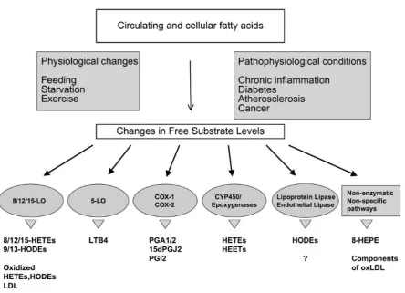
7
differentiation and p roliferation. The d istribution
and abundance of t he ligands a lso depend o n a
variety o f pathophysiological situations a ssociated
with hyperlipidemia, hypertension, diabetes, chronic
inflammation,
c ancer, and a therosclerosis. I t is
important to note that some of these endogenous lipid
mediators also signal through the classic cell surface
G-protein-linked r eceptors and t herefore have m any
PPAR-independent effects (11).
Fig. 1. Endogenous pathways for PPAR ligand production (Adapted from Michalik L, et al. 2006).
Among synthetic ligands, compounds of the
fibrate
family
(clofibrate,
f
enofibrate,
and
bezafibrate)
and
their derivatives have been widely used to characterize
PPAR functions (43). Taken together, mouse models
suggest that PPAR functions to increase fatty-acid use
in t he f asting state and t hat i n the pathophysiologic
context o f a high-fat diet, PPAR -induced f atty-acid
catabolism m ight p revent hypertriglyceridemia.
Consistent with this prediction, an activated variant of
PPAR (Leu162Val) is associated with low serum TG
levels and reduced adiposity (46).
PPAR is expressed in adipocytes, macrophages,
and m uscle, where it r egulates development, lipid
homeostasis, and g lucose m etabolism. Endogenous
PPAR agonists i nclude f atty a cids and e icosanoids
(47,48,49). The PPAR genetic program includes target
genes involved in the uptake of glucose in muscle
(c-Cbl associated protein and glucose transporter 4), lipid
metabolism (scavenger receptor, adipocyte-fatty-acid–
binding protein, lipoprotein lipase, fatty-acid–binding
protein, a cyl-CoA s ynthetase, and CYP4B1), and
energy expenditure ( glycerol k inase and uncoupling
proteins 2 and 3) (50,51,52,63,54,55,56,57,58).
the
finding
that PPAR ligands inhibit the formation
of a therosclerotic l esions i n LDL-re
ceptor–deficient
mice (62).
The landmark
finding
that the thiazolidinedione
class of insulin sensitizers, including rosiglitazone and
pioglitazone, function as h
igh-affinity
PPAR agonists
has validated t he e
fficacy
o f PPAR m odulation in
treating the MetS (63).
PPAR is expressed ubiquitously and is activated
by f atty a cids and c omponents of very-low-density
lipoprotein ( VLDL) (43,47). PPAR t arget genes
control -oxidation in murine brown fat (long-chain
and very-long-chain acyl-CoA s ynthetase, l ongchain
and very-long-chain acyl-CoA dehydrogenase, and
acyl-CoA oxidase), energy expenditure ( uncoupling
proteins 1 and 3 ), and lipid storage ( macrophage
adipose differentiation–related protein) ( 64,65). In
the pathophysiological context o f a high-fat diet,
PPAR c ould function to i ncrease adipose fatty-acid
catabolism and m ay p lay a role i n VLDL – i nduced
lipid a ccumulation in a therosclerotic f oam c ells. A
high-affinity
synthetic PPAR agonist has been shown
to i ncrease HDL and decrease LDL, T G, and f asting
insulin in obese rhesus monkeys. These studies suggest
that therapeutic activation of PPAR has the potential
to decrease diet-induced obesity w ithout a ctivating
the PPAR dependent adipogenic program (66).
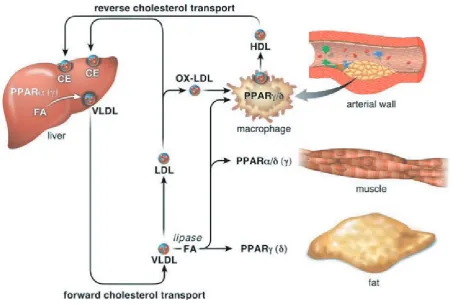
PPARs AND CONTROL OF METABOLISM
Lipids a re e ssential for energy homeostasis,
reproductive and o rgan physiology, and numerous
aspects o f cellular biology. T hey a re a lso linked t o
many pathological processes, such as obesity, diabetes,
heart disease, and inflammation. T
o meet the different
demands from a variety of tissues, the human body has
evolved a s ophisticated lipoprotein transport system
to deliver cholesterol and fatty acids to the periphery
(Fig. 2) (67).
9
Disturbances in this system are integral components
of lifethreatening diseases, best exemplified by the
metabolic syndrome, which refers to patients who
are insulin-resistant (hyperinsulinemic), dyslipidemic
(elevated TG and decreased HDLC levels) (68).
Lipids and their derivatives have a role in the
genetic control of their own systemic transport,
cellular uptake, storage, mobilization, and use. Fine
tuning of these metabolic processes is a hallmark of
healthy organisms. Lipid homeostasis depends on
factors that are able to transduce metabolic parameters
into regulatory events representing the fundamental
components of the general control system (69).
The identification of fatty acids as endogenous
ligands for PPARs has provided a unique approach to
study lipid homeostasis at the molecular level (68).
Liver is the key site of metabolic integration
where fatty acids are mobilized and, depending on
the body’s needs, either stored or used as an energy
source. In the fasting state, the fuel sources of the
body shift from carbohydrates and fats to mostly fats,
and fatty acids that were stored during feeding are
released from the adipocyte and taken up by liver.
There they are either reesterified to TGs and assembled
into VLDL or broken down through -oxidation
and used to generate ketone bodies. Earlier studies
have demonstrated that in the liver, PPAR directly
regulates genes involved in fatty acid uptake [fatty
acid binding protein (FABP)], -oxidation (acyl-CoA
oxidase) and -oxidation (cytochrome P450). Gene
targeting studies confirmed that PPAR is essential for
the up-regulation of these genes caused by fasting or
by pharmacological stimulation with synthetic ligands
such as the fibrates (70,71,72).
Fasting also results in severe hypoglycemia,
hypoketonemia, and elevated plasma levels of
nonesterified fatty acid, indicating a defect in fatty
acid uptake and oxidation caused by dysregulation
of these genes (73,74). In line with these observations,
the fibrate class of drugs including fenofibrate and
gemfibrozil, which are synthetic ligands for PPAR ,
lower serum TGs and slightly increase HDL cholesterol
levels in patients with hyperlipidemia (75), most
likely due to induction of fatty acid oxidation through
activation of PPAR . PPAR has also been shown to
down-regulate apolipoprotein C-III, a protein which
inhibits TG hydrolysis by LPL. This activity of PPAR
ligands further contributes to the lipid-lowering effect
(68).
Apolipoprotein A-V (apoA-V) is now recognized
as a key regulator of serum TG levels. Administration
of the PPAR agonist caused a 50% decrease in TG that
reversed at washout. Serum apoA-V concentrations
increased 2-fold, correlated inversely with TG, and
were reversible at washout. The apoA-V/apoC-III
ratio increased 2-fold, with this increase also reversible
at washout. These data demonstrate for the first time
that a PPAR agonist increases circulating apoA-V
protein levels and the apoA-V/apoC-III ratio (76).
Unlike its function in the adaptive response
to fasting, the role of PPAR in cardiovascular
pathogenesis appears to be detrimental.
Cardiac-specific PPAR overexpression increases fatty acid
oxidation and concomitantly decreases glucose
transport and use, a phenotype similar to that of the
diabetic heart, indicate that PPAR senses fatty acids
and induces their use, and thus plays a causative
role in cardiomyopathy. The net effect, however, of
fibrate intervention in cardiovascular disease is likely
beneficial because systemic TG reduction should result
in less fat accumulation in the heart and at the vessel
wall (68).
Adipocytes are the main site for lipid storage
and modulate the levels of lipids in the blood stream
in response to hormonal signals. PPAR has high
expression in this tissue and has been shown to
potentiate adipocyte differentiation from fibroblasts
(54). The PPAR 2 isoform prevents lipotoxicity by (a)
promoting adipose tissue expansion, (b) increasing the
lipid-buffering capacity of peripheral organs, and (c)
facilitating the adaptive proliferative response of
-cells to insulin resistance (77).
PPAR also promotes cholesterol efflux through
the induction of a transcriptional cascade involving
the nuclear sterol receptor LXR and its downstream
target ABCA1, a membrane transporter that is
important for HDL-mediated reverse cholesterol
transport (61,78,79,80). In this view, one would predict
that in the absence of proportionately increased
ox-LDL, pharmacological activation of PPAR should
shift the balance from lipid loading to lipid efflux and
improve the status of the atherosclerotic lesion (68).
Exercise increases fatty acid oxidation (FAO),
improves serum HDLC and TG, and upregulates skeletal
muscle PPARδ expression. In parallel, PPARδ
agonist-upregulated FAO would induce fatty-acid uptake (via
peripheral lipolysis), and inluence HDLC and TG-rich
lipoprotein particle metabolism, as suggested in preclinical
models (8β). In their report, Oliver et al demonstrate that
a selective PPARδ agonist increases ABCA1 expression
and cholesterol eflux from cells and increases HDLC in
primates. PPARδ agonists may provide a new approach
to the treatment of cardiovascular disease by promoting
reverse cholesterol transport (66).
Molecular and functional analyses suggest that
PPARδ activation reduces hepatic glucose output by
increasing glycolysis and the pentose phosphate shunt and
promoting fatty acid synthesis in the liver. This uncovered
hepatic activity thus constitutes the earliest component
of the regulatory mechanism by which PPARδ regulates
insulin sensitivity, in addition to its known function in fatty
acid -oxidation (8γ).
Coupling increased hepatic carbohydrate
catabolism with its ability to promote -oxidation in
muscle allows PPARδ to regulate metabolic homeostasis
and enhance insulin action by complementary effects
in distinct tissues. The combined hepatic and peripheral
actions of PPARδ suggest new therapeutic approaches to
treat type II diabetes (8γ).
It is now evident that PPARs, which are activated by
various lipid molecules, function in distinct target tissues
and coordinately regulate different metabolic pathways.
PPARα and PPARδ potentiate fatty acid use in liver and
muscle, respectively, whereas PPAR promotes lipid
storage in adipocytes. In this dynamic system, lipids are
shuttled between these tissues according to the needs of
the body by lipoproteins. In this view, lipoproteins not
only deliver energy substrates but also carry endogenous
activators for
these receptors (68).
PPARs AND OBESITY
The cluster of medical sequelae, collectively referred to as
the MetS, poses one of the most serious threats to public
health that our society faces.Why does obesity carry with it
a stereotyped collection of medical problems? That is, why
is adipose tissue unable to store excess calories in a safe
way? The answer to this question is not clear, but it may
relect the reality that fat is not simply a storage depot, but
rather a dynamic tissue that constantly communicates with
other key tissues in the body, including liver,muscle and the
appetite centers in the brain. In the past decade, tremendous
advances in understanding this system of signals and
sensors have been made, and the emergence of PPARs as
key regulators of obesity and metabolism shedding light on
how problems begin and how they may be therapeutically
approached (41).
The initial suggestion that PPAR stimulated
adipogenesis was based on the abservation that
overexpression of PPAR in cells was by itself suficient
to induce adipocyte differentiation (54). Consistent with
this, PPAR increases the expression of genes that promote
fatty acid storage, whereas it represses genes that induces
lipolysis in adipocytes (84).
PPAR- is a master regulator in the formation of fat
cells and their ability to function normally in the adult
(85). PPAR is induced during adipocyte differentiation,
and forced expression of PPAR in nonadipogenic cells
effectively converts them into mature adipocytes (54). In
addition, PPAR knockout mice fail to develop adipose
tissue (86,87,88). Consistent with these indings, humans
with dominant-negative mutations in a single allele PPARG
(the gene encoding PPAR ) have partial lipodystrophy
and insulin resistance (89,90,91). In vitro studies suggest
that PPAR is the ultimate effector of adipogenesis in a
transcriptional cascade that also involves members of the C/
EBP transcription factor family (9β,9γ).
Fat cells develop from a ibroblast-like preadipocyte
to a mature, lipid-enriched adipocyte. The underlying
transcriptional regulatory network that controls the
maturation of adipocytes has been the focus of intense
research and is reviewed elsewhere (94).
The highest levels of PPAR are expressed in
adipose tissue (βγ,β4). In 1994, Spiegelman and colleagues
discovered that expression and activation of PPAR was
suficient to induce adipogenesis (54). The essential role of
PPAR in adipogenesis has been clearly demonstrated in
functional and genetic knockdown experiments (86,87).
(55), fatty-acid transport protein (95), and oxidized
LDL receptor 1 (96), which all favor adipocyte uptake
of circulating fatty acids; phosphoenolpyruvate
carboxykinase (97,98), glycerol kinase (58), and the
glycerol transporter aquaporin 7 (99), which promote
recycling rather than export of intracellular fatty
acids. Together, these pathways lead to the net flux of
fatty acids from the circulation and other tissues into
adipocytes. Although increased fat storage would be
expected to boost the size of adipocytes, TZD treatment
actually leads to smaller adipocytes (100). This is
partly due to increased adipocyte differentiation,
leading to new smaller cells. In addition, TZDs induce
the coactivator PPAR -coactivator 1a (PGC-1 ), which
promotes mitochondrial biogenesis (101), leading to
an increase in fatty-acid oxidation that further protects
against adipocyte hypertrophy (102).
At a molecular level, how does activation of
PPAR , a protein that is mainly present in adipose
cells, lead to systemic insulin sensitization? Two
plausible mechanisms should be considered. First,
pharmacologic activation of PPAR in adipose tissue
improves its ability to store lipids, thereby reducing
lipotoxicity in muscle and liver. This model involves
activation of genes encoding molecules that promote
a combination of lipid storage and lipogenesis, such
as aP2 (fatty-acid binding protein), CD36 (receptor
for lipoproteins), lipoprotein lipase (hydrolysis of
lipoproteins), FATP-1 (fatty acid transporter), glycerol
kinase, and SREBP-1 and SCD-1 (regulators of sterol
and fatty acid synthesis, respectively). Activation
of this metabolic pathway causes body-wide lipid
repartitioning by increasing the TG content of adipose
tissue and lowering free fatty acids and TG in the
circulation, liver and muscle, thereby improving insulin
sensitivity (58, 103). Second, PPAR -specific drugs alter
the release of signaling molecules from fat, including
leptin, TNF , resistin and adiponectin, which by virtue
of serum transport have far-reaching metabolic effects
in other tissues. For example, PPAR agonists inhibit
the expression of TNF and resistin, which both
promote insulin resistance (58,104,11105,106). On the
other hand, PPAR agonists stimulate the production
of adiponectin, which promotes fatty acid oxidation
and insulin sensitivity in muscle and liver. As a result,
hepatic glucose production is reduced and muscle
glucose use is increased (107,108,109). This pathway is
mediated by adiponectin receptors and downstream
AMP-activated protein kinase (110,111), making the
intracellular AMP-activated kinase pathway another
potential pharmacologic target for type 2 diabetes
mellitus. Besides the aforementioned mechanisms, it
is possible that PPAR directly modulates the insulin
signal transduction pathway in adipose tissue by
increasing the expression of intracellular proteins such
as c-Cbl-associated protein (CAP), which stimulates
glucose transport (51).
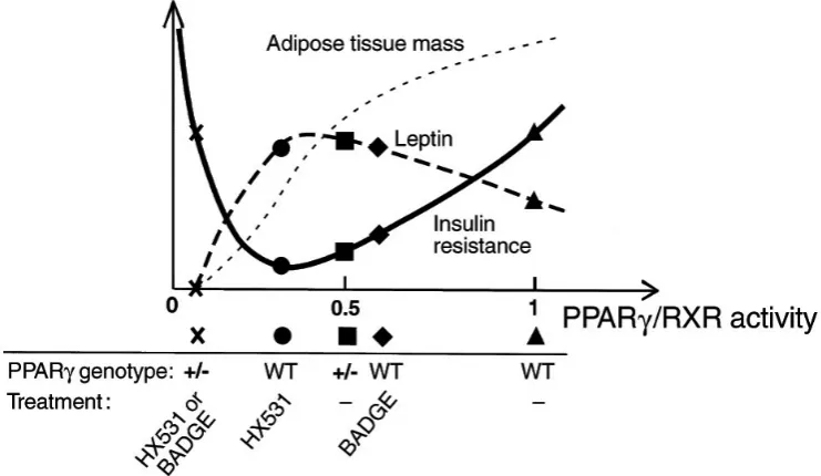
There may be an appropriate level of PPAR /RXR
activity for insulin sensitivity. This raise the possibility
that there may be a hitherto unrecognized U-shaped
relationship between PPAR /RXR activity and insulin
resistance within physiologically “normal” limits
(Figure 3) (112).
Yamauchi et al have shown herein that PPAR /RXR
promotes fat storage in the body by a combination of direct
induction of molecules involved in TG accumulation and
suppression of leptin gene expression as well as inactivation
of PPARα−signaling pathways. In times of fasting, this
PPAR /leptin/PPARα network maximizes energy storage,
which is quite advantageous for survival. In times of feast,
which are the norm in industrialized nations nowadays,
however, this network causes excessive adiposity, insulin
resistance, and obesity related diseases such as diabetes.
Thus appropriate antagonism of PPAR /RXR, which
simultaneously leads to appropriate agonism of leptin and
PPARα, may be a logical approach to protection against
obesity and related diseases such as type β diabetes (11β).
PPAR , function as an important adipocyte
determination factor. In contrast, TNFα inhibits
adipogenesis, cause dedifferentiation of mature adipocytes,
and reduces the expression of several adipocyte – speciic
genes. Reduced PPAR gene expression is likely to
represent an important component of the mechanism by
which TNFα exerts its antiadipogenic effects (11γ).
Insulin receptor substrate (IRS)-1 and IRS-β have
dominant roles in the action of insulin (114), but other
substrates of the insulin receptor kinase, such as Gab1,
c-Cbl, SHβ-B and APS, are also of physiological relevance
(115,116,117,118). Although the protein downstream
of tyrosine kinases-1 (Dok1) is known to function
as a multisite adapter molecule in insulin signaling
(119,1β0,1β1), its role in energy homeostasis has remained
unclear. Hosooka et al show that Dok1 regulates adiposity.
Dok1 promotes adipocyte hypertrophy by counteracting
the inhibitory effect of ERK on PPAR and may thus
confer predisposition to
diet-induced obesity. (1ββ).
The nuclear receptor corepressors NCoR and
SMRT repress gene transcription by recruiting a histone
deacetylase complex.Their roles in PPAR action
have been controversial. Recent evidence, however,
suggests that NCoR and SMRT repress PPAR -mediated
transcriptional activity on speciic promoters in the
adipocyte. In addition, by repressing PPAR action, these
corepressors inhibit the ability of adipocyte differentiation
to proceed (1βγ).
PPARδ initially received much less attention than
the other PPARs because of its ubiquitous expression and
the unavailability of selective ligands. However, genetic
studies and recently developed synthetic PPARδ agonists
have helped reveal its role as a powerful regulator of
fatty acid catabolism and energy homeostasis (9,ββ). The
PPARδ agonist GW501516 was shown to lower plasma
TG levels in obese monkeys while raising high-density
lipoprotein levels, prompting the initiation of clinical
trials to assess its eficacy in hyperlipidemic patients (66).
Studies in adipose tissue and muscle have begun to uncover
the metabolic functions of PPARδ. Transgenic expression
of an activated form of PPARδ in adipose tissue produces
lean mice that are resistant to obesity, hyperlipidemia and
tissue steatosis induced genetically or by a highfat diet.
The activated receptor induces genes required for fatty
acid catabolism and adaptive thermogenesis. Interestingly,
the transcription of PPAR target genes for lipid storage
and lipogenesis remain unchanged. In parallel,
PPARδ-deicient mice challenged with a high-fat diet show
reduced energy uncoupling and are prone to obesity.
Together, these data identify PPARδ as a key regulator of
fat-burning, a role that opposes the fat-storing function of
PPAR (64). Thus, despite their close evolutionary and
structural kinship, PPAR- and PPARδ regulate distinct
genetic networks. The thermogenic function of PPARδ is
markedly similar to that of the nuclear cofactor PGC-1α
(1β4,1β5). Indeed, PPAR strongly associates with
PGC-1α both in cultured cells and in tissue, so it is possible
that many metabolic effects of PGC-1α may be mediated
through PPARδ(41).
Upon agonist stimulation of PPARδ, KLF5
was deSUMOylated, and became associated with
transcriptional activation complexes containing both
the liganded PPARδ and CREB binding protein (CBP).
This activation complex increased the expression of
Cpt1b, Ucpβ and Ucpγ. Thus, SUMOylation seems to
be a molecular switch affecting function of KLF5 and
the transcriptional regulatory programs governing lipid
metabolism (1β6).
PPARs AND CONTROL OF INFLAMMATORY
RESPONSES
In addition to the regulation of lipid metabolism, PPARs
and LXRs play roles in influencing inflammatory
and immune responses. PPARs can be activated by
eicosanoids, which are produced by metabolism
of arachidonic acids and other long-chain PUFAs
during inflammatory responses (36,48,49,59,129). For
example, ligands for PPAR are leukotriene LTB4 and
8(S)-hydroxyeicosatetraenoic acid (HETE), whereas
15deoxy-prostaglandin J2 (15d-PGJ2), 15- HETE, and
13-hydroxyoctadecadienoic acid (HODE) act as ligands
for PPAR . Interestingly, the expression of PPARs is
differentially regulated by factors that control the
development of immune responses. PPAR expression
is dramatically upregulated in macrophages and
T cells during the inflammatory response and can
be induced in vitro by interleukin (IL)-4 and other
immunoregulatory molecules (130,131) In contrast,
IFN and lipopolysaccharide (LPS) repress the
expression of PPAR (132). PPAR is highly expressed
in elicited peritoneal macrophages, while low levels
of PPAR are present (133). The opposite pattern is
observed in primary human monocytes (134). PPAR
and PPAR have been shown to inhibit the expression
of proinflammatory genes, suggesting that they might
inhibit inflammatory responses in vivo. Activation of
PPAR resulted in the induction of genes involved in
fatty acid oxidation with the subsequent degradation
of fatty acids and fatty acid derivatives like LTB4.
In addition, the response to LTB4 and arachidonic
acid was prolonged in mice lacking the PPAR gene
as compared with wild-type mice (135). However,
some in vivo studies show proinflammatory effects
for PPAR ligands, such as an increase in the plasma
levels of TNF during endotoxemia (136) and in the
production of monocyte chemoattractant protein
(MCP-1) by endothelial cells (137). Thus, PPARs and
LXRs negatively regulate transcriptional programs
involved in the development of inflammatory
responses (138).
PPAR activators reduced cytokine-induced
expression of VCAM-1 and ICAM-1 in human
carotid artery endothelial cells in culture. Pasceri
et al (139) reported that fibrates reduced C-reactive
protein (CRP)–induced expression of monocyte
chemoattractant protein (MCP)-1 in human umbilical
vein endothelial cells. With regard to clinical studies,
Marchesi et al observed that 3-month therapy with
fenofibrate in hyperTGmic patients decreased
VCAM-1 and ICAM-VCAM-1 levels in the fasting state.
A molecular rationale for suppression of IL-1–
induced CRP transcription is provided by the fact that
fenofibrate upregulates I- B protein expression. This
reduces nuclear translocation of p50–NF- B, resulting
in decreased amounts of nuclear p50–NF- B and
CCAAT/enhancer binding protein- complexes, the
major determinants of CRP transcription. These results
provide strong evidence for a direct suppressive effect
of fenofibrate on CRP expression, independent of
cholesterol lowering and atherogenesis (140,141)
.Endothelial dysfunction associated with the
metabolic syndrome and other insulin-resistant states
is characterized by impaired insulin-stimulated nitric
oxide production from the endothelium and decreased
blood flow to skeletal muscle. Thus, improvement
in insulin sensitivity leads to improved endothelial
function (142).
Teissier et al extend our insight into the connections
between PPARs, LDL metabolism, and oxidative stress
(143). The oxidized form of LDL (ox-LDL) promotes
inflammation in part via ox-LDL uptake by scavenger
receptors and subsequent nuclear factor B activation.
They find that synthetic PPAR agonists induce the
production of reactive oxygen species (ROS) in a
PPAR –dependent manner by inducing NADPH
.
Moreover, these investigators offer the intriguing
notion that ROS interact with LDL to activate PPAR
and subsequently limit inflammation, as indicated by
PPAR-dependent repression of inducible nitric oxide
synthase (iNOS) gene transcription (145).
Several reports (146,147,148) have indicated that
PPAR agonists reduce macrophage inflammatory
responses, such as the elaboration of cytokines, nitric
oxide, and matrix metalloproteinases (MMPs). In
contrast, other reports have indicated that PPAR
agonists stimulate the expression of proinflammatory
receptors and cytokines (59,60,149,150,151). Finally,
there are even reports that PPAR agonists have no
impact at all on macrophage inflammatory cytokine
production (150,152).
PPAR generally inhibits inflammatory response
genes by negatively interfering with the NF- B, STAT,
and AP-1 signaling pathways in a DNA-binding
independent manner (153). Indeed, PPAR modulates
chemokine gene expression by inhibiting expression
of MCP-1 and its receptor CCR2 in monocyte/
macrophages (154,155). PPAR also plays a role in
the regulation of CXC chemokine pathway. CXCR2
upregulation by PPAR leads to the acquisition of
IL-8/Gro responsiveness, as measured by enhanced
reactive oxygen species (ROS) formation (156).
PPAR expression in human ECs has been
demonstrated by reverse transcription polymerase
chain reaction (157) and more definitively using
Western blotting and immunohistochemistry (158).
Subsequent data suggested that PPAR activation can
influence target genes and processes that are of central
relevance to endothelial biology. One such example is
the role of chemokines (chemoattractant cytokines),
signals for inflammatory cell recruitment (158).
Recently, Huang et al demonstrated that the
PPAR ligands, either 15-deoxy-∆ prostaglandin J2
(15d-PGJ2) or ciglitazone, increased endothelial nitric
oxide (•NO) release without altering endothelial nitric
oxide synthase (eNOS) expression. However, the
precise molecular mechanisms of PPAR -stimulated
endothelial •NO release remain to be defined.
Superoxide anion radical (O2•-) combines with •NO
to decrease •NO bioavailability. NADPH oxidase,
which produces O2•-, and Cu/Zn-superoxide
dismutase (Cu/Zn-SOD), which degrades O2•-,
thereby contribute to regulation of endothelial cell
•NO metabolism. These findings further elucidate
the molecular mechanisms by which PPAR ligands
directly alter vascular endothelial function (159).
CCAAT/enhancer-binding proteins (C/EBPs)
upregulate transcription of various inflammatory
cytokines and acute phase proteins, such as interleukin
(IL)-1 , IL-6, TNF , and cyclooxygenase-2. Recent
studies have demonstrated that PPAR is present
expression of these genes. Interestingly, PPAR gene
promoter has tandem repeats of C/EBP-binding motif,
and C/EBP- plays a pivotal role in transactivation of
PPAR gene. Recent findings strongly suggest that C/
EBP- is negatively autoregulated via transactivation
of PPAR . This feedback mechanism probably
downregulates transcription of inflammatory cytokines
and acute phase proteins, and modulates inflammatory
responses in the early process of atherosclerosis (160).
PPAR controls the inflammatory status of the
macrophage. Deletion of PPAR from foam cells
increased the availability of inflammatory suppressors,
which in turn reduced atherosclerotic lesion area by
more than 50%. Lee et al propose an unconventional
ligand-dependent transcriptional pathway in which
PPAR controls an inflammatory switch through its
association and disassociation with transcriptional
repressors. PPAR and its ligands may thus serve as
therapeutic targets to attenuate inflammation and
slow the progression of atherosclerosis (161).
PPAR , but not PPAR , is the major nuclear
VLDL sensor in the macrophage, which is a crucial
component of the atherosclerotic lesion. In addition
to -oxidation and energy dissipation, activation of
PPAR by VLDL particles induces key genes involved
in carnitine biosynthesis and lipid mobilization
mediated by a recently identified TG lipase, transport
secretion protein 2 (also named desnutrin, iPLA2 , and
adipose TG lipase), resulting in increased fatty acid
catabolism. Unexpectedly, deletion of PPAR results
in derepression of target gene expression, a phenotype
similar to that of ligand activation, suggesting that
unliganded PPAR suppresses fatty acid utilization
through active repression, which is reversed upon
ligand binding. This unique transcriptional mechanism
assures a tight control of the homeostasis of
VLDL-derived fatty acid and provides a therapeutic target for
other lipid-related disorders, including dyslipidemia
and diabetes, in addition to coronary artery disease
(162).
Ligand activation of PPAR in ECs has a potent
antiinflammatory effect, probably via a binary
mechanism involving the induction of antioxidative
genes and the release of nuclear corepressors. PPAR
agonists may have a potential for treating inflammatory
diseases such as atherosclerosis and diabetes (163).
5
mediated pathway in the antiinflammatory activities
of PPAR (164).
PPARs AND HYPERTENSION
The quest for better understanding for the
pathophysiological basis of hypertension and
atherosclerosis is ongoing. The complexity of
hypertension and atherosclerosis and of the underlying
mechanisms is becoming increasingly apparent (165).
Tordjman et al (166) explore the role of the
candidate gene, PPAR , in the regulation of blood
pressure and atherogenesis. PPAR is believed
to impart direct protection in the vessel wall by
intervening at essentially every level of the atherogenic
process: inflammation, monocyte recruitment and
adhesion, cholesterol transport, plaque formation,
and thrombosis, mostly through downregulation
of NF- B and AP-1 (167,168,169). In contrast to the
plethora of data regarding the atherogenic process,
the role of PPAR in the regulation of vascular tone
and blood pressure is still unclear. Several studies
conducted in different rat models of hypertension
yielded inconsistent results (170,171).
Evidence that PPAR regulates the expression
of the RAS starting with its rate-limiting step, renin.
Based on the findings of the current study and on
some published data, we suggest that, in the setting
of an activated RAS and high endogenous levels of
Ang II, PPAR activation could be detrimental. In this
context, PPAR activation is likely to promote even
higher levels of Ang II, increase oxidative stress, raise
blood pressure, and ultimately hasten atherosclerosis.
Further investigations aimed at delineating the
relationships between the transcription factor PPAR
and the components of the RAS and their respective
molecular effectors are needed (166).
Evidence demonstrate that short-term clofibrate
treatment modulates vasomotor tone, producing effects
that are consistent with antagonism of AT1-mediated
but promotion of AT2-mediated renal and systemic
hemodynamic effects of angiotensin II. These effects
appear to involve nitric oxide production and are
accompanied by concomitant changes in the expression
of AT receptors – increased expression of AT2 receptors
but diminished expression of AT1 receptors. PPAR
ligands also protect against pathological damage
especially resulting from angiotensin II hypertension
(172).
Endothelin-1 (ET-1) is a vasoactive peptide that
causes vasoconstriction and vasodilation by binding
to ETA receptors on vascular smooth muscle cells
and ETB receptors on endothelial cells, respectively
(173,174,175).
The role of cytochrome P450 (CYP450) hydroxylase
metabolites in the maintenance of arterial pressure is
complex because of their contrasting hypertensive and
antihypertensive properties (176). Clofibrate, a PPAR
agonist, has been shown to reduce arterial pressure in
Dahl salt-sensitive rats on high salt diet by inducing
the genes that code for CYP4504A (CYP4A) enzymes
in the renal cortex (177,178). CYP4A is the enzyme
responsible for the synthesis of 20-HETE. Interestingly,
20-HETE has actions to reduce sodium transport and,
like ETB receptor activation, has been implicated in
salt-dependent hypertension (179,180,181,182,183).
Therefore, chronic PPAR agonist treatment reduces
salt-dependent hypertension produced by ETB
receptor blockade in male and female Sprague–
Dawley rats. This suggests a possible relationship
between ETB receptor activation and the maintenance
of CYP4A protein expression in the kidney of rats fed
a high-salt diet (184).
Docosahexaenoic acid (DHA), PPAR activator,
reduces blood pressure in some hypertensive models by
unclear mechanisms. PPAR activator DHA attenuated
the development of hypertension, corrected structural
abnormalities, and improved endothelial dysfunction
induced by Ang II. These effects are associated with
decreased oxidative stress and inflammation in the
vascular wall (180).
In the absence of dexamethasone, fenofibrate
lowered fasting TG and cholesterol but unexpectedly
increased systolic blood pressure by ambulatory
monitoring. These data suggest that PPAR activation
in humans does not correct insulin resistance induced
by glucocorticoids and may adversely affect blood
pressure (185).
The current study by Tordjman et al is of prime
importance, because the investigators have successfully
focused our attention on a controversy that involves
PPAR . This central molecule which action had been
considered until now beneficial and a prime therapeutic
target, may in fact turn out to be a candidate gene for
hypertension and for atherosclerosis and, thus, a foe
to human health. More in-depth research is required
to establish if, when, and how PPAR might indeed
be involved in the generation of high blood pressure
and atherosclerosis in humans, issues that remain, at
present, unresolved (165).
PPAR
may represent a link among obesity,
metabolic dysfunction, and activity of either the
circulating or tissue renin-angiotensin system in
hypertension. There is no doubt that the mechanisms
regulating the renin-angiotensin system by PPAR
will prove to be quite complex given the positive
stimulation on renin transcription reported in this
issue, along with the negative impact on the AT1
receptor reported previously. That TZDs are generally
thought to modestly lower blood pressure suggests
that a delicate balance must exist between the effects of
PPAR on the renin-angiotensin system (renin versus
AT1 receptor) and other vasoconstrictors, such as ET-1
(186).
Among the transcription factor binding sites in
the enhancer is the hormone response element (HRE).
Several members of the nuclear hormone receptor
superfamily, including retinoic acid receptor and RXR,
have been shown to bind to the HRE and to regulate
renin gene expression (187). In addition, vitamin D
has been reported to negatively modulate renin gene
expression through a vitamin D receptor– dependent
mechanism, which may involve the HRE (187,188).
Because the HRE is homologous to a PPRE, and
PPAR , retinoic acid receptor- , RXR , and vitamin
D receptor are all members of the same subfamily of
ligand-activated transcription factors, it should not
be surprising that PPAR may have to be included
among those transcription factors thought to regulate
renin expression.
PPAR activation by TZDs causes a
down-regulation of AT1 receptor gene expression via a
PPAR -dependent mechanism in vascular smooth
muscle cells. Therefore, PPAR may play a role in the
regulation of Ang II action (189).
PPARs AND THE METABOLIC SYNDROME
Metabolic syndrome (MetS) is a cluster of metabolic
abnormalities, which is characterized by abdominal
obesity, insulin resistance, dyslipidemia, elevated blood
pressure, and a proinflammatory and prothrombotic
milieu (200,201).
MetS appears to affect a significant proportion
of the population. While up to 80% of the almost
200 million adults worldwide with diabetes will die
of CVD, people with MetS are also at increased risk,
being twice as likely to die from and three times as
likely to have a heart attack or stroke compared to
people without the syndrome (192). Subjects with
Whether or not it is accepted that MetS is a specific
disease entity or just a constellation of symptoms, the
prevalence of this condition is increasing worldwide
and patients need to tackle these risk factor through
either lifestyle or pharmacological approaches, in
order to reduce the odds of developing diabetes and
cardiovascular disease (CVD) (194).
PPARs are intimately involved in nutrient
sensing and the regulation of carbohydrate and lipid
metabolism. PPAR and PPAR appear primarily to
stimulate oxidative lipid metabolism, while PPAR
is principally involved in the cellular assimilation of
lipids via anabolic pathways. These may nevertheless
have much greater significance for the public health
burden in the abnormal lipid and carbohydrate
metabolism of the MetS (195).
One of the hallmarks of MetS is depressed levels
of HDLC, which occurs in 37% of patients with MetS
(194). Whereas HDLC is not the primary target of
lipid-modulating therapy, it is recognized as an important
secondary target of therapy, and, thus, treatments that
raise HDLC may be important in reducing CVD risk
(197).
The identification of PPARs as molecular targets
for drugs to treat hypertriglyceridemia and type 2
diabetes mellitus has fueled interest in their biology
and potential as targets to treat the metabolic
syndrome (198). In keeping with their roles as lipid
sensors, ligand-activated PPARs turn on feed-forward
metabolic cascades to regulate lipid homeostasis via
the transcription of genes involved in lipid metabolism,
storage, and transport. Additionally, PPARs may
suppress
inflammation
through
mechanisms
involving the release of antiinflammatory factors or the
stabilization of repressive complexes at inflammatory
gene promoters (161,199).
7
Hypertriglyceridemia and abdominal obesity are
key components of the MetS. They may result from
an inability of adipose tissue to sequester fatty acids
appropriately for storage (202). Instead, fatty acids
are deposited as ectopic fat in skeletal muscle (203),
liver (204), and other organs (205). It is thought that
such fat accumulation is linked to impaired metabolic
function of the tissue (206,207). Obesity-related insulin
resistance involves the release of mediators, such as
FFAs, TNF , or resistin from adipocytes and decreased
production of adiponectin, all of which impair insulin
action in skeletal muscle (207).
Adipose tissue as an endocrine organ also releases
proinflammatory mediators that promote vascular
damage and atherosclerosis. TNF inhibits insulin
signaling contributing to insulin resistance and activates
multiple mechanisms of inflammation via NF- B (208).
Leptin can alter insulin action and has recently been
recognized to be an important mediator of
obesity-related hypertension (209). Angiotensinogen, the
precursor of angiotenin II, a key mediator of vascular
injury, can be produced and secreted by adipose tissue
(210). Plasminogen activator inhibitor 1 (PAI-1) is
typically increased in the obesity/insulin-resistance
state and plays an important role in atherothrombosis
(211,212). In contrast, excessive visceral adipose tissue
has been shown to be associated with decreased
adiponectin levels (111), an important hormone that
exerts antidiabetic (109,110) and antiatherogenic
functions (213,214). Adiponectin activates
AMP-activated protein kinase, which promotes skeletal
muscle glucose uptake and suppresses hepatic glucose
production (110). Importantly, adiponectin also inhibits
NF- B activation, thus, attenuating inflammation (215).
Visfatin, a growth factor with insulin mimetic action,
was recently cloned from fat (216). Unlike adiponectin,
plasma levels of visfatin increase in parallel with
visceral fat in both mice and humans (216), so the
role of visfatin in insulin resistance needs additional
investigation. Taken together, these observations
suggest that the adipocyte is an integral coordinator
of the relationship among obesity, diabetes, and CVD
(207).
Interestingly, it is possible that the intra –
myocellular lipid (IMCL) impact on insulin sensitivity
might be modulated by the oxidative disposal of
long-chain fatty acyl-CoAs, depending on the efficiency
of the regulation of its flux and metabolism within
the mitochondrion. These latest findings represent a
fascinating and promising novel understanding linking
small abnormalities of tissue energy metabolism to a
steady tendency toward both weight gain and ectopic
fat accumulation – particularly in skeletal muscle – as
a common denominator inducing both obesity and
insulin resistance (203).
The PPAR agonist GW501516 attenuated
multiple metabolic abnormalities normally associated
with the MetS in humans, and this was probably
due to an increase in skeletal muscle fatty acid
oxidation. Presently, the individual components of the
MetS are treated separately; i.e., statins are used for
elevated cholesterol, fibrates are used to reduce TG,
and metformin and thiazolidinediones are used for
hyperglycemia. The wide range of beneficial effects
suggested by the response to GW501516 calls for a
larger study in patients to evaluate the clinical efficacy
of PPAR agonists for the treatment of hyperlipidemia,
liver fat accumulation, obesity, and insulin resistance
(217). Thus, PPAR is pivotal to control the program
for fatty acid oxidation in the skeletal muscle, thereby
ameliorating obesity and insulin resistance through its
activation in obese animals (128).
Accelerated atherosclerosis is a major cause of
morbidity and death in insulin-resistant states such as
obesity and the MetS, macrophages from obese (ob/ob)
mice have increased binding and uptake of oxidized
LDL, in part due to a posttranscriptional increase in
CD36 protein. Macrophages from ob/ob mice are also
insulin resistant, as shown by reduced expression and
signaling of insulin receptors. Defective macrophage
insulin signaling predisposes to foam cell formation
and atherosclerosis in insulin-resistant states and
that this is reversed in vivo by treatment with PPAR
activators (218).
Obesity and insulin resistance, the cardinal
features of MetS, are closely associated with a state of
low-grade inflammation (219,220). In adipose tissue
chronic overnutrition leads to macrophage infiltration,
resulting in local inflammation that potentiates insulin
resistance (220,222).
that macrophage polarization towards the alternative
state might be a useful strategy for treating T2D (223).
PPARs AS THERAPEUTICS TARGETS FOR MetS
Because these nuclear receptors are activated by
extracellular signals and control multiple gene targets,
PPARs can be seen as nodes that control multiple inputs
and outputs involved in energy balance, providing
insight into how metabolism and the vasculature may
be integrated. The ongoing clinical use of fibrates,
which activate PPAR , and thiazolidinediones, which
activate PPAR , establishes these receptors as viable
drug targets, whereas considerable in vitro animal
model and human surrogate marker studies suggest
that PPAR activation may limit inflammation and
atherosclerosis. Together, these various observations
have stimulated intense interest in PPARs as therapeutic
targets and led to large-scale cardiovascular end-point
trials with PPAR agonists (224).
The current clinical approach to MetS is to focus
on appropriate management of accompanying risk
factors. While priority should be given to management
of underlying risk factors with therapeutic lifestyle
changes, associated major risk factors should be
treated according to evidence – based medicine goals
and principles, and appropriate clinical attention
should be given to the presence of emerging risk
factors. Clearly, there are multiple targets for therapy
to reduce the high risk of the MetS. While no single
treatment for the MetS as a whole yet exist, it is well
established that lifestyle changes, for example, changes
in diet and increased physical exercise, form the first
– line strategy of intervention (194).
In MetS, a condition of impaired fatty acid
metabolism in adipose tissue generally results in the
increased release of free fatty acids into the circulation.
This in turn leads to multiple abnormalities in the
circulating lipoprotein profile including low HDL, high
TG, and VLDL remnants, with average LDL. The triad
of elevated TG, reduced HDL and small dense LDL,
along with concomitant increased in TG – rich remnant
particles, comprises the atherogenic dyslipidemia
of MetS. MetS, with or without progression to T2D,
is therefore a major atherogenic factor. CHD risk
reduction in MetS requires not only aggressive LDL
– C lowering but also management of each aspect of
dyslipidemia, including lowering TG levels, increasing
HDL – C levels, and increasing the size of the average
LDL – C particle. Based on available evidence, LDL
patients with atherogenic dyslipidemia with elevated
TG, non – HDL – C represents a secondary target of
treatment after the LDL – C goal is achieved. The
tertiary goal in these patients is to raise HDL – C when
it is reduced after attaining goals for LDL – C and non
– HDL – C (194).
PPAR activation can improve metabolic parameters
like glucose and lipid levels but also alter directly
vascular responses by regulating target genes including
those encoding adhesion molecules, The ATP – binding
cassette transporter 1 (ABCA 1), lipoprotein lipase,
cytokines and chemokines (226). PPAR activation has
many anti – atherosclerotic effects with disctinct and
overlapping targets. For instance, lipoprotein lipase
acts on circulating lipoproteins to activate PPAR and
intiates PPAR – dependent positrive feedback loop
for TG – rich lipoprotein catabolism (44). Exogenist
PPAR agonist such as fenofibrate reduce TNF
– induced activation of NF-
(227), a transcription
factor integrating inflammation and atherosclerosis.
There have been indications that PPAR is involved
in the pathogenesis of insulin resistance. In this
regard, PPAR knockout mice are protected from diet
– induced insulin resistance probably because of of
inhibition of PPAR - dependent fatty acid oxidation
(227). By contrast, genetic defects in PPAR can
recapitulate all the salient features of MetS in humans
(96). Therefore, PPARs are likely to be involved in the
development of MetS and, accordingly, can serve as
potential therapeutic targets for the prevention and
treatment of MetS (228).
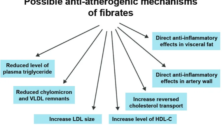
9
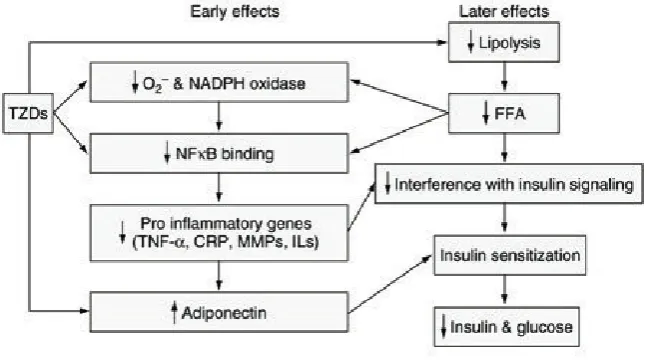
Fig. 4. Effects of fibrates with the potential to protect against cardiovascular disease (Adapted from Barter PJ, 2008).