The Protective Effect of Field Mint Leaves in Reducing Stomach Ulcer in
Rats Induced by Aspirin
Vanitha Ratha Krishnan,1 Januarsih A. Rachman,2 Abdul Hadi Hassan3
1Faculty of Medicine Universitas Padjadjaran, 2Department of Anatomy and Cell Biology Faculty
of Medicine Universitas Padjadjaran, 3Department of Pathology Anatomy Faculty of Medicine Universitas Padjadjaran/Dr. Hasan Sadikin General Hospital Bandung
Abstract
Background: Stomach mucosal wall erosion is caused by the imbalance of the aggressive factors and mucosal defensive factors due to the common causes such as the side effect of consuming non-steroidal
anti-inflammatory drugs. Field mint (Menthaarvensis) leaves have been used as an alternative option to cure and
prevent the gastric problems. The aim of this study was to analyze the protective effect of Field mint leaves infusion in reducing stomach ulcer in rats induced by Aspirin.
Methods: The experimental study was conducted at Histology Laboratory of Faculty of Medicine, Universitas
Padjadjaran, Bandung. Sixteen rats were divided into 4 groups randomly: group I (control negative group), group II (control positive group, given 90mg/day Aspirin, group III (the treatment group, given 5cc of Field mint leaves infusion and 90 mg Aspirin) and group IV (the treatment group, given 5.6µg of Misoprostol and 90 mg Aspirin). Mucosal wall erosions were determined by using microscope. Data were analyzed using non-parametric Kruskal-Wallis test and Mann-Whitney U-test (CI 95% and p-value<0.05)
Results: Group II had high score of mucosal wall erosions after given only aspirin. In group III and IV, the
score of mucosal wall erosions were low. However there was no difference in score of mucosal wall erosions
between group III-IV (p<0.05)
Conclusions: Field mint (Menthaarvensis) leaves infusion is able to prevent stomach mucosal wall erosions
induced by Aspirin as misoprostol does. [AMJ.2015;2(3):363–68]
Keywords: Aspirin, field mint (Menthaarvensis), stomach mucosal wall erosions
Correspondence: Vanitha Ratha Krishnan, Faculty of Medicine, Universitas Padjadjaran, Jalan Raya Bandung-Sumedang Km.21, Jatinangor, Sumedang, Indonesia, Phone: +628170204955 Email: [email protected]
Introduction
Stomach ulcer is resulted from disparity of antagonistic gastric luminal factors such as acid and pepsin and defensive mucosal barrier function. Many environmental and host factors can lead to an ulcer formation. This is caused by increasing the gastric acid secretion or weakening the mucosal barrier. One of the
most significant causes of the ulcer is the use of Non-Steroidal Anti-Inflammatory Drugs
such as Aspirin. Aspirin causes an inhibition
of cyclooxygenase enzyme (COX) which
inhibits endogenous prostaglandin especially
Prostaglandin E1 (PGE1), Prostaglandin E2 (PGE2) and Prostaglandin analogue (PGI2).
Thus, mucosal defense is in breached. Prostaglandin is accountable in regulating
mucous cell secretion and since COX inhibits
prostaglandin causing the barrier function is disrupted. The decreased Prostaglandin inhibits basal bicarbonate secretion from mucosa and augmentation of acid production.1-4
Traditional medicine remains widespread in many countries due to its affordability and availability. For example is the use of Field mint plant which is a constant plant belonging to the Mentha genus. This mint plant is found
worldwide such as in Europe, Asia and North
America. Its mutual names include Field mint, Wild mint or Corn mint. Field mint is
well-known by its scientific name Mentha
many advantages. Field mint tea is mainly used as an invigorating drink and condiment.5-9
Moreover, Field mint leaves have various medicinal properties in preventing gastrointestinal disorders. Field mint oil can be used for both intrinsic and extrinsic indications.
The food and drug administration (FDA) approved the oil of mint a “Generally Regarded as Safe” (GRAS) status. Mint leaves have many benefits such as rich of antioxidants, for
example phenols, vitamins including vitamin A, beta carotene and vitamin-C.6 Thus, the present study was undertaken to determine
the protective effects of 5 cc/day Field Mint
leaves infusion in the experimental erosion of stomach mucosal layer in rats induced by Aspirin. 5-9
Methods
In this study, the subjects used were 16 of
12 weeks white male Wistar rats with 200
grams each adapted for one week. The rats were obtained from the Pharmacological Laboratory, Faculty of Medicine, Universitas Padjadjaran. The materials used in this study
were fresh Field mint (Mentha arvensis)
leaves. The leaves were purchased from
the local vegetable supermarket (Jatos) in Jatinangor and were identified. The leaves were shade dried, finely powdered and made into an infusion. A total of 30 grams Field mint (Mentha arvensis) leaves powder was added with 500ml of water and an additional of extra water 2 times of the material weight (8ml) in a
special pan to make the infusion. Then, heated
over a water bath for 15 minutes, calculated from the temperature in the pot at 900 C, and
stirring was done occasionally. The infusion
was filtered while still hot.39 The leaves were given in the form of 5cc/ rat infusion per oral.
This particular dosage was given to attain an optimum effect of the study.
Drugs used in this study were Aspirin and Misoprostol. This study was an experimental
laboratory study using a simple randomization design plan. A total of 16 rats were used as experimental animals and grouped randomly into 4 groups using random allocation method as follows: Group I which was the negative control group was given aqueous solution. Group II which was the positive control group
was given aspirin solution (90mg). Group III,
the treatment group was given aspirin solution
(90mg) and field mint (Mentha arvensis) leaves infusion (5cc/rat) and Group IV, also a
treatment group was given Aspirin solution
(90mg) and Misoprostol (3.6µg).
Before the study, the rats were adapted to the laboratory environment for one week. The rats were given food in the form of pellets and also water as needed. The rats were weighed every day and at the end of adaptation period and were observed closely to make sure they are healthy. Next, all rats went on
a fast. Over the next five days, every morning
each treatment group was given a respective
drug: Treatment group III was given 5cc/rat field mint (Mentha arvensis) leaves infusion and treatment group IV was given 3.6µg of Misoprostol. One hour later, group III and IV were given 90 mg Aspirin. At the end, the rats
went on a fast for 24 hours. Finally, all rats
were sacrificed with anesthesia using diethyl
ether. The rats were dissected by laparotomy to obtain the gaster.
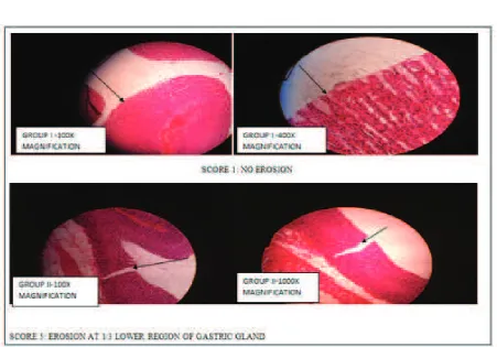
The gastric mucosal layer (pyloric part) was then observed microscopically using the 100x and 400x magnifications after a fixation and
staining. The depth of the mucosal wall erosion were observed and noted in each group. The
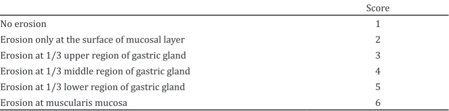
extend of mucosal wall erosion were classified according to Wattimena (1982) (Table 1).10
The statistical analysis was conducted by using a computer. The statistical test showed
CI 95% and p-value<0.05 (significant) and p-value <0.01 (highly significant). The
microscopic data were analyzed by Kruskal-Wallis test, to assess the median difference among those various groups. If the
Kruskal-Table 1 Scoring of the Extend of Mucosal Wall Erosion
Score
No erosion 1
Erosion only at the surface of mucosal layer 2
Erosion at 1/3 upper region of gastric gland 3
Erosion at 1/3 middle region of gastric gland 4
Erosion at 1/3 lower region of gastric gland 5
Figure 1 Microscopic Observation of Stomach Mucosal Erosion in Group I (Control Group) and Group II (Aspirin Group)
Wallis test is significant, the Mann-Whitney
test was used to assess the median differences
between two groups: Group I-II; Group I-III; Group I-IV; Group II-IV; Group III-IV.
Results
The microscopic results were obtained by microscopic evaluation to the stomach mucosal layer on the histological slide using
the 100X and 400X light microscope. Data
were collected during the evaluation by observing the erosions of the mucosal layer of the rat`s stomach. The severity of gastric mucosal erosions was as illustrated below
(Figure 1 and Figure 2).
The microscopic Wattimena score data collected from all four groups were analyzed by using non parametric Kruskal-Wallis test to asses statistical differences between various
groups of animals with CI 95% and α = 5%. Ho rejected when p-value < 0.05 and accepted when p-values > 0.05. From the statistical test,
it is shown that there was a difference between
those groups (Table 2).
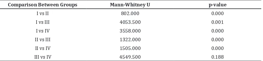
The microscopic data collected were then analyzed by using Mann-Whitney U test to asses statistical difference between two
groups of animals with CI 95% and α = 5%. Ho rejected when p value < 0.05 and accepted
when p value > 0.05. From the statistical test,
there were differences between group I-II,
group I-III, group I-IV, group III, group II-IV. However, there was no difference between group III-IV (Table 3).
Discussion
It was proven from the results of the present
investigation that the leaves infusion of field mint (Mentha arvensis) possesses a protective
effect in aspirin- induced acute ulcer. It has
shown a significant reduction in the gastric
lesions of the aspirin treated groups of animals. Although the etiology of the gastric ulcer is unknown in most of the cases, it is generally accepted that they are resulted from an imbalance between aggressive factors and the maintenance of mucosal wall integrity through the endogenous defense.
From the microscopic results as stated above, it seems that aspirin can cause the
erosions of the stomach mucosal layer. Various factors are identified to be the risk factors and
predisposing factors for the stomach mucosal wall erosion due to aspirin consumption. These factors have a close relation with the pathogenesis of Aspirin-induced gastric ulcer. The disturbance in mucosal resistance allows the injury by endogenous factors such
Table 2 Effects of Field mint Leaves Infusion on Aspirin Induced in Gastric Mucosal Erosions
Wattimena Score)
Group Wattimena Score p
1 2 3 4 5 6 Median Range 0.000
Group I 89% 10% 1% 0% 0% 0% 1 2
Group II 12% 21% 20% 16% 24% 7% 3 5
Group III 71% 18% 10% 1% 0% 0% 1 3
Group IV 61% 28% 9% 2% 0% 0% 1 3
Note : n = 100 areas of observation in each group; group I (control); group II (Aspirin); group III (FLI); group IV (Misoprostol); p < 0,05 (significant); p<0.01 (highly significant)
Table 3 Statistical Comparison among Groups
Comparison Between Groups Mann-Whitney U p-value
I vs II 802.000 0.000
I vs III 4053.500 0.001
I vs IV 3558.000 0.000
II vs III 1322.000 0.000
II vs IV 1505.000 0.000
III vs IV 4549.500 0.188
as acid, pepsin, and bile salts. Furthermore, aspirin administration for acute ulcer model
of animals in this study was sufficiently
induced mucosal erosions of the stomach even if it took four hours for ulcer induction before
sacrificing. This was proven by the significant results of theAspirin group (Group II) mucosal
wall erosions compared to the control group
(Group I) which only received aqueous
solution and daily normal diet pellet.
After ingestion of NSAIDs, the structural damage to the stomach surface epithelium occurs. The oxygen derived free radicals plays a big impact in the mechanism of aspirin induced gastric ulcers. Aspirin induces the reactive oxygen metabolites in rats, which contributes to mucosal injury. The potential antioxidant protective effect of Field mint leaves on gastric mucosal tissue is a topic of high current interest.11-13
The Aspirin group (Group II) showed a significant increase in ulceration whereas in the Field mint leaves infusion group (Group III), there was a significant reduction of the
aspirin effect. Field mint leaves are used as herbal remedies in folk medicine. Menthol,
caryophyllene and flavonoids which are the
active agents in Field mint leaves are known to possess a potent antiulcer activity. It also reduces gastric acid secretion induced by histamine in rats. Field mint oil comprises vitamins A and C, omega-3 fatty acids, and minerals including potassium, manganese, iron, magnesium, calcium, and copper which increase the production of mucosal barrier in stomach mucosal wall.
Field mint leaves infusion inhibited the increase of the stomach mucosal wall lesions in Aspirin induced lesions in rats. Field mint leaves infusion pretreatment offered a protection against the action of aspirin showed that the presence of some antioxidant phytoconstituents might have the gastric mucosa from free radical-induced damage. The effective scavenging of free radicals by
Vitamin C and Carotene may have reduced
the progressive ulceration. Field mint leaves
contain flavonoids which can reduce gastric
tissue histamine content. This fact in turn may have inhibited gastric secretions. Carotene is a natural fat-soluble pigment which has antioxidant activity in vitro demonstrated that can take up singlet oxygen scavenge peroxyl radicals and inhibit lipid peroxidation.11-13
In the present study, it was found that
animals in the Control group (Group I) also
developed some erosions through microscopic observation. This could be due to the
normal physiological changes in the gastric mucosal cells regulation as a response to the environmental factors in the laboratory such as temperature. The cells of the gastrointestinal tract have a rapid turnover rate, which makes its mucosa become one of the most rapidly proliferating tissues in the human body. Under normal circumstances, cell populations within the gastrointestinal tract are maintained at a dynamic state because the cell loss due to the exposure of the gastric mucosa to pH, osmolarity and temperature is balanced by a continuous cell renewal.
The other possible reason of the control
group (Group I) having mucosal wall erosions was due to its non-inflamed prior mucosal condition. Inflamed gastric tissue is more
resistant to aspirin-induced damage than
non-inflamed gastric tissue. Physiologically, acid
secretions in the stomach are under control of nervous and endocrine systems. The pH can
reach a minimum level of 0.8. Fasting for long
periods of time can cause acidic conditions which leads to mucosal wall erosions. During the study, the rats went on a fast for 24 hours
prior to sacrifice.
Furthermore, the gastric mucosal erosions of rats belonged to the Field mint leaves
Infusion Group (Group III) did not show a significant result when it was compared to the Misoprostol group (Group IV).The reason
for this condition was due to the very similar mechanism of action of both Field mint leaves and Misoprostol which has an antioxidant effect to free radicals. For these results, it can
be concluded that the 5 cc/day of Field mint
leaves infusion had an adverse protective effect toward aspirin-induced gastric erosions in rats. The study limitation was that the concentration of mint leaves infusion used was only one so the different effect from different concentration cannot be found. Further study should be conducted using several concentration with larger sample size.
References
1. Katzung BG, Masters SB, Trevor AJ. Basic and clinical pharmacology. 11th ed. New
York: McGraw-Hill Companies Inc; 2009. p. 584.
2. Mitchell J. Non-steroidal anti-inflammatory analgesic drugs. AnaesthesiaUK.
2006 [Cited 2012 April 21]. Available
from: http://www.frca.co.uk/article.
aspx?articleid=100633.
Cambridge: Cambridge University Press; 2011.
4. Gabriel SE, Jaakkimainen L, Bombardier
C. Risk for serious gastrointestinal
complications related to use of nonsteroidal
anti-inflammatory drugs: a meta-analysis. Ann Intern Med. 1991; 115(10):787–96. 5. Pandey AK, Rai MK, Acharya D. Chemical
composition and antimycotic activity of
the essential oils of corn mint (Mentha arvensis) and lemon grass (Cymbopogon flexuosus) against human pathogenic fungi. Pharmaceutical Biology. 2003; 41(6):421–5.
6. Moerman D. Native American ethnobotany: a database of Native American foods, drugs,
dyes, and fibers of Native American peoples
derived from plants. Dearborn Michigan:
University of Michigan Dearborn; 2003. 7. Dwivedi D, Khandelwal G, Patidar RK,
Singh V. Antimicrobial activity of mentha
arvensis against clinical isolates of human cariogenic pathogens- an in-vitro study.
IJPSR. 2012;3(5):1355–60.
8. Dorman HJ, Koşar M, Kahlos K, Holm
Y, Hiltunen R. Antioxidant properties
and composition of aqueous extracts from Mentha species, hybrids, varieties,
and cultivars. J Agric Food Chem. 2003; 51(16):4563–9.
9. Akram M, Uzair M, Malik NS, Mahmood A, Sarwer N, Madni A, et al. Mentha arvensis
Linn.: a review article. J Med Plants Res. 2011; 5(18):4499–503.
10. Wattimena JR. L’hypoproteinemiae
experimentale chez le rat, exploritation pharmacocinetique et pharmacologie du modele. These doctor d`etat es screces pharmaceutique. Montpellier: Universite
De Mountepellier; 1982.
11. McCord JM. The evolution of free
radicals and oxidative stress. Am J Med. 2000;108(8):652–9.
12. Becker JC, Domschke C, Pohle T. Current approaches to prevent NSAID-induced
gastropathy – COX selectivity and beyond. Br J Clin Pharmacol. 2004; 58(6):587–600.
13. Rahman K. Studies on free radicals, antioxidants, and co-factors. Clin Interv