Colorectal cancer among young native Indonesians: A clinicopathological and
molecular assessment on microsatellite instability
Aru W. Sudoyo,1 Bethy Hernowo,2 Ening Krisnuhoni,3 Ary H. Reksodiputro,1 Daldiyono Hardjodisastro,4 Evlina S. Sinuraya5
1 Division of Hematology, Department of Internal Medicin, Cipto Mangunkusumo Hospital-Faculty of Medicine, University of
Indonesia, Jakarta, Indonesia
2 Department of Pathology Anatomy, Faculty of Medicine, University of Pajajaran, Bandung, Indonesia 3 Department of Pathology Anatomy, Faculty of Medicine, University of Indonesia, Jakarta, Indonesia
4 Division of Gastroenterology, Department of Internal Medicin, Cipto Mangunkusumo Hospital-Faculty of Medicine, University
of Indonesia, Jakarta, Indonesia
5 Laboratory Pathology Anatomy, Dharmais Cancer Hospital, Jakarta, Indonesia
Abstrak
Tujuan: Untuk mengevaluasi karakter klinikopatologis kanker kolorektal pada pasien muda Indonesia dan menilai kaitannya dengan ekspresi protein MLH1, MSH2, dan SMAD4, dan membandingkannya dengan pasien kanker kolorektal usia di atas 60 tahun.
Metode: Data rekam medis pasien kanker kolorektal usia di bawah 40 tahun dan usia di atas 60 tahun , dikumpulkan dari 3 rumah sakit: Jakarta, Makasar, dan Bandung. Kelompok etnis dipilih dari suku bangsa Jawa, Makasar
(Sulawesi Selatan, dan Minangkabau (Sumatera Barat) yang dikonirmasi berdasarkan kuesioner. Pada spesimen
tumor dilakukan pemeriksaan histopatologi, gradasi tumor, serta pemeriksaan imunohistokimia untuk penentuan ekspresi protein MLH1 dan MSH2 untuk menilai mutasi instabilitas mikrosatelit. Ekspresi protein SMAD4 diperiksa untuk memastikan bahwa jaringan tumor tidak berasal dari instabilitas mikrosatelit.
Hasil: Telah dikumpulkan 121 penderita kanker kolorektal dari etnis Sunda, Jawa, Makasar, dan Minangkabau. Derajad keganasan antara pasien muda dan pasien tua berbeda secara bermakna (p = 0.001). Pewarnaan imunohistokimia untuk protein MSH2 dan MLH1 yang dilakukan pada masing-masing 92 dan 97 pasien, menunjukkan tidak terdapat perbedaan bermakna dalam hal ekspresi MLH1 dan MSH2 dan gradasi tumor, yang berarti tidak ada hubungan antara instabilitas mikrosatelit dan derajad tumor.
Kesimpulan: Karakter kliniko patologi kanker kolorektal pada penduduk asli Indonesia, tidak berbeda antara pasien usia muda (< 40 tahun) dan pasien usia tua (>60 tahun) pada kelompok etnis yang sama. Juga tidak terdapat perbedaan dalam ekspresi protein MSH2 dan MLH1, yang merupakan indikator instabilitas mikrosatelit. (Med J Indones 2010; 19:245-51)
Abstract
Aim: To obtain clinicopathological characteristics of colorectal cancer among young native Indonesians and to assess MLH1, MSH2, and SMAD4 protein expressions, comparing them with a matched population of colorectal cancer patients aged 60 years old and older.
Methods: Medical records of colorectal cancer patients aged 40 years or younger and 60 years or older from several hospitals in three Indonesian cities – Jakarta, Makassar, and Bandung - were reviewed. The “native” ethnic groups were selected from those originating from Java, Makassar (South Celebes), Miinangkabau (West Sumatra). Ethnicity
of 121 colorectal carcinoma patients was conirmed by fulilling requirements in a questionnaire. Tumor specimens
of those patients underwent evaluation for histopathology, tumor grading as well as immunohistochemical analysis to assess MLH1, MSH2 protein expressions to detect microsatellite instability mutation pathway and SMAD4 protein
expression to reconirm that the specimens were not microsatellite instability origin.
Results: There were 121 colorectal carcinoma cases of Sundanese, Javanese, Macassarese and Minangkabau ethnic group. This study indicated that colorectal cancer has statistically different grade (p = 0.001) between the young and the older patients. Immunohistochemical staining for MSH2 protein and MLH1 were done for 92 and 97 specimens
respectively. There was no signiicant difference between the expressions of MLH1 and MSH2 on tumor grading,
indicated there was no correlation between microsatellite instability and tumor grading in this study.
Conclusion: Colorectal cancer in young native Indonesian patients (40 years old or less) was not different in clinicopathological characteristics compared to older patients (60 years old or more) in similar ethnic groups. There was also no difference in MSH2 and MLH1 protein expressions, important indicators of microsatellite instability and . (Med J Indones 2010; 19:245-51)
Colorectal cancer ranks as the 10th most common cancer in the world, including Indonesia.1,2 The incidence varies among different ethnic groups or population.3
There are two types of colorectal cancer, i.e. sporadic and familial / hereditary, and the sporadic type occurs in most of cases (85%). In developed countries, young colorectal cancer patients (less than 40 years) are mostly of the hereditary type (hereditary non-polyposis colorectal cancer, HNPCC), that is 5% of all colorectal cancer population. The diagnosis of HNPCC is established by using the Amsterdam I and II criteria.4,5
In developed countries, the incidence of colorectal cancer increases sharply after the age of 50 years; whereas only 3% are found among those patients less than 40 year of age and complied with the Amsterdam / Bethesda modiication criteria for HNPCC (hereditary non-polyposis colon cancer) with the following characteristics: 1) right-sided location; 2) a lower pathologic stage; 3) less tendency of metastasis; and 4) a better prognosis.6 In Asia and Africa, colorectal cancer cases at young age are also reported with a higher incidence,7,8 which may reach 4-5 times that of developed countries, but neither of those reports explain about biologic characteristics of colorectal cancer.
There are two main pathways in the development of colorectal cancer. First, chromosomal instability (CIN) pathway consists of aneuploidy disorders and mutations of several genes, i.e. APC, Ki-ras, DCC, SMAD and p53. In developed countries, such pathway is found in more than 90% sporadic colorectal cancer.9-14 The second pathway involves microsatellite instability (MIN), which mainly consists of mutations in mismatch repair genes, represented by MSH2, MLH1, MSH3, PMS1 and PMS2.15-18 In developed countries, MIN pathway is found in most of young colorectal cancer patients which usually has a good prognosis.
In Indonesia, we notice a different pattern. Data derived from the Department of Pathology Anatomy, Faculty of Medicine, University of Indonesia, in the period 1996 to 1999 revealed that there were 35.2% colorectal cancer cases of young age (less than 40 years). While the data of Ministry of Health reveals the incidence of colorectal cancer under 45 years of age in 4 major cities of Indonesia, i.e. 47.85%, 54.5%, 44.3% and 48.2% in Jakarta, Bandung, Makasar and Padang, respectively.19 Compared to developed countries, there is higher incidence of young colorectal cancer patients in Indonesia. In addition, the young Indonesian patients
with colorectal cancer are frequently admitted to the hospitals with a more progressive disease and do not have well-response to chemotherapy.20 This will greatly bring inluences on productivity and family’s inancial problem regarding that the patients are still at the productive age. Consequently, it is deemed necessary to further investigate the characteristics of colorectal cancer in these young patients.
The present study is focused on native Indonesian patients; and in this case, we selected the Javanese, Sundanese, Macassarese, and Minangkabau ethnic groups based on anthropological studies i.e. the pattern of modern human migration toward Indonesian archipelago. The selection of these ethnic groups is important since there will be a shift paradigm of cancer treatment strategies in the future, which will not only based on population approach but also include individual genetic proile based for strategic cancer prevention, early diagnosis and prompt treatment.
METHODS Patients
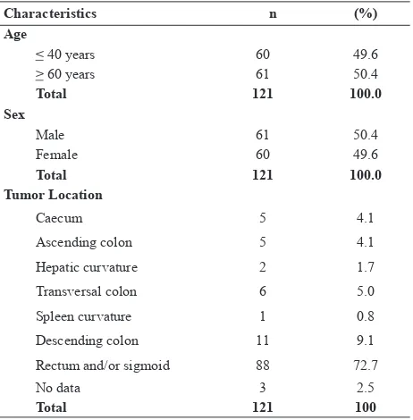
The present study began with perusal of 121 medical records in patients with colorectal cancer aged 40 years or less and aged 60 years or more who underwent surgical resection at the Department of Surgery, Cipto Mangunkusumo Hospital, Dharmais Cancer Hospital, Gatot Subroto Central Army Hospital, Hasan Sadikin District Hospital, and Dr. Wahidin Distric Hospital. Subsequently, we interviewed the patients or their relatives to trace ethnicity data and other data such as whether their disease may be categorized into hereditary colorectal cancer in keeping with the Amsterdam / Bethesda Criteria.6,19,21 Data of 118 colorectal cancer specimens was obtained from archives at Department of Pathology Anatomy, School of Medicine in three well-known Universities, i.e. University of Indonesia, University of Padjadjaran and University of Hasanuddin. All specimens underwent evaluation for histopathology, tumor grading as well as immunohistochemical analysis.
Immunohistochemical staining
to detect microsatellite instability mutation pathway using hMLH1 and hMSH2 antibody and SMAD4 protein expression to reconirm that the specimens were not microsatellite instability origin using SMAD4 monoclonal antibody.22,12
The immunohistochemical staining approach was selected based on previous studies.46,157 In brief, there were 3 sections for each cases in order to perform histopathology evaluation, MLH1 and MSH2 protein expression with 4 μm-thick sections were sliced from archival formalin-ixed parafin-embedded tissue blocks, kept overnight at 35-40oC temperature in the incubator, deparafinized, dehydrated using a graded series of ethanol solutions and treated by a 100 ml epitope retrieval system. Subsequently, 30% of hydrogen peroxide/methanol was administered for 5 min. The slides were rinsed with phosphate-buffered saline and then blocked with blocking solution for 10 min at room temperature. The were then incubated with hMLH1, hMSH2 antibody solution (1:100 dilutions; (Zymed Laboratories Inc., San Francisco, USA) or SMAD4 (B-8), sc-7966 (Santa Cruz Biotechnology Inc., CA, USA) for 45 min at room temperature. After the sections were incubated with biotinylated secondary antibody for 10 min, bound biotinylated antibody was then tested by HRP-streptovidin complex method (Histostain-S; Zymed Laboratories Inc., CA, USA). Diaminobenzidine was used as the chromogen.
Immunohistochemical analysis
Microscopic analysis was performed by two pathology anatomy experts of two different institutions. Stroma of tissues, normal epithelium and lymph follicles were important internal control marker for such analysis. The analysis was expressed as positive (+) result, negative with positive internal control (0/+) and suspect negative result for either tumor with negative result or negative result in internal control marker (0/0). Positive results included more than 20% expression of protein MSH2 or MLH1, which were then categorized as follows: 20-50%, 50-80% and more than 80%. Analysis of MSH2 or MLH1 protein expression was performed to demonstrate whether the colorectal cancer in young patients follows the microsatellite instability pathway
has the same extent as mentioned in the literatures. Evaluation of SMAD4 protein was performed to conirm that microsatellite instability pathway was not involved in carcinogenesis of those tumor tissues.12 Unexpressed SMAD4 protein, which was interpreted as mutation of DPC gene may also conirm the existence of CIN pathway. However, parts of them were included as non-CIN and non-MIN pathway.12
Statistical analysis
Comparison of the proportion of mismatch repair genes expressions between young (< 40 years) and old (> 60 years) patients with colorectal cancer was evaluated by using the X2-test with p < 0.05 considered as signiicant statistic value. A correlation test was performed to evaluate the relationship between mismatch repair genes expressions and clinical manifestation, i.e. the diagnosis, type of cancer and tumor location. Inter-observer validity was measured based on k (“kappa”) – statistics. We used kT = kS = 1 (goodness of it test) of 95%conidence interval [CI]) to evaluate the signiicance of agreement, and kT = kS (goodness of it test) with 95% CI to evaluate the signiicance of difference of two agreements. The following guidelines strength of agreement were applied: k value and strength of agreementof: 1) k < 0.2 = bad; 2) k value of 0.21 – 0.40 = intermediate; 3) k value of 0.41 – 0.60 = moderate; 4) k value of 0.61 – 0.80 = good; and 5) kvalue of 0.80 – 1.00 = excellent. SPSS ver. 10.05 for Windows was used for data analyses.
RESULTS
Histopathological result and grading
Of 118 histopathological specimens available, the most frequent histopathological type was adenocarcinoma and most of them were grade 1. Based on the age group, signet ring cell carcinoma and mucinous adenocarcinoma were signiicantly (p = 0.000) more frequent in young patients compared to old patients. There was statistically signiicant difference in grade (p = 0.001) between the young and old patients, in which grade 3 was more frequently found in young patients.
Immunohistochemical staining result for MSH2 and MLH1 protein
Of the 121 data collected, immunohistochemical staining for MSH2 protein and MLH1 were done for 92 and 97 specimens respectively. The other specimens were not stained because of limited or inadequate specimens. The results of immunohistochemical staining for MSH2 and MLH1 protein are shown in Figure 1 and Figure 2.
As shown in Table 2 and Table 3, more than 50% specimens showed positive staining for MSH2 but 83.5% showed negative staining for MLH1. The MSH2 and MLH1 staining were done simultaneously in 88 specimens, but the expression pattern of MLH1 was not parallel with MSH2. Fifty ive percent (41 of 74)
Characteristics n (%)
Age
< 40 years 60 49.6
> 60 years 61 50.4
Total 121 100.0
Sex
Male 61 50.4
Female 60 49.6
Total 121 100.0
Tumor Location
Caecum 5 4.1
Ascending colon 5 4.1
Hepatic curvature 2 1.7
Transversal colon 6 5.0
Spleen curvature 1 0.8
Descending colon 11 9.1
Rectum and/or sigmoid 88 72.7
No data 3 2.5
Total 121 100
Table 1. Characteristic of study subjects cases with negative MLH1 expression showed positive MSH2 staining.
Based on the age group, MSH 2 and MLH1 expressions demonstrated no signiicant difference. There was no statistically signiicant difference in MSH2 and MLH1 expressions between the right and left colon.
However, specimens with negative MSH2 expression were more likely to be located in the right colon and MLH1 was negative for all right-sided colorectal cancer cases. MLH1 and MSH2 protein expressions also showed no signiicant difference for tumor location when the specimens were compared based on patient age group. Nevertheless, the young patients were more likely to have negative expressions for both MLH1 and MSH2 protein expressions. There was no signiicant difference between the expressions of MLH1 (p = 0.674) and MSH2 (p = 0.412) on tumor grading, which may be interpreted as there was no correlation between microsatellite instability and tumor grading in this study. The k (Kappa) statistics for MSH2 was 0.67 (95% CI: 0.48-0.87) and the k- statistics for MLH1 was 0.67 (95% CI:0.64-0.90). It demonstrated reliable inter- and intra-observer validity.
DISCUSSION
Microsatellite instability is one of two main pathways which is important in the development of colorectal cancer. This pathway mainly consists of mutations in mismatch repair genes, represented by some genes such as MSH2 and MLH1.15,16 Previous studies demonstrate that it is frequently found in young colorectal cancer patients who usually have hereditary type (hereditary
Distribution n (%)
Negative staining (< 20%) 40 43.5
Positive staining 20 – 50% 12 13.0
Positive staining 50 – 80% 19 20.7
Positive staining > 80% 21 22.8
Total 92 100.0
Table 2. The distribution of MSH2 protein expression
Distribution n (%)
Negative staining (< 20%) 81 83.5
Positive staining 20 – 50% 10 10.3
Positive staining 50 – 80% 5 5.2
Positive staining > 80% 1 1.0
Total 97 100.0
non-polyposis colorectal cancer, HNPCC).4,5 In Indonesia, we found a different pattern, demonstrating a higher incidence of young colorectal cancer patients who have more progressive disease and do not show good-response in chemotherapy, which will greatly inluence productivity and family’s inancial problem. 19 However, little is known about the biologic characteristic of colorectal cancer. Therefore, we think that it is necessary to investigate further especially on the microsatellite instability in colorectal cancer.
Colorectal cancer is one of most cancer that closely associated with environment factor.25 A study in the United States demonstrated that there was a signiicant difference of disease-free survival associated with non-cancer related health conditions between the white and Afro-American colorectal cancer patients.26
In the present study, ethnicity data was easily-traced because almost all of the subjects were domiciled near to their place of birth. However, the present study was not aimed to make any correlation or comparison among those ethnic groups (Javanese, Sundanese, Macassarese and Minangkabau). Those ethnic groups were selected in order to obtain a subject population that represented the native Indonesian population. According to anthropologic and genetic review of the experts in some literatures,27-32 the selected ethnic groups are included in one “family”, which has similar genetic characteristics. Such similarities in geographic, culture and phenotype were assumed to be a valid representative of a population that will bring advantages for other studies in the future.
Tumor location, histopathology and grading
Overall data result of tumor location indicates similar data with the overseas data or data in the developed countries, i.e. rectum as the dominant area.2,33 Similar result also occur for histopathology data revealing adenocarcinoma, the main histopathology type, which is similar to the overseas data.27 Furthermore, signet ring cell carcinoma and mucinous adenocarcinoma were signiicantly dominant in the young patients compared to the old patients.
Tumor grading indicates that grade 3 was signiicantly dominant in the young patients with colorectal cancer compared to the old patients. Histopathological grading acts as a guideline marker of tumor aggressiveness and in turn, it can be associated with tumor prognosis and/ or therapeutic choice.34
The patterns of MSH2 and MLH1 protein expression of the young and old colorectal cancer patients The present study demonstrates that there is no signiicant difference in the pattern of MSH2 and MLH1 protein between the young and old patients of colorectal cancer. This may provide an impression that the carcinogenesis of colorectal cancer in the young patients is similar to the old patients, which is not in accordance with the pattern of hereditary type (sporadic cancer). In both groups of colorectal cancer patients, there were normal MSH2 protein expression and mutated MLH1 protein expression, which indicates microsatellite instability.
Microsatellite instability has been reported by Liu et al as 58% (of 31 cases) in colorectal cancer patients aged less than 35 years. However, among those cases, there were 5 cases which experienced germline mutation.35 In 2003, Kamory et al found that there was 54% (20 of 37 cases) colorectal cancer patients who had no expression of mismatch repair genes because of somatic inactivation. By conducting a promoter methylation assay, they noticed that such somatic inactivation was caused by methylation of promoter genes.36
Surprisingly, most of population of the young and old patients with colorectal cancer showed negative staining of MLH1, i.e. 83.5% (81 of 97 cases). Such negative staining suggests that there is no protein expression of mismatch repair gene MLH1 and that there is microsatellite instability caused by defect in the mismatch repair gene, which may be induced by reagent fault or technical error in staining procedures. Some studies indicate that there is a possibility of negative immunohistochemical staining caused by other mutations.37-39 Some studies have reported that, MLH1 may not be 100% match with the microsatellite instability and although the immunohistochemical staining and microsatellite instability are quite sensitive for screening evaluation, we may need additional PMS2 and MSH6 in immunohistochemical staining to encounter the microsatellite instability.39,40 Recently, it is noticed that in many cases of sporadic colorectal cancer, hMLH1 expression is frequently disappear or perish, which is not only thorough mutation process but also through epigenetic silencing process by promoter hypermethylation.28,42 Therefore, it may explain the discrepancy of MSH2 and MLH1 staining in the present study.
SMAD4 evaluation
expression and to add power of this study by recognizing any chromosomal instability (CIN). The SMAD4 was not expressed in specimens of the present study, either in the young or old patients.
It may be concluded that carcinogenesis of colorectal cancer in the young and old patients in such population does not occur through the microsatellite instability pathway. Nevertheless, the negative staining of MLH1 should be interpreted as the presence of microsatellite instability, which may be confusing if it is not associated with the process of DNA methylation, which is obviously mentioned in the literature.
REFERENCES
1. Evans WE, McLeod HL. ABC of colorectal cancer – Epidemiology. BMJ 2000; 321: 805-8.
2. McLaughlin JR, Boyd NF. Epidemiology of cancer. In: Tannock IF, Hill RP, editors. The Basic Science of Oncology. 3rd ed. New York: McGraw-Hill, 1998. 3. Potter JD. Colorectal cancer: of molecules and populations.
J Natl Cancer Inst. 1999; 91: 916-32.
4. Roh SA, Kim HC, Kim JS, Kim JC. Characterization of mutator pathway in younger-age-onset colorectal adenocarcinomas. J Korean Med Sci 2003;18:387-91. 5. Zorluoglu A, Yilmazlar T, Ozguc H, Bagcivan E, O G. Colorectal
cancers under 45 years of age. Hepatogastroenterology 2004;51:118-20.
6. Vasen H. Clinical diagnosis and management of hereditary colorectal cancer syndromes. J Clin Oncol 2000;18:81s-92s.
7. Squire JA, Whitmore GF, Phillips R. Genetic basis of
cancer. In: Tannock IF, Hill RP, editors. The basic science of oncology. 3rd ed. New York: McGraw-Hill; 1998. p.48-78. 8. Chung YFA, Weud M, H0 J, Nyam D, Leong A, Ho YH, et
al. Young age is not a poor prognostic marker in colorectal cancer. Br J Surg 1998;85:1255-9.
9. Ministry of Health of Indonesia.Annual Cancer Report. Directorate General of of Medical Services and the Indonesian Society of Pathologists. Jakarta; 1995.
10. Cassidy J, J T, Twelves C. Xelox (capecitabine plus
oxaliplatin): active irst line therapy for patients with
metastatic. J Clin Oncol 2004; 22:2084-91.
11. Cunningham D, Humblet Y, Siena S. Cetuximab (C225) alone or in combination with irinotecan (CPT-11) in patients with epidermal growth factor receptor (EGFR)-positive, irinotecan-refractory metastatic colorectal cancer (MCRC). Proceedings of The Annual Meeting American Society of Clinical Oncology; 2003; San Diego; USA:252.
12. Lenz H, Mayer R, Gold P. Activity of cetuximab in patients with colorectal cancer refractory to both irinotecan and oxaliplatin (abstract 3510). Proceedings of The Annual Meeting American Society of Clinical Oncology; 2004; New Orleans:248. 13. Hurwitz H, Fehrenbacher L, Novotny W, Cartwright T,
Hainsworth J, Heim W, et al. Bevacizumab plus irinotecan,
luorouracil, and leucovorin for metastatic colorectal cancer.
N Engl J Med 2004;350:2335-42.
14. Vogelstein B. Genetic alterations during colorectal tumor development. N Engl J Med 1988;319:525-32.
15. Fearon EC, Vogelstein B. A genetic model for colorectal carcinogenesis. Cell 1990; 61:759-67.
16. Grem J. From the laboratory to the clinic: The limitations of
rationally designed luoropyrimidine-based therapy and the
problem of apoptosis-impaired cancer cells. In: Bleiberg H, Kemeny N, Rougier P, Wilke H, editors. Colorectal cancer: a clinical guide to therapy. 2nd ed. London: Martin Dunitz, 2002. p.399-417.
17. Seitz J. 5-luorouracil/leucovorin versus capecitabine in patients
with stage III colon cancer. Semin Oncol. 2001;1 :41-4. 18. Vasen H, Wijnen JT, Morreau H. What does genetic testing
tell and not tell in familial (nonpolyposis) colorectal cancer. Am Soc Clin Oncol. 2000: 37-43.
19. Cravo ML. Validation and simpliication of Bethesda guidelines
for identifying apparently sporadic forms of colorectal carcinoma with microsatellite instability. Cancer 1999;85:779-85. 20. Hamilton SR, Vogelstein B, Kudo S, Riboli E, Nakamura
S, Hainaut P, et al. Carcinoma of the colon and rectum. In: Stanley HR, Aaltonen LA, editors. Pathology and genetics: Tumours of the digestive system. New York: World Health Organization, 2001:105-119.
21. Bisset D, Ahmed F, McLeod HL, Cassidy J. Optimal
strategies for the use of the oral luoropyrimidines. In:
Cunningham D, Haller DG, Miles A, editors. The effective management of colorectal cancer. London: Aesculapius Medical Press; 2000. p.51-62.
22. Halvarsson B, Lindblom A, Rambech E, Lagerstedt K, Nilbert M. Microsatellite instability analysis and/or immunostaining for the diagnosis of hereditary nonpolyposis colorectal cancer? Virchows Arch 2004;444:135-41. 23. Gertig DM, Hunter DJ. Genes and environment in the
etiology of colorectal cancer. Cancer Biol. 1998;8:285-98. 24. Wudel LJ, Jr, Chapman WC, Yu S, Stain SC. Disparate
outcomes in patients with colorectal cancer. Arch Surg. 2002;137:550-6.
25. Koentjaraningrat. Pendahuluan. In: Manusia dan kebudayaan di Indonesia. Jakarta: Penerbit Djambatan, 1999: p.1-36. 26. Setianingsih I, Williamson R, Marzuki S, Harahap A, Tamam
M, Forrest S. Molecular basis of B thalassemia in Indonesia: application to prenatal diagnosis. Mol Diagn. 1998;3:11-20. 27. Yusuf I, Djojosubroto MW, Ikawati R, Lum K, Kaneko A,
Marzuki S. Ethnic and geographical distributions of CYP2C19 alleles in the populations of southeast asia. In: Marzuki S, Verhoef J, Snippe H, editors. Tropical Diseases: From Molecule to Bedside. New York: Kluwer Academic; 2004: p.37-46. 28. Setianingsih I, Williamson R, Daud D, Harahap A,
Marzuki S, Forrest S. Phenotypic variability of Filipino B-Tahalssemia/ HbE patients in Indonesia. Am J Hematol. 1999;62:7-12.
29. Cavalli-Sforza LL, Cavalli-Sforza F. The great human diasporas: the history of diversity and evolution. 2nd ed. Reading, Massachussetts: Perseus Books, 1995:
31. Slingerland JM, Tannock IF. Cell proliferation and cell death. In: Tannock IF, Hill RP. The Basic Science of Oncology. New York: McGraw-Hill;1998. p.134-65.
32. Gibbons A. The peopling of the Paciic. Science
2001;291:1735-42.
33. Ding Y-C, Wooding S, Harpending HC, Chi H-C, Li H-P, Fu Y-X, et al. Population structure and history in East Asia. Proc Natl Acad Sci 2000;97:14003-6.
34. Cavalli-Sforza LL, Piazza A, Menozzi P, Mountain J. Reconstruction of human evolution: bringing together genetic, archaeological, and linguistic data. Proc Natl Acad Sci. 1988;85:6002-6.
35. Bellwood P. Prehistory of the Indo-Malaysian Archipelago. Revised ed. Sydney: Academic Press, 1997:
36. Cloutier B. People of South East Asia. cited 2 Agustus 2010: screens. Available from: URL: http://berclo.net/ page00/00en-sea-people.html.
37. Sugimoto C, Kitamura T, Guo J, Al-Ahdal MN, Shchelkunov SN, Otova B, et al. Typing of urinary JC virus DNA offers a
novel means of tracing human migrations. Proc Natl Acad Sci. 1997;94:9191-6.
38. Setianingsih I. Alpha Thalassemia in Indonesia: Phenotypes and Molecular Defects. In: Marzuki S, Verhoef J, Snippe H, editors. Tropical Diseases: From Molecules to Bedside. New York: Kluwer Academic, 2003. p. 47-56.
39. Burchard EG, Ziv E, Coyle N, Gomez SL, Tang H, Karter AJ, et al. The Importance of Race and Ethnic Background in Biomedical Research and Clinical Practice. N Engl J Med. 2004;348:1170-5.
40. Schwartz RE. Racial proiling in medical research. N Engl
J Med 2001;344:1392-3.
41. Pääbo S. Genomics and society: The human genome and our view of ourselves. Science 2001;291:1219-20. 42. Wijnen JT, Khan PM, Vasen H, van der Klift H, Mulder A,
van Leeuwen-Cornelisse I, et al. Hereditary nonpolyposis colorectal cancer families not complying with the Amsterdam
Criteria show extremely low frequency of