52
V. SIMPULAN DAN SARAN
A. Simpulan
Berdasarkan hasil penelitian maka dapat disimpulkan sebagai berikut :
1. Sabun A dan sabun B tidak menunjukkan penghambatan pertumbuhan
terhadap keseluruhan sampel isolat Staphylococcus aureus yang diisolasi dari daerah Babarsari, Sleman, DIY dibandingkan dengan kontrol S. aureusATCC 6538.
2. Faktor etnis berpengaruh terhadap daya penghambatan Sabun A dan B
terhadap Staphylococcus aureus sampel Babarsari, Sleman, Yogyakarta. 3. Jangka waktu penggunaan sabun dan aktifitas hobi seperti berenang dan olah
raga tidak memperlihatkan pengaruh terhadap penghambatan Staphylococcus aureusdi Babarsari.
B. Saran
1. Perlu dilakukan penentuan standar kadar triclosan murni dan pengukuran
kadar triclosan yang terkandung dalam sabun A dan sabun B terhadap
2. Perlu adanya penelitian lanjutan tentang pengukuran Konsentrasi Hambat
Minimum untuk mengetahui penghambatan sabun mandi cair berbahan aktif
triclosan terhadapStaphylococcus aureusdari daerah Babarsari, Yogyakarta. 3. Perlu dilakukan penelitian yang berkesinambungan tentang penetapan batas
53
penghambatan triclosan terhadap pertumbuhan S. aureus untuk melihat tingkat resistensi S. aureus dari daerah Babarsari, Yogyakarta.
4. Perlu dilakukan penggolongan isolat berdasarkan morfologi dan pengujian
sifat biokimia.
5. Perlu penentuan jumlah probandus untuk untuk menentukan jumlah kelompok
54
DAFTAR PUSTAKA
Adolfson-Erici, M., Peterson, M., Parkkonen, dan Sturve, J., 2002. Triclosan, a commonly used bactericide found in human milk and in the aquatic environment in Sweden. Chemosphere 46 (10): 1485–1489.
Akpomie, O. O., Akponah, E., dan Okorawhe, P., 2012. Amylase production potentials of bacterial isolates obtained from cassava root peels. Agricultural Science Research Journal 2: 95-99.
American Public Health Association, 1996. Recommended Methods for the Microbiological Examination of Foods. 2nd Ed., APHA Inc., New York.
Anjarwati, D. I.,danDharmawan, A. B., 2010. Identifikasi Vancomisin Resistant Staphylococcus aureus (VRSA) Pada Membran Stetoskop Di Rumah Sakit Margono Soekarjo Purwokerto. Mandala of Health Vol. 4, no. 2: 57-69
Anonim, 2003. Dilema Triclosan dan Produk Antibakteri.
http://www.kalbe.co.id/index.php?mn=news&tipe=detail&detail=16444 , 17 Oktober 2010.
Anonim, 2005. Soap and Detergent Association. Chemistry : Soap and Detergent.
http://www.sdahq.org/cleaning/chemistry, 17 Oktober 2010.
Anonim, 2006. Antimicrobial products in the home: The evolving problem of antibiotic resistance. Upton Allen Paediatric, Child Health. 11 Vol. 3 : 169 -173.
Anonim, 2008. Preventing Colds: Washing Your Hands Is More Effective Than Taking Vitamin,
www.sciencedaily.com, 17 Oktober 2010.
Anonim 2010 c, WHO global strategy for containment of antimicrobial resistance. http://www.who.int/csr/resources/publications/drugresist/en/EGlobal_Strat.pdf. 11 September 2012
Anonim, 2010d. Selective and Differential Media. kcfac.kilgore.edu/micro/Selective% 20&%20Differential%20Media.doc, 18 Oktober 2010
Anonim, 2010d. Blood Agar, http://www.austincc.edu/microbugz/blood_agar_test.php, 20 Oktober 2010
Anonim, 2011. Use Antibiotics Rationaly. http://ino.searo.who.int/LinkFiles/Home_WHD11-Messages-11_03_31-FAQs.pdf. 9 September 2012
Bamber, A. I. dan Neal, T. J. (1999). An assessment of triclosan susceptibility in methicillin-resistant and methicillin-sensitiveStaphylococcus aureus. Hospital Infection Journal. 41: 107–9.
Bannerman, T. L., dan Peacock, S. J., 2007. Manual of Clinical Microbiology 9th Edition. Washington D. C.
Barel, A. O., Paye, M., dan Maibach, H. I., 2001. Handbook of Cosmetic Science and Technology. Marcell Dekker Inc., New York.
Berger-Bächi, B., dan Rohrer, S., 2002. Factors Influencing Methicillin Resistance in
Staphylococci. Archives of Microbiology 178 :165–171.
Bowersox, J., 1999. Experimental Staph Vaccine Broadly Protective in Animal Studies. NIH. Bowersox, J., 2007. Experimental Staph Vaccine Broadly Protective in Animal Studies. NIH.
Breed, R. S., Murray, E.G.D., dan Nathan, R. S., 2001. Bergey’s Manual of Determinative of Bacteriology. Seventh Edition. The Williams and Wilkins Company. Baltimore.
Breakwell, D., Smith, K., Robinson, R., Woolverton, C., dan MacDonald, B., 2012. Colony Morphology Protocol. http://www.microbelibrary.org/component/resource/laboratory-test/3136-colony-morphology-protocol, 11 Januari 2012.
Bridson, E.Y., 1998. The Oxoid Manual Eighth Edition, Oxoid Ltd., Basingstoke
Carter, A. P., Clemons, W. M., Brodersen, D. E., Morgan-Warren, R. J., Wimberly, B. T., dan Ramakrishnan, V., 2000. Functional insights from the structure of the 30S ribosomal subunit and its interactions with antibiotics.Nature 407: 340–8
Cappucino, J. G. danSherman, N., 2005. Microbiology: A Laboratory Manual. 7th ed. Pearson Education Inc.USA.
Cavvalo, J. D., Fabre, R., Leblanc, F., Nicholas-Chanoine, M. H., Thabaut, A., 2000. Antibiotic Susceptibility and Mechanism of Beta-Lactamase Resistance in 1310 Strains of
Pseudomonas aeruginosa: a French Multicentre Study. JournalofAntimicrobial and Chemotherapy46: 133-136
Chambers, H.F., 2001. The changing epidemiology of Staphylococcus aureus.Emerging Infectious DiseasesJournal7 (2): 178–82.
Chen, Y., Pi, B., Zhou, H., Yu, Y., dan Li, L. M., 2009. Triclosan Resistance in Clinical Isolates ofAcinetobacter baumannii. Medicinal Microbiology Journal 58: 1086-1091.
Chang, S., Sievert, D. M., Hageman, J. C., Boulton, M. L., Tenover. F. C., Downes, F. P., Shah, S., Rudrik, J. T., Pupp. G. R., Brown. W. J., Cardo, D., dan Fridkin, S. K., 2003. Infection with Vancomycin-ResistantStaphylococcus aureus Containing thevanAResistance Gene.New England Journal of Medicine 348:1342-1347
Chuanchen, R., Beinlich K., Hoang T.T., Becher A., Karkhoff-Schweizer, R.R., dan Schweizer, H.P. 2001. Cross-Resistance Between Triclosan and Antibiotic in Pseudomonas aeruginosais Mediated by Multidrug Eflux Pump : Exposure of a Susceptible Strain to Triclosan Select nfxB Mutants Overexpressing MexCD-Opr. American Journal of Infection Control 31: 124–7.
Cheung, A. L., Koomey, J. M., Butle,r C. A., Projan, S. J., dan Fischetti, V. A. 1992. Regulation of Exoprotein Expression in Staphylococcus aureus, by a Locus (sar) Distinct from agr.
Proceedings of the National Academy of SciencesUSA. Vol 89: 6462-6466.
Cheung, A. L., Eberhardt, K., Chung, E., Yeaman, M. R., Sullam, P. M., Ramos, M., danBayer, A. S. 1994. Diminished virulence of a sar-/agr- mutant of Staphylococcus aureus in the rabbit model of endocarditis. Journal of Clinical Investigation 94: 1815-1822.
Cheung, A. L., Schmidt, K., Bateman, B., dan Manna, A. C. 2001. SarT, a repressor of alpha-hemolysin in Staphylococcus aureus. Journal of Infection and Immunity 69: 4749-4758. Cole, E. C., Addison, R., Rubino, J., Leese, K., Dulaney, P., Newell, M., Wilkins, J., Gaber, D.,
Wineinger, T., dan Criger, D., 2003. Investigation of Antibiotic and Antibacterial Agent Cross-Resistance in Target Bacteria from Homes of Antibacterial Product Users and Non-Users, Journal of Applied Microbiology 95:664-676.
Cookson, B. D., Farrelly, H., Stapleton, P., Garvey, R. P. J. danPrice, M. R. (1991). Transferable resistance to triclosan in MRSA. Lancet 337, 1548–9
Davin-Regli, A., Bolla, J. M., James, C., Lavigne, J. P., Chevalier, J., Garnotel, E., Molitor, A., dan Pagès, J. M., 2008. Membrane Permeability and Regulation of Drug "Influx and Efflux" in Enterobacterial Pathogens, Current Medical Chemistry Targ9: 750-759. Deora, R., Tseng, T., dan Misra, T. K., 1997. Alternative transcription factor sigmaSB of
Staphylococcus aureus: characterization and role in transcription of the global regulatory locus sar. Journal of Bacteriology. 179: 6355-6359.
Forbes, B.A., Sahm , D.F., dan Weissfeld, A.S., 1998. Diagnostic Bacteriology.Tenth Edition.
Mosby Inc, Missouri.
Foster, T. J., O’Reilly, M., Phonimdaeng, P., Cooney, J., Patel, A. H.,dan Bramley, A. J., 1990. Genetic Studies of Virulence Factors of Staphylococcus aureus-properties of Coagulase and Gamma-toxin, Alpha-toxin, Beta-toxin and Protein A in the Pathogenesis of S. aureusInfections. Molecular Biology of Staphylococci VCH Publishing, NewYork Fraise, A.P. 1998. Tropical Treatment for Eradiction of MRSA. CME Bulletin of Medical
Microbiology 2: 10-1
Futagawa-Saito, K., M. Suzuki, M., Ohsawa, S., Ohshima, N., Sakurai, W., Ba-Thein, T., dan Fukuyasu. 2004. Identification and prevalence of an enterotoxin-related gene, se-int, in Staphylococcus intermedius isolates from dogs and pigeons. Journal of Applied Microbiology, 96: 1361–1366.
Gabbar, K. M. A., 1992. Procedures in Veterinary Microbiology, Field Document No.6, 2ndEdition. Central Veterinary Diagnosis Laboratory, Tando Jam.
Ganiswara, S.G., 1995. Farmakologi dan Terapi. Bagian Farmakologi Fakultas Kedokteran Universitas Indonesia, Jakarta.
Gaspersz, V., 1994, Metode Perancangan Percobaan, Penerbit CV Armico, Bandung.
Giraudo, A. T., Raspanti, C. G., Calzolari, A., danNagel, R. 1994. Characterization of a Tn551-mutant of Staphylococcus aureus defective in the production of several exoproteins. Canada Journal of Microbiology 40: 677-681.
Grahams, P. L., Lin, S. X., dan Larson, L. E., 2006. A US Population-Based Survey of
Staphylococcus aureus Colonization. Annals of Interntional Medicine. 144(5): 318-25. Griffin, D.H., 1981, Fungal Physiology, John Wiley and Sons, Inc., New York.
Grubbs, J.,R. 2002. The Effects of Triclosan Derivatives against the Growth of Staphylococcus Aureus. Liberty University. Amerika Serikat
Gunn, B. A., Dunkelberg, W. E., dan Creitz J. R., 1972. AmericanJournal of Clinical Pathology. 57: 236-238.
Hanssen, A. M., Kjeldsen, G., dan Sollid, J. U. E., 2004. Local Variants of Staphylococcal
Harris, L. G., Foster S. J., dan Richards R. G., 2002. An Introduction to Stsphylococcus aureus for Identifying and Quantifying Staphylococcus aureus Adhesins in Relation to Adhesion to Biomaterial: Review. European Cell and Material Vol. 4 2002 p: 39-60
Heath, R. J., Yu, Y. T., Shapiro, M. A., Olson, E., dan Rock, C. O., 1998. Broad Spectrum Antimicrobial Biocides Target the FabI Component of Fatty Acid Synthesis. Journal of Biological Chemistry273:30316-30320.
Heath, R. J., Li, J., Roland, G. E.,danRock, C. O. 2000. Inhibition of the Staphylococcus aureus
NADPH-dependent enoyl-acyl carrier protein reductase by triclosan and hexachlorophene. Journal of Biological Chemistry. 275, 4654–4659
Hedge, P.P., Andrade, A.T. , dan Bhat, K., 2006. Microbial Contamination of "In Use" Bar Soap in Dental Clinics. Indian Journal of Dental Research, India. 17 ( 2 ) : 70-3
Hermawan, A., Hana, W., dan Wiwiek, T. 2007. Pengaruh Ekstrak Daun Sirih (Piper betle L.) Terhadap Pertumbuhan Staphylococcus aureus dan Escherichia coli dengan Metode Difusi Disk. Universitas Erlangga.
Honeyman A.L., Friedman H., dan Bendinelli M. 2001. Staphylococcus aureus Infection and Disease. Plenum Publishers. New York.
Isenberg, 1992. Clinical microbiology procedures handbook, vol. 1. American Society for Microbiology,Washington, D.C.
Jawetz, M. 2001. Melnick, & Adelberg’s Medical Microbiology. McGraw-Hill Companies Inc.,
San Fransisco.
Jay, J. M. 1992. Modern Food Microbiology. 4 Ed. Champman and Hall. London. Jevons M. P., 1961. Celbenin-resistant staphylococci.British Medical Journal;1:124-5.
Jones, R. N., 2000. Detection of Emerging Resistance Pattern within Longitudinal Surveillance System: Data Sensitivity and Microbial Susceptibility. MYSTIC Advisory Board. Meropenem Yearly Susceptibility Test Information Collection. Journal of Antimicrobial and Chemotheraphy. 46: 1-8
Jutono, J.S., Hartadi, S., Kabirun, S., Darmosuwito, S., dan Soesanto, 1980, Pedoman Praktikum Mikrobiologi Umum Untuk Perguruan Tinggi, Departemen Mikrobiologi Fakultas Pertanian Universitas Gadjah Mada,Yogyakarta.
Khan, T.S. dan R. Rind, 2001. Isolation and Characterization of Bacterial Species from Surgical and Non-Surgical Wounds Located on Body Surface of Buffaloes, Cattles, Sheep and Goats. Pakistan Journal. of Biological Science., 4 (6): 696-702.
Kim, T.J., Na, Y.R. dan Lee, J.I. 2005., Investigations Into the Basis of Chloramphenicol and Tetracycline Resistancein Staphylococcus intermediusIsolates from Cases of Pyoderma in Dogs. Journal of Veterinary Medicine B 52, 119–124.
Kumar, S. S., dan KAmaraj, M., 2011. Antimicrobial Activity of Cucumis anguria L. by Agar Diffusion Method. Botany Research International 4 (2): 41-42
Kusmayati dan Agustini, N. W. R. 2007. Uji Aktivitas Senyawa Antibakteri dari Mikroalga
(Porphyridium cruentum). Biodiversity. 8, 1 : 48-53.
Lear, C. J., Maillard, J. Y., Dettmar, P. W., Goddard, P. A., dan Russell, A. D., 2002. Chloroxylenol- and Triclosan- Tolerant Bacteria from Industrial Sources. Journal of Industrial Microbiology and Biotechnology29:238-242.
Lestari, E. S., Severin, J. A., dan Verbrugh, H. A., 2009. Antimicrobial resistance among pathogenic bacteria in Southeast Asia: a review. Department of Medical Microbiology and Infectious Diseases. Rotterdam.
Lindberg, M., Jönsson, K., Müller, H., Jonsson, H., Signas, C., Höök, M., Raja, R., Raucc ,I. G., dan Anantharamaiah, G. M., 1990.. Molecular Biology of Staphylococci. VCH Publishing, New York.
Lindsay, J. A., Ruzin, A., Ross, H. F., Kurepina, N., dan Novick, R. P., 1998. The Gene for Toxic Shock Toxin is Carried by a Family of Mobile Pathogenicity Islands in
Staphylococcus aureus.Molecular Microbiology 29, 2: 527–43
Liu, G. Y., Essex, A., Buchanan, J.T., Datta, V., Hoffman, H. M., Bastian, J. F., dan Fierer, J.,2005. Staphyloxanthin Plays a Role in the Fitness of Staphylococcus aureus and its Ability to Cope with Oxidative Stress. Journal of Infection and and Immunity 74, 8: 4950–3
Madigan, M.T., Martinko, J.M., dan Parker, J., 2000, Brock Biology of Microorganisms, 9th Edition, Prentice-Hall Inc., New Jersey.
Madigan M.T. dan Martinko J.M., 2005. Brock Biology of Microorganisms 11th ed., Prentice Hall, New Jersey.
Manna, A. C., Bayer, M. G., dan Cheung, A. L., 1998. Transcriptional analysis of different promoters in the sar locus in Staphylococcus aureus. Journal of Bacteriology 180: 3828-3386.
Manious, A. G., Hueston, W. J., Everett, C. J., Diaz, V. A., 2006. Nasal carriage of
Staphylococcus aureus and Methicillin-resistant S. aureus in Unted States, 2001-2002.
Annals of Family Medicine.4(2): 132-7.
Maibach, H., dan Aly, R:, 1981. Skin Microbiology: Relevance to Clinical Infection. Springer-Verlag, New York,
McBride, M. E., 1984. Microbial Flora of In-Use Soap Product. Journal of Applied and Environmental Microbiology 8: 338-341
McMurry, L.M., Oethinger , M., dan Levy, S.B., 1998. Triclosan Targets Lipid Synthesis. Nature 394, 6693: 531–532.
McMurry, L. M., McDermott, P. F., dan Levy, S. B., 1999. Genetic Evidence that InhA of
Mycobacterium smegmatis is a Target for Triclosan. Journal of Antimicrobial Agent and Chemotheraphy 43:711-713.
Morin, R. B. dan Gorman, M. 1995. Kimia dan Biologi Antibiotik β-lactam (Chemistry and Biology β-lactam Antibiotics. Edisi III. Diterjemahkan oleh Mulyani S. IKIP Semarang Press. Semarang.
Morfeldt, E., Janzon, L., Arvidson, S., dan Löfdahl, S., 1988. Cloning of a chromosomal locus (exp) which regulates the expression of several exoprotein genes in Staphylococcus aureus. Journal of Molecular Genetics and Genomics 211: 435-440.
Murti, A.P. 2009. Sekitaran Kota Babarsari, http://pumpkinsquad.blogspot.com/ 2009/02/yogyakarta-keberadaan-beberapa.html, 20 Oktober 2010
Navarro, L., 2005. Time and Extent Application Ammandement of Triclosan. Ciba Specialty Chemical Corporation, United State of America.
Neal, J. M., 2006., At a Glance Farmakologis Medis Edisi 5. Penerbit:Erlangga
Ngo, K. 2005. Antibacterial Soap: Unnecessary and Harmful, San Diego Oceans Foundation, San Diego.
Noor, N. N,. 2008.Epidemiologi. Rineka Cipta. Jakarta
Notoatmodjo, S., 2007. Kesehatan Masyarakat Ilmu & Seni. Rineka Cipta, Jakarta.
Manyaran 01 di Kota Semarang. Program Pendidikan Sarjana Kedokteran Universitas Diponegoro, Semarang.
Obied, A. l., 1983.Field investigation, clinical and laboratory findings of camel mastitis. M.Sc. thesis, University of Khartoum.
Okeke, I. N., Laxminarayan, R., Bhutta, Z. A., Duse, A. G., Jenkins, P., O’Brien, T. F., dan Pablas-Mendez, A., 2005. Antimicrobial resistance in developing countries. Part I: recent trends and current status. Lancet 5:481-493
Otto, M., 2009. Staphylococcus epidermidis, the ‘Accidental’ Pathogen. Macmillan Publishers, Amerika Serikat.
Parikh, S. L., Xiao, G., dan Tonge, P. J., 2000. Inhibition of InhA, Enoyl Reductase from
Mycobacterium tuberculosis by Triclosan and Isoniazid. Journal of Biochemistry
39:7645-7650.
Pelczar, M.J. dan Chan, E.C.S. 1988. Dasar-Dasar Mikrobiologi.Penerbit Universitas Indonesia Press. Jakarta.
Poole, K. (2007) Efflux Pumps as Antimicrobial Resistance Mechanisms. Annals of Family Medicine39:162- 176.
Prescott , L.M., Harley, J.P., dan Klein, D.A. 2002. Microbiology. 5th Ed. McGraw-Hill, Boston. Rachal, T., K. Leonard, L., Martinez, J. G., Breaux, A., Corbin R., dan Nathaniel. 2009. Prevalence of SCCmec types in methicillin resistant Staphylococcus intermedius in healthy pets from Southeastern United States. Journal of Infection Disease and Immunity 1 : 006-010.
Recsei, P., Kreiswirth, B., O’Reilly, M., Schlievert, P., Gruss, A., dan Novick, R. P.,1986. Regulation of exoprotein gene expression in Staphylococcus aureus by agr. Journal of Molecular Genetics and Genomics 202: 58-61.
Ruiz, M.L.V., Silva, P.G., dan Laciar, A.L. 2009. Comparison of Microplate, Agar Drop and Well Diffusion Plate Methods for Evaluating Hemolytic Activity of Listeria monocytogenes.African Journal of Microbiology Resistance. 3 (6): 319-324.
Russell, A.D. 2004. Whither Triclosan. Journal of Antimicrobial and Chemotherapy. 53 (5): 693–695.
Sahm, D. F., Karlowsky, J. A., Kelly, L. J., Critchley, I. A., Jones, M. E., Thornsberry, C., Mauriz, Y., Kahn, J., 2001. Need for Annual Surveilance of Antimicrobial Resistance in
Streptococcus pneumoniaein the United States: 2-year longitudinal Analysis. Journal of Antimicrobial and Chemotherapy. 45: 1037-1042
Schmidt, K. A., Manna, A. C., Gill, S., dan Cheung, A. L., 2001. SarT, a repressor of alpha-hemolysin inStaphylococcus aureus. Journal of Infection and Immunity 69: 4749-4758.
Schwarz, S., Lange, C. dan Werckenthin, C. 1998., Molecular Analysis of the Macrolide-lincosamide Resistance Gene Region of a Novel Plasmid from Staphylococcus hyicus. Journal of Medicinal Microbiology 47: 63–70.
Schwarz, S., 2000. Molecular Analysis of the Translational Attenuator of a Constitutively Expressed erm(A) Gene from Staphylococcus intermedius. Journal of Antimicrobial and Chemotherapy 46: 785–788.
Setiabudy dan Gan. 1995. Pengantar Antimikroba dalam buku Farmakologi dan Terapi. Edisi keempat Jakarta: Fakultas Kedokteran Universitas Indonesia, Jakarta.
Sjahrurachman, Agus, 1996., Resistensi Bakteri terhadap Aminoglikosida. Cermin Dunia Kedokteran No. 108. Pusat Penelitian dan Pengembangan P.T. Kalbe Farma, Jakarta. Slater-Radosti, C., Van Aller G., dan Greenwood, R., 2001. Biochemical and genetic
characterization of the action of triclosan on Staphylococcus aureus. Journal of Antimicrobial and Chemotherapy 48 (1): 1-6.
Shaw, K.J., Rather, P.N., Hare, R.S. dan Miller, G.H. 1993., Molecular Genetics of Aminoglycosides Resistance Genes and Familial Relationships of Aminoglycosides-modifying Enzymes. Microbiology Review 57: 138–163.
Shaikh, S. N., 1999. Bacteriological studies on the uteri of the slaughtered goats. M.Sc (Hons) Thesis, Department of Microbiology, Sindh Agriculture University Tando Jam.
Sharp, S. E., danSearcy, C. 2006. Comparison of Mannitol Salt Agar and Blood Agar Plates for Identification and Susceptibility Testing of Staphylococcus aureus in Specimens from Cystic Fibrosis Patients.Journal of Clinical Microbiology 44 (12): 4545–4546
Shield, P., dan Cathcart, L., 2011. Motility Test Medium Protocol. www.onlinemicrobiologyprotocol.com/motility_test. 11September 2012.
Smith, A. C., dan Hussey, M. A., 2005. Gram Stain Protocol
www.onlinemicrobiologyprotocol.com/gram_staining.11 September 2012
Stapleton, P.D. dan P.W. Taylor. 2002. Methicillin resistance in Staphylococcus aureus:
Stone R. V., 1935. Distribution of Elastin in Hamsters and the Turnover Rates of Different Elastin Pools. Proceedings of TheSocietyfor Experimental Biology and Medicine 33:185-187.
Sulistiyo, 1971. Farmakologi dan Terapi, EKG, Yogyakarta.
Suller, M. T. E., danRussell, A. D. 2000, Triclosan and antibiotic resistance in Staphylococcus aureus, Journal of Antimicrobial and Chemotherapy.46 (1): 11-18.
Tabak, M., Scher, K., Hartog, E., Romling, U., Matthews, K. R., Chikindas, M. L., dan Yaron, S., 2007. Effect of Triclosan on Salmonella typhimurium at Different Growth Stages and in Biofilms. FEMS Microbiology Letter267:200-206.
Tegmark, K., Karlsson, A., danArvidson, S., 2000. Identification and characterization of SarH1, a new global regulator of virulence gene expression in Staphylococcus aureus. Molecular Microbiology 37: 398-409.
Todar, K. 2012. Regulation and Control of Metabolism in Bacteria.
http://textbookofbacteriology.net/regulation_5.html.11 September 2012.
Ward, W. H. J., Holdgate, G. A., Rowsell, S., McLean, E. G. ,. Paupit, R. A., Clayton, E., Nichols, W. W.,Colls, J. G., Minshull, C. A., Jude, D. A., danMistry, A., 1999. Kinetic and Structural Characteristics of the Inhibition of enoyl(acyl carrier protein) reductase by Triclosan.Biochemistry 38:12514–12525.
Waters, A. E., Contente-Cuomo,T., Buchhagen, J., Liu, C. M., Watson, L., Pearce, K., Foster, J., Bowers, J., Driebe, E. M., Engelthaler, D. M., Keim, S. P., dan Price, L. B., 2011. Multidrug-Resistant Staphylococcus aureus in US Meat and Poultry. Clinical Infectious Diseases.Arizona
Wilson, B. A., 2009. The Fate and Effect of Triclosan in The Lower Hudson River Estuary. UMI Microform, United State of America
Winn, Jr., W., 2006. Konemann’s Color Atlas and Diagnostic Text of Microbiology, 6th ed., p. 945–1021. Lippencott Williams & Wilkins Publishers, Philadelphia, PA.
Whitt, D. D., danSalyers, A. A., 2002. Bacterial Pathogenesis: A Molecular Approach(2nd ed.). ASM Press, United State of America
Webber, M. A., Coldham, N. G., Woodward, M. J. dan Piddock, L. J. V., 2008. Proteomic Analysis of Triclosan Resistance in Salmonella enterica serovar Typhimurium. Journal of Antimicrobial and Chemotherapy62:92-97.
Woeste, S., dan Demchick, P., 1991. New Version of the Negative Stain. Journal of Applied Environtment Microbiology. 57(6): 1858-1859
World Health Organization, 2004. Guidelines for Drinking-water Quality 3rd Edition., Geneva. Yazdankhah, S. P., Scheie, A. A., Hoiby, E. A., Lunestad, B. T., Heir, E., Fotland, T. O.,
Nartestad, K., dan Kruse, H., 2006. Triclosan and Antimicrobial Resistance in Bacteria: an Overview. Journal of Microbial Drug Resistance12:83-90
65 Lampiran 1. Kuisioner
A. Form Kuisioner
LEMBAR KUISIONER
POLA PENGHAMBATAN SABUN MANDI CAIR BERBAHAN AKTIF TRICLOSAN TERHADAP PERTUMBUHAN Staphylococcus aureus DI
DAERAH BABARSARI, SLEMAN, YOGYAKARTA
( Kenny Simon / Fak. Teknobiologi UAJY / 060801005 / HP : 081904240690)
A. Data Pribadi
1. Umur : ……….
2. Jenis Kelamin : ……….
3. Pekerjaan : ……….
4. Asal Daerah : ……….
5. No. HP : ……….
6. Daerah Aktivitas : ……….
B. Informasi Penggunaan Sabun
1. Sabun mandi yang digunakan : ………..
2. Jenis sabun (*) : ( Cair / Batang )
3. Alasan pemilihan sabun : ……….
4. Lama pemakaian sabun : ………..
5. Sabun mandi lain yang digunakan : ………...
6. Intensitas mandi (*) : - 2 kali sehari
- 3 kali sehari
- Lainnya (…… kali sehari)
7. Konsentrasi sabun : air (*) : - 75% : 25% - 50% : 50% - 25% : 75%
- Lainnya ( ……. : …….. )
C. Keterangan Pelengkap
1. Waktu penggunaan antibiotika terakhir : ………
2. Sedang menggunakan produk antibakteri lain : ( ya / tidak ) (*) 3. Sedang dalam terapi pengobatan kulit : ( ya / tidak ) (*) 4. Sedang menggunakan produk perawatan kulit : ( ya / tidak ) (*)
5. Intensitas melakukan renang : ………
6. Intensitas mandi dengan air panas : ………
66 Lampiran 2. Tabel Analisis Statistik
Tabel 8. Hasil ANAVA Sabun A Berbahan Aktif Triclosan Terhadap Pembentukan Zona Hambat Isolat Staphylococcus aureusDaerah Babarsari.
Tabel 9. Hasil Analisis DMRT Sabun A Berbahan Aktif Triclosan Terhadap Pembentukan Zona Hambat Isolat Staphylococcus aureus Daerah Babarsari.
Perlakuan N
Subset for alpha = 0.05
1 2 3 4 5 6
Sampel 3 3 3.00000
Sampel 9 3 3.00000
Sampel 18 3 3.00000
Sampel 8 3 3.25000 3.25000
Sampel 1 3 3.66667 3.66667
Sampel 2 3 3.75000 3.75000
Sampel 4 3 5.00000 5.00000 5.00000
Sampel 14 3 5.33333 5.33333 5.33333 5.33333
Sampel 10 3 5.75000 5.75000 5.75000 5.75000
Sampel 11 3 6.25000 6.25000 6.25000
Sampel 12 3 6.33333 6.33333 6.33333
Sampel 5 3 6.58333 6.58333 6.58333
Sampel 6 3 6.58333 6.58333 6.58333
Sampel 7 3 6.83333 6.83333 6.83333
Sampel 15 3 6.91667 6.91667 6.91667
Sampel 16 3 7.25000 7.25000 7.25000
Sampel 17 3 7.75000 7.75000
Sampel 13 3 8.08333
Kontrol 3 13.25000
Sig. .078 .053 .094 .072 .083 1.000
Keterangan :
a. Rata-rata kelompok pada himpunan bagian yang sama telah ditunjukkan b. Menggunakan rata-rata ukuran sampel yang sesuai = 3,000
Sumber Jumlah Kuadran Derajat Bebas (df) Rata-Rata Tengah F Sig.
Diantara Kelompok 330.557 18 18.364 9.904 .000
Di dalam Kelompok 70.458 38 1.854
Tabel 10. Hasil ANAVA Sabun B Berbahan Aktif Triclosan Terhadap Pembentukan Zona Hambat Isolat Staphylococcus aureusDaerah Babarsari.
Tabel 11. Hasil Analisis DMRT Sabun B Berbahan Aktif Triclosan Terhadap Pembentukan Zona Hambat Isolat Staphylococcus aureus Daerah Babarsari.
Keterangan :
a. Rata-rata kelompok pada himpunan bagian yang sama telah ditunjukkan b. Menggunakan rata-rata ukuran sampel yang sesuai = 3,000
Sumber Jumlah Kuadran Derajat Bebas (df) Rata-Rata Tengah F Sig.
Diantara Kelompok 267.035 18 14.835 16.126 .000
Di dalam Kelompok 34.958 38 .920
Total 301.993 56
Perlakuan N
Subset for alpha = 0.05
1 2 3 4 5 6 7 8 9
Sampel 6 3 .00000
Sampel 5 3 1.33333 1.33333
Sampel 8 3 1.58333 1.58333 1.58333
Sampel 7 3 2.25000 2.25000 2.25000
Sampel 4 3 2.66667 2.66667 2.66667
Sampel 3 3 3.33333 3.33333 3.33333
Sampel 9 3 3.33333 3.33333 3.33333
Sampel 13 3 3.91667 3.91667 3.91667
Sampel 1 3 4.75000 4.75000 4.75000
Sampel 2 3 4.83333 4.83333 4.83333
Sampel 11 3 5.25000 5.25000 5.25000
Sampel 10 3 5.33333 5.33333 5.33333
Sampel 12 3 5.33333 5.33333 5.33333
Sampel 14 3 5.58333 5.58333 5.58333
Sampel 17 3 5.83333 5.83333
Sampel 16 3 5.91667 5.91667
Sampel 18 3 6.00000 6.00000
Sampel 15 3 6.83333
Kontrol 3 9.41667
68 Lampiran 3. Hasil Uji Kemurnian
A A
B
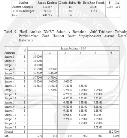
Gambar 12. Hasil Seleksi Pada Mannitol Gambar 13. Uji Gelatinase Salt Agar
Keterangan: A : Koloni kuning, Keterangan : A: Hidrolisa gelatin positif
perubahan warna (warna putih
disekitar koloni. pada medium).
B: Produksi Asam.
A A
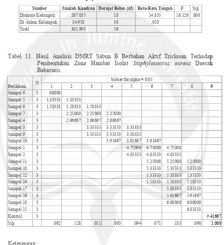
A
Gambar 14. Hasil Pengecatan Gram Gambar 15. Hasil Pengecatan Negatif
(Perbesaran 10x100) (Perbesaran 10x100)
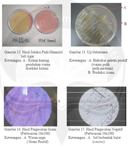
Gambar 16. Penampakan Morfologi Keterangan : A : Koloni kekuningan,
Bentuk bulat halus
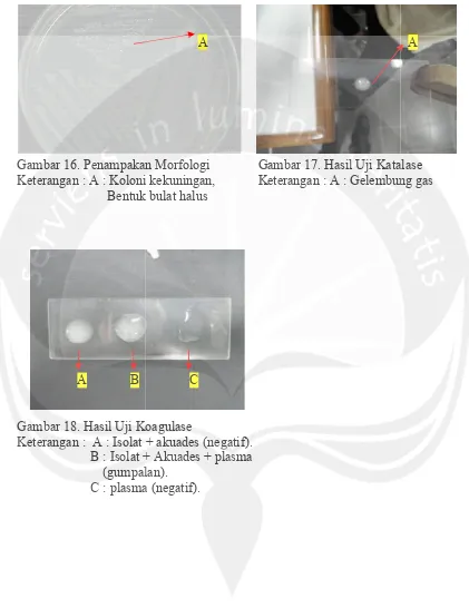
A B
Gambar 18. Hasil Uji Koagulase Keterangan : A : Isolat + akuades (ne
B : Isolat + Akuades + plasma (gumpalan).
C : plasma (negatif).
A
. Penampakan Morfologi Gambar 17. Hasil Uji Katalase Keterangan : A : Koloni kekuningan, Keterangan : A : Gelembung gas
Bentuk bulat halus
C
Hasil Uji Koagulase
Keterangan : A : Isolat + akuades (negatif). B : Isolat + Akuades + plasma
(gumpalan). C : plasma (negatif).
A
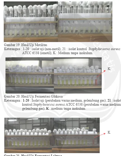
Gambar 19. Hasil Uji Motilitas
Keterangan : 1-20 : isolat uji (non-motil), 21 : isolat kontrol Staphylococcus aureus
ATCC 6538 (amotil), K : Medium tanpa inokulum.
K
Gambar 20. Hasil Uji Fermentasi Glukosa
Keterangan : 1-20: Isolat uji (perubahan warna medium, gelembung gas), 21: isolat kontrol Staphylococcus aureusATCC 6538 (perubahan warna medium, gelembung gas), K : medium tanpa inokulum.
K
Gambar 21. Hasil Uji Fermentasi Laktosa
K
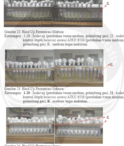
Gambar 22. Hasil Uji Fermentasi Maltosa
Keterangan : 1-20 : Isolat uji (perubahan warna medium, gelembung gas), 21 : isolat kontrol Staphylococcus aureusATCC 6538 (perubahan warna medium, gelembung gas), K : medium tanpa inokulum.
K
Gambar 23. Hasil Uji Fermentasi Sukrosa
Keterangan : 1-20: Isolat uji (perubahan warna medium, gelembung gas), 21: isolat kontrol Staphylococcus aureusATCC 6538 (perubahan warna medium, gelembung gas), K: medium tanpa inokulum.
K
Gambar 24. Hasil Uji Peptonisasi Susu
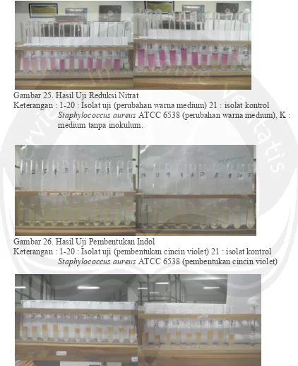
Gambar 25. Hasil Uji Reduksi Nitrat
Keterangan : 1-20 : Isolat uji (perubahan warna medium) 21 : isolat kontrol
Staphylococcus aureusATCC 6538 (perubahan warna medium), K : medium tanpa inokulum.
Gambar 26. Hasil Uji Pembentukan Indol
Keterangan : 1-20 : Isolat uji (pembentukan cincin violet) 21 : isolat kontrol
Staphylococcus aureusATCC 6538 (pembentukan cincin violet)
Gambar 27. Hasil Uji Voges-Proskauer
Keterangan : 1-20 : Isolat uji (perubahan warna medium) 21 : isolat kontrol
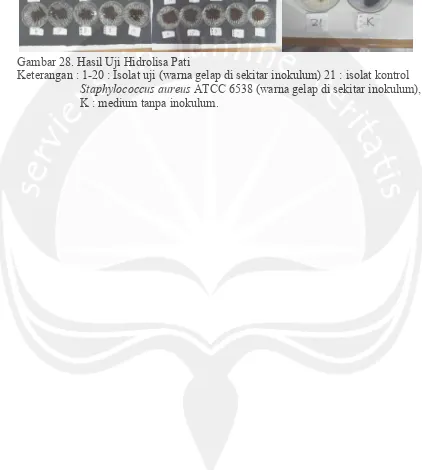
Uji Hidrolisa Pati
Gambar 28. Hasil Uji Hidrolisa Pati
Keterangan : 1-20 : Isolat uji (warna gelap di sekitar inokulum) 21 : isolat kontrol
74
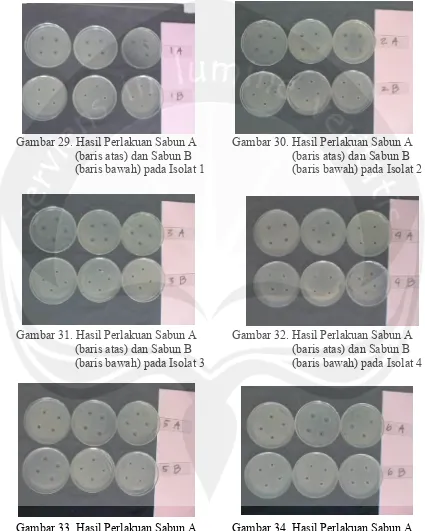
Lampiran 4. Gambar Pembentukan Zona Hambat
Gambar 29. Hasil Perlakuan Sabun A Gambar 30. Hasil Perlakuan Sabun A (baris atas) dan Sabun B (baris atas) dan Sabun B (baris bawah) pada Isolat 1 (baris bawah) pada Isolat 2
Gambar 31. Hasil Perlakuan Sabun A Gambar 32. Hasil Perlakuan Sabun A (baris atas) dan Sabun B (baris atas) dan Sabun B (baris bawah) pada Isolat 3 (baris bawah) pada Isolat 4
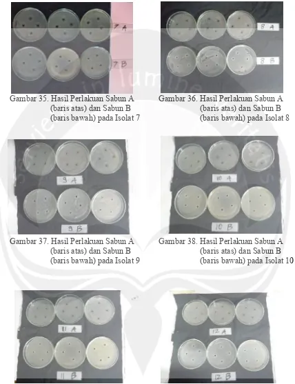
Gambar 35. Hasil Perlakuan Sabun A Gambar 36. Hasil Perlakuan Sabun A (baris atas) dan Sabun B (baris atas) dan Sabun B (baris bawah) pada Isolat 7 (baris bawah) pada Isolat 8
Gambar 37. Hasil Perlakuan Sabun A Gambar 38. Hasil Perlakuan Sabun A (baris atas) dan Sabun B (baris atas) dan Sabun B (baris bawah) pada Isolat 9 (baris bawah) pada Isolat 10
Gambar 41. Hasil Perlakuan Sabun A Gambar 42. Hasil Perlakuan Sabun A (baris atas) dan Sabun B (baris atas) dan Sabun B (baris bawah) pada Isolat 13 (baris bawah) pada Isolat 14
Gambar 43. Hasil Perlakuan Sabun A Gambar 44. Hasil Perlakuan Sabun A (baris atas) dan Sabun B (baris atas) dan Sabun B (baris bawah) pada Isolat 15 (baris bawah) pada Isolat 16
Gambar 47. Hasil Perlakuan Sabun A Gambar 48. Hasil Perlakuan Sabun A (baris atas) dan Sabun B (baris atas) dan Sabun B (baris bawah) pada Isolat 19 (baris bawah) pada Isolat 20
Gambar 49. Hasil Perlakuan Sabun A (baris atas) dan Sabun B (baris bawah) pada Isolat Isolat Kontrol
(Staphylococcus aureus