Vol 3, No 2, April - June
1994
Endothelial Cell CultureEndothelial Cell
Culture From Human
Umbilical
Cord Vein
Sri Bekti Subakir, Dewi Irawati soerya Santoso, Minarma Siagian, Maging IGAF
Abstrak
Sel endotel tne,negang peranan penting dalam berbagai prosesfisiologis tnaupun patologis. Fungsi sel endotel pada peristiwa biologik dapat diketahui lebih baik bila sel endotel dapat diisolasi dan dikultur secara invitro. Keberhasilan mengultur sel indotel di daerah tropis bergantung pada cara ,nengatasi kontaminasi bakteri dan jamur. Telah dilakukan penelitian mengisolasi dan mengultur sel endotel dari v. utnbilikalis tali pusat mempergunakan ntetode Jaffe dengan modifikasi. Dari 50 tali pusat yang dipergunakàn, 35 nenberikan kubur sel yang konfluen. Dari hasil penelitian ini nrungkin dapat dikenbangkan penelitian lebihioniut ,irng"rai fungsi sel endotel yang sangat penting pada beberapa proses fisiologis rnaupun patologis.
Abstract
Endothelial cells play an i,nportant role in various physiologic and pathologic processes. Their function in biologic eyents can be
better understood if these endothelial cells can be isolated and cultured in vitro. The success of endothelial cell cultire in the tropics
depends
was nade to isolate and culture endothelial cellsfrom hu)nanuntbilica
a outof 50 umbilical corcl vein satuples cultured, 35 produced
confluen
of further siudies on the funciions of endothelial cells in physiologic and pathologic processes.Keywords : Huutan utnbilical vein endothelial cell culnre, Endothelial cell isolation, Bacterial an4 fungal contanùnation, Confluent growlh.
65
INTRODUCTION
Endothelial cells play a major role in various
physio-logic processes, such as hemostasis, vascular
permea-bility, and
other vascular responses to physiologic orpathologic
stimuli,
Abnormalitiesin
endotelial cell structure and function can cause vascular diseases,such as thrombosis, atherosclerosis, and vasculitis.l'2 These various functions can be studied
if the
cells can be isolated and cultured in vitro.Endothelial cells can be isolated and cultured
from bovine aorta, human umbilical cord vein, pul-monary artety, etc.3 Many investigators
from
other countries have reported success in isolating and cultu-ring endothelial cells. Long-term in vitro cell culturein
tropical countriesis
challenging because of the problem of fungal and bacterial contamination.3This investigation hopes to pave the way for other
investigators interested
in
studying endothelial cellfunctions through cell culture.
METHODS
Endothelial cells can be easily isolated from human umbilical cord veins
of neonates. Collagenase
can selectively digest the subendothelial basement mem-brane leaving the internal elastic layer intact, This loosens the endothelial cells without fibroblast conta-mination.In
this study, the umbilical cords were obtainedfrom neonates born in the Budi Kemuliaan Maternity
Hospital, Jakarta. They were at term,of normal births,
with no intrapartum infection, and with clear amniotic
66
Subakir et al.fluid. The mothers consented to donate 20 - 25 cm of
their newborns' umbilical cord. The cord was severed
from the placenta immediately after birth.
The cord was inspected and all areas with clamp
marks 'were cut
off.
It
was then placedin
a sterile container filled with cord buffer (0.14 M NaCI,0.004M KCL,0.001
M
phosphate buffer, pH7.4,0.011 Mglucose) and held at 4oC until processlrrg.a To control bacterial contamination during the process of
obtain-ing the cord,
15 mgof
gentamycin and 100 mgof
ampicillin were added to every 100 ml of cord buffer.After the clamp marks have been discarded, the
umbilical cord vein
was cannulatedwith
a
bluntneedle. The vein was then perfused with 50 ml of cord
buffer at 370C to wash out any
blood left in
the vein. The other end of the vein was also cannulated with a polyethylene tubing. The cord was perfused again and allowed to drain, to insure the solution runs out of thetubing. The polyethylene tube was clamped and the
umbilical vein infused with 5 ml of 0,4 % collagenase
(Clostridium histolyticum, type
I,
Sigma). The umbili-cal cord with polyethylene tube and clamps are thenwrapped
in
aluminiumfoil
and incubatedfor
l5
minutes at37oC.Only carefully selected umbilical cords should be used. Cords damaged by clamps should be discarded, since
it
can cause contamination to the fibroblast cul-ture. The collagenase incubation time must not exceed 15 minutes, or the basement membrane can be digested and the underlying structures disrupted.After
incubation, the collagenase solution con-taining the endothelial cells was flushed top to bottom and perfusing the vein withl0
- 20 ml of cord buffer. The effluent was collected in a sterile concical centri-fuge tube containing 0.5 - I .0 ml fetal calf serum (FCS, Sigma). The FCSwill
neutralize andhalt furtherendo-thelial digestion by collagenase. The cells were sedi-mented at 250 g for 5 minutes. The precipitate was resuspended by trituration in 2 ml of culture medium
(TC 199),
to
which antibiotics, an antifungal drug, and FCS have been added. Each 80ml of TC
199 was given 20 ml FCS, 15 mg gentamycin, 100 mg ampicil-lin, and 0.4 ml fungizone. The antibiotics and antifu-ngal were twice the usual dose.All
culture medium was passed through a 2 mu sterile filter.The cell suspension was cultured in a T-25 flask
(Falcon Plastics) lined with O.27o gelatinsolution. The cells were fed with a complete change of fresh culture
medium after
24 hours and twice
in
the followingweek. Endothelial cell growth was monitored and pho-tographed every day through a phase contrast
micro-scope. Although growth factors can simulate cell
repli-cation
in
culture, they will not be needed if cell prolife-ration is sufficiently profuse. Generally, theendothe-Med J Univ Indon
lial cells
will
achieve confluent growth ( a monolayergrowth covering the entire surface of the flask base ) on day 3 or 4.)
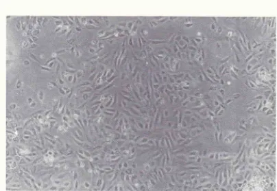
RESULTS
There were 50 umbilical cord vein samples received.
Endothelial cells were successfully isolated from 42 samples, from which 35 confluent growths were pro-duced. Isolation was less successful in 8 samples and
the cultures failed to yield confluent growths.
Con-fluent monolayers were achieved on day 4 (Figure
l).
Bacterial contamination occurred to 7 cultures on day 2 and to 4 cultures after day 3. Fungal
contamina-tion appeared later, after day 6. Only 10 cultures
sur-vived more than
6
dayswith only
1 fungal conta-mination.DISCUSSION
The failure of endothelial cells to grow in tissue culture is mainly due to contamination of fibroblasts or blood cells.
At
the risk of flushing away some endothelial cells not adhering to the T-flask surface along with the fibroblasts and blood cells, a complete change of fresh culture medium after 60 minutes of culture can reduce the possibility of contamination by those cells.In this trial,0.4 % collagenase with an incubation
time of
15 minutes is applied. Jaffe usedO.l
7o col-lagenase and 10 minutes of incubation; The range mayvary from O.L
-
O.47ofor
collangenase and 10-
30 minutes for the incubation time.6Endothelial cell morphology is identified through
a phase contrast microscope. The cells were homo-genous, closely opposed, large, and polygonal with an oval, centrally located nucleus. The findings (figure 2)
were consistent
with
endothelial cells cultured byJaffe. Ideally,
a
transmission electron microscope should be used to identify these endothelial cells.When confluent
cell growth was
achieved, the cells were harvested with 0.2% tryspsin-O.O5%F,DTAand subcultured.
On day
I
of
the subculture, cell
growth was identicalto the primary
endothelial cellculture.
These endothelial cells were found to be capable
of
migrating towards an appropriate stimulus. They can therefore be used in an angiogenesis assay.Cultured endothelial cells are 6i6-qcopically
dis-tinct from
cultured fibroblasts. Cultulêd fibroblasts grow close to one another in parallel arrays withVoI 3, No 2, Anril - June 1994
Figure 1. The tnorphology of hwnan endothelial cells at conJluence stage (100 x)
[image:3.595.118.522.389.667.2]Endothelial Cell
Culture
6768
Subakir et al.and
spindle-shaped cells.Unlike
endothelial cells, fibroblasts do not have the ability of migrating duringan angiogenic reaction.
No additional growth f,actor was required, since the cultures yielded dense, confluent cell growth able
to
surviveup
to 7
-
8
days. The endothelial cells probably produce sufficient growth factor by autocrineor paracrine secretion.
It seems that double doses of antibiotics and anti-fungal drugs are needed to control bacterial and fungal contamination
in
the tropics. These high doses werenot found to be detrimental to cell culture growth.
Acknowledgements
This research is supported by the Special Programme
of
Research, Development and Research Training in Human Reproduction, World Health Organization.The authors wishes
to
expresstheir gratitude
to theMed J Univ Indon
Department of Pathology, Faculty
of
Medicine, Uni-versityof
Indonesiafor
the use of the tissue culture laboratory.REFERENCES
l.
Thorgeirson G, Robertson AL. The vascular EndotÈelium Pathobiologic Significance. Am.J. Pathol. 197 8; 93803-472.
Patton HD et al. Textbook of Physiology 2lst ed. I-ondon:Saunders Company 1989; 862-3
3.
Iaffe EA. Biology of Endothelial cells. Boston: MartinusNijhoff Publisher 1984; l-12
4.
laffe EA, Nachman RL, Becker CG, Minick CR. Culture ofHuman Endothelial Cells Derived from Umbilical Veins. J.
Clin. Invest. 197 3 ;52:27 45 -5 6
5.
Simionescu N, Simionescu M. Endothelial Cell Biology in Health and Disease. New York: Plenum Press 1988; 123-436.
Thilo-Komer DGS, Heinrich D, Temme H. Endothelial cells in culture. In: Thilo-Korner DGS, Giesen Feg, Freshney RIeditors. The Endothelial cell a pluripotent control cell ofthe