Development of Visualisation Suite for a Low Cost Electroencephalography (EEG) System
TAN JUN WEI
This Report Is Submitted In Partial Fulfilment of Requirements For The Bachelor Degree in Electronic Engineering (Computer Engineering)
Fakulti Kejuruteraan Elektronik dan Kejuruteraan Komputer Universiti Teknikal Malaysia Melaka
ii
UNIVERSTI TEKNIKAL MALAYSIA MELAKA
FAKULTI KEJURUTERAAN ELEKTRONIK DAN KEJURUTERAAN KOMPUTER
BORANG PENGESAHAN STATUS LAPORAN
PROJEK SARJANA MUDA II
Tajuk Projek : DEVELOPMENT OF VISUALISATION SUITE FOR A LOW COST ELECTROENCEPHALOGRAPHY (EEG) SYSTEM
Sesi Pengajian :
1 4 / 1 5
Saya ……….………..
mengaku membenarkan Laporan Projek Sarjana Muda ini disimpan di Perpustakaan dengan syarat-syarat kegunaan seperti berikut:
1. Laporan adalah hakmilik Universiti Teknikal Malaysia Melaka.
2. Perpustakaan dibenarkan membuat salinan untuk tujuan pengajian sahaja.
3. Perpustakaan dibenarkan membuat salinan laporan ini sebagai bahan pertukaran antara
institusi pengajian tinggi.
4. Sila tandakan ( √ ) :
SULIT*
*(Mengandungi maklumat yang berdarjah keselamatan atau kepentingan Malaysia seperti yang termaktub di dalam AKTA RAHSIA RASMI 1972)
TERHAD** **(Mengandungi maklumat terhad yang telah ditentukan oleh organisasi/badan di mana penyelidikan dijalankan)
TIDAK TERHAD
Disahkan oleh:
__________________________ ___________________________________
(TANDATANGAN PENULIS) (COP DAN TANDATANGAN PENYELIA)
Tarikh: ……….. Tarikh: ………..
TAN JUN WEI
iii
“I hereby declared that this report entitled Development of Visualisation Suite for a Low Cost Electroencephalography (EEG) System is a result of my own except for notes that
have been cited clearly in the references”
Signature : ………..
Student Name : TAN JUN WEI
iv
“I hereby declared that I have read this report and in my opinion, this report is sufficient in term of the scope and quality for the award the Bachelor of Electronics Engineering
(Computer Engineering) with honours”
Signature : ………..
Student Name : DR LOW YIN FEN
v
vi
ACKNOWLEDGEMENT
vii
ABSTRACT
viii
ABSTRAK
ix
TABLE OF CONTENT
CHAPTER CONTENT PAGE
PROJECT TITLE
CONFIRMATION OF REPORT STATUS DECLARATION
SUPERVIOR’S CONFIRMATION DEDICATION
ACKNOWLEDGEMENT ABSTRACT
ABSTRAK
TABLE OF CONTENT LIST OF FIGURE
LIST OF ABBREVIATION LIST OF APPENDIX
i ii iii iv v vi vii viii
x CHAPTER I II CONTENT INTRODUCTION
1.1. Problem Statement 1.2. Objectives
1.3. Scope of Work
LITERATURE REVIEW
2.1. Electroencephalography 2.1.1. Electroencephalography 2.1.2. History of Encephalography 2.1.3. Application of Encephalography 2.2. Low cost electroencephalography 2.3. Graphical User Interface (GUI) 2.3.1. GUI of Emotiv EPOC 2.3.2. GUI of EEGLAB 2.3.3. GUI of EEGTESTING 2.4. Feature and function of GUI
2.4.1. Feature and function of GUI Emotiv EPOC 2.4.2. Feature and function of GUI in EEGLAB 2.4.3. Feature and function of GUI in EEGTESTING 2.5. EEG electrode placement
2.6. EEG Recording Techniques
xi CHAPTER III IV CONTENT METHODOLOGY
3.1. Overview of the system
3.2. Flow Chart of the project implementation 3.3. Load Emotiv libraries into MATLAB 3.4. Identify the feature and function of GUI
3.5. Implement signal processing method and function into MATLAB .m file
3.6. Design GUI for visualization purpose using MATLAB 3.6.1. Open a New GUI in MATLAB
3.6.2. Set the GUI Figure Size
3.6.3. Layout the Simple GUIDE GUI
3.6.3.1. Adding components into GUI Layout 3.6.3.2. Aligning the GUI components
3.6.3.3. Labelling the GUI components 3.6.4. Saving the GUI Layout
3.6.5. Set the background picture of GUI 3.7. Integrate MATLAB .m file into GUI 3.8. Perform test with real subject
3.9. Debug
RESULT AND DISCUSSION
4.1. Complete GUI (Graphical User Interface) 4.2. 2D (two-dimensional) Plotting
4.3. 3D (three-dimensional) Surface Plotting 4.4. FFT (Fast Fourier Transform) Plotting
xii
V
4.5. Gyroscope Plotting
4.6. Recording function in GUI
CONCLUSION AND RECOMMENDATION
5.1. Future recommendations 5.2. Market Potential of project
REFERENCE APPENDIX
53 56
60
60 61
xiii
LIST OF FIGURE
NUMBER 2.1 2.2 2.3 2.4 2.5 2.6 2.7 2.8 2.9 2.10 2.11 2.12 2.13 2.14 2.15 2.16 3.1 3.2 3.3 TITLE
MindWave™ Headsets by Neuro Sky EPOC headset by Emotiv
EEG display on TestbenchTM software Gyro display on TestbenchTM software
Data Packets display on TestbenchTM software FFT display on TestbenchTM software
Data recording playback on TestbenchTM software User Interface of EEGLAB software running under Linux Scroll activity in EEGLAB tools
Welcome page
Selections of sample results and other records page Brainwaves pattern
Data scrolling in EEGLAB
International 10-20 Electrode Placement System
Electrode cap with electrodes placed after 10-20 electrode placement system
Equipment for EEG recording: amplifier unit, electrode cap, conductive jelly, injection, and aid for disinfection
System Diagram of the project Flow Chart of the project
Libraries file added into MATLAB path
xiv 3.4 3.5 3.6 3.7 3.8 3.9 3.10 3.11 3.12 3.13 3.14 3.15 3.16 3.17 3.18 3.19 3.20 3.21 3.22 4.1 4.2 4.3 4.4 4.5 4.6 4.7 4.8 4.9 4.10
Coding of loading libraries into MATLAB Assigning number of data channel
Coding of 2D, 3D and FFT plotting to display the number of channel
Coding of Gyroscope x and y plotting to display the number of channel
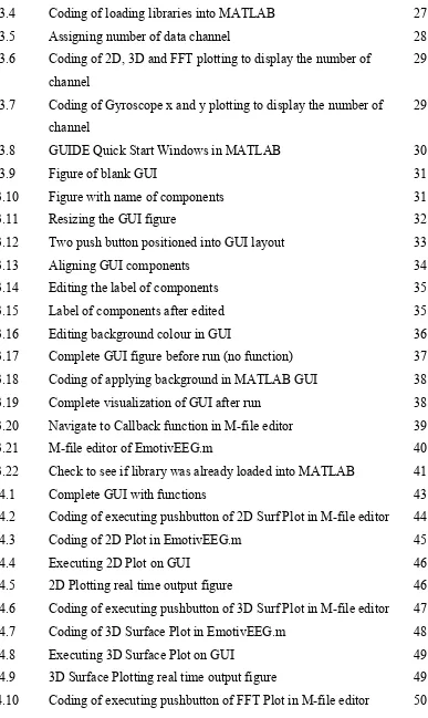
[image:14.612.139.526.66.715.2]GUIDE Quick Start Windows in MATLAB Figure of blank GUI
Figure with name of components Resizing the GUI figure
Two push button positioned into GUI layout Aligning GUI components
Editing the label of components Label of components after edited Editing background colour in GUI
Complete GUI figure before run (no function) Coding of applying background in MATLAB GUI Complete visualization of GUI after run
Navigate to Callback function in M-file editor M-file editor of EmotivEEG.m
Check to see if library was already loaded into MATLAB Complete GUI with functions
Coding of executing pushbutton of 2D Surf Plot in M-file editor Coding of 2D Plot in EmotivEEG.m
Executing 2D Plot on GUI
2D Plotting real time output figure
Coding of executing pushbutton of 3D Surf Plot in M-file editor Coding of 3D Surface Plot in EmotivEEG.m
Executing 3D Surface Plot on GUI
3D Surface Plotting real time output figure
Coding of executing pushbutton of FFT Plot in M-file editor
xv 4.11 4.12 4.13 4.14 4.15 4.16 4.17 4.18 4.19 4.20 4.21 4.22 4.23 4.24
Coding of FFT Plot in EmotivEEG.m Executing FFT Plot on GUI
FFT Plotting real time output figure
Coding of Gyroscope in UpdateDataGyro(self) function Coding of executing pushbutton of Gyro Plot in M-file editor Coding of Gyro Plot in EmotivEEG.m
Executing Gyro Plot on GUI
Gyroscope Plotting real time output figure
Coding of recording EEG signal function in Gyro Plot in EmotivEEG.m
Coding of executing pushbutton of Recording (10s) in M-file editor
Coding of executing pushbutton of Recording (20s) in M-file Recording function running in MATLAB command window EEG data saved as .mat file after record function complete Matrix data within the recorded .mat file
xvi
LIST OF ABBREVIATION
GUI - Graphical User Interface
EEG - Electroencephalography
GYRO - Gyroscope
API - Application Program Interfacing
xvii
LIST OF APPENDIXES
APPENDIX
A B C D E
TITLE
Project Planning (Gantt Chart)
Source Code of implemented functions (.m file) Source Code of GUI Editor (.m file)
INOTEK Poster
INOTEK Certificate of Achievement
PAGE
1
CHAPTER I
INTRODUCTION
An electroencephalography (EEG) is a system that measures and records the electrical activity of the human brain. It provides evidence of how the brain functions over time and used to be the first line method in the evaluation of brain disorders for medical industry nowadays. An electroencephalogram also able to detect abnormalities in the brain waves or electrical activity of the brain. During the procedure, electrodes consisting of small metal discs with thin wires are pasted on the scalp. The electrodes detect tiny electrical charges that result from the action of the brain cells. The charges are amplified and appear as a graph on a computer screen or as a recording that may be printed out on paper then the reading will be interpreted.
2
In this project, a visualization suite that provides real time information will be developed for a low-cost EEG system. The development of this project consists of designing a graphical user interface (GUI) to show the brain’s signal activity on laptop/desktop by connecting EEG device (bio-amplifier) for signal amplification. The EEG device will be connected either wireless or wired to laptop/desktop. A reliable and sensitive visualization suite with real-time information is expected to be developed from low-cost EEG.
1.1 Problem Statement
3
1.2 Objectives
The main objectives of this project are:
to develop a visualization suite that able to show real-time information from a low cost EEG system,
to implement simple signal processing method using MATLAB software.
1.3 Scope of work
4
CHAPTER II
LITERATURE REVIEW
2.1 Electroencephalography
2.1.1 Electroencephalography
5
2.1.2 History of Electroencephalography
In 1875. Richard Catton who regarded as the first scientist to study and investigate the brain potentials. During his investigation, The results (presented in 1875) showed that ‘‘weak currents of changing directions pass through the multiplier when the electrodes are placed at two points of the external surface, or one electrode on the gravy matter and one on the surface of skull.’’ This observation can be considered as the discovery of electroencephalographic activity. In 1890, Adolf Beck, physician of and professor of physiology from University of Lwów Galicia, had investigated the spontaneous activity of the brain of rabbits and dogs. He discovered the rhythmical oscillations of neural activity and “alpha blocking” which is the disappearance of rhythmical oscillations when the eyes were stimulated with light [3]. Then, his co-worker Napoleon Cybulski presented the electroencephalogram in a graphical form by applying a galvanometer with a photographic attachment. He also the first person who observe epileptic EEG activity in a dog effected by an electric stimulation [4]. In 1929, Hans Berger recorded the first electroencephalogram (EEG) from surface of human scalp [5]. Eventually, in 1935, the major fields of today’s clinical electroencephalography was born by the research from F. Gibbs and H. Davis [3].
2.1.3 Application of Electroencephalography
6
better brain allocation. Besides, EEG can determine the position and strength of neural activity in different regions of brain [2].
Brain computer interface (BCI) is a communication system that recognizes user’s command only from his or her brainwaves and reacts according to them. In this application, the personal computer (PC) and subject must be trained so that simple task can be executed which consist of desired motion of an arrow then displayed on the screen of PC through subject’s imaginary of the motion of human’s left or right hand. As conclusion of imaging process, certain characteristic of the brainwaves that shown can be used for user’s command recognition [2].
Another related application is Neuro-feedback, a type of biofeedback for the brain, which practitioners say can address a host of neurological ills among them attention deficit hyperactivity disorder, autism, depression and anxiety. This application allowing patients to alter their own brain waves through practice and repetition. The procedure is controversial, expensive and time-consuming. Brain cells communicate with one another, through a constant signal of electrical impulses. Their patterns show up on an electroencephalogram (EEG), as brain waves with different frequencies. A major attraction of the technique is the hope that it can help patients avoid drugs, which often have side effects.Besides, patients practice routines that seem more like exercising a muscle [7].
Based on research from R. Bickford (1987), the clinical applications of the EEG in humans and animals are used to [8]:
Monitor alertness, coma and brain death
Locate areas of damage following head injury, stroke, tumor, etc.
Test afferent pathways (by evoked potentials)
Monitor cognitive engagement (alpha rhythm)
Produce biofeedback situations, alpha, etc.;
Control anaesthesia depth
7
Test epilepsy drug effects
Assist in experimental cortical excision of epileptic focus Monitor human and animal brain development
Test drugs for convulsive effects
Investigate sleep disorder and physiology
2.2. Low cost electroencephalography
Multi-electrode, medical grade EEG system have already been used in the medical industry such as hospitals and laboratories for a long time. Fortunately, the invention of low-cost electroencephalography also known as a low-cost bio amplifier makes it available to take this technology from the medical industry to informal environments for example schools and homes. The price of this device is cheaper compared to the medical EEG system is easy to use [9].