SELECTED BIOMARKERS; IN VIVO AND IN SITU
APPLICATIONS
KHUSNUL YAQIN
GRADUATE SCHOOL
BOGOR AGRICULTURAL UNIVERSITY BOGOR
STATEMENT ABOUT DISSERTATION AND INFORMATION
SOURCE
I herewith declare that the dissertation titled “Selected Biomarkers: In Vivo and In Situ Applications” is made by myself under direction of the supervisor committee and has not been proposed in any form to other university. The sources that derived or cited from published and unpublished journal articles or books or other authors have been acknowledged and the list of references is listed at the end of each chapter of this dissertation.
Bogor, 8th of September 2008
ABSTRAK
KHUSNUL YAQIN. Biomarker; Aplikasi In Vivo dan In Situ. Dibimbing oleh BIBIANA WIDIYATI LAY, ETTY RIANI, dan ZAINAL ALIM MASUD.
Penelitian in vivo dilakukan untuk menguji toksisitas dimethoate terhadap sistem neuro-immune kerang biru Mytilus edulis dengan menggunakan dua biomarker yaitu aktivitas kolinesterase dan fagositosis. Efek dose-dependent
terjadi pada kolinesterase kerang biru, M. edulis yang dipapar dengan konsentrasi diemthoate yang berbeda yaitu 0,00; 7,88; 15,75; 31,50 dan 63,00 µg/l selama 14 hari. Pengaruh dimethoate yang signifikan terhadap aktivitas kolinesterase terjadi pada konsentrasi 31,50 dan 63,00 µg/l. Sebaliknya respon hormetik tampak pada aktivitas fagositosis yaitu terjadi reduksi aktivitas fagositosis pada konsentrasi yang terendah yang diikuti oleh stimulasi pada konsentrasi yang tertinggi. Mentransformasikan biomarker tingkat rendah seperti aktivitas kolinesterase ke tingkat yang lebih tinggi seperti tingkah laku merupakan penelitian yang menarik dalam bidang ekotoksikologi. Oleh karena itu telah dilakukan penelitian untuk mengevaluasi pengaruh trichlorfon terhadap sistem saraf dan tingkah laku M. edulis dengan menggunakan aktivitas kolinesterase dan laju penyaringan. Hasil dari penelitian tersebut menunjukkan bahwa insang adalah organ yang mengandung aktivitas kolinesterase yang paling sensitif dibandingkan dengan organ yang lain. Sensitivitas laju penyaringan untuk mendeteksi dampak buruk trichlorfon berada pada rentang sensitivitas aktivitas kolinesterase M. edulis. Selanjutnya percobaan dekotantaminasi menunjukkan bahwa inhibisi aktivitas kolinesterase dan reduksi laju penyaringan akibat paparan pestisida dapat pulih ke tingkat yang normal kecuali pada aktivitas kolinesterase kaki dan mantel. Prosedur Pearson dan Backward diaplikasikan untuk mengetahui derajat korelasi antara aktivitas kolinesterase pada setiap organ dan laju penyaringan. Hasil penelitian tersebut menunjukkan bahwa aktivitas kolinesterase pada mantel adalah yang paling berkorelasi dengan aktivitas penyaringan. Dalam kaitannya dengan kerang hijau Perna viridis, untuk meningkatkan kompetensinya sebagai “sentinel organism” dilakukan karakterisasi enzim kolinesterase P. viridis. Pendekatan substrat dan inhibitor yang spesifik menunjukkan bahwa kolinesterase P. viridis
terdiri atas dua jenis enzim. Enzim tersebut adalah asetilkolinesterase yang specifik dan butirilkolinesterase yang tidak spesifik. Penerapan aktivitas kolinesterase pada insang P. viridis pada hot spot biomonitoring di tiga perairan pantai di Indonesia menunjukkan bahwa enzim ini merupakan suatu biomarker yang berdayaguna. Hasil biomonitoring menunjukkan bahwa P. viridis yang berasal dari daerah yang diasumsikan bersih di Pangkajene Kepulauan (Pangkep), Sulawesi Selatan mempunyai tingkat aktivitas kolinesterase yang paling tinggi dibandingkan dengan P. viridis dari dua lokasi di Teluk Jakarta (Cilincing dan Kamal Muara). Hasil penggunaan enzim sebagai biomarker dipertegas oleh respon fagositosis kerang hijau. Terdapat stimulasi aktivitas fagositosis kerang yang dikumpulkan di dua lokasi di Teluk Jakarta dibandingkan dengan kerang hijau yang berasal dari Pangkep.
SUMMARY
KHUSNUL YAQIN. Selected Biomarkers; In Vivo and In Situ Applications. Under direction of BIBIANA WIDIYATI LAY, ETTY RIANI, and ZAINAL ALIM MASUD.
Selected biomarkers, cholinesterase (ChE) and phagocytic activities from blue mussels, Mytilus edulis have been used to detect the effects of neuro-immunotoxicity of organophophate (OP) pesticide, dimethoate. The serial dilutions of dimethoate concentrations which were 0.00, 7.88, 15.75, 31.50 and 63.00 µg/l, showed dose-dependent effects on the ChE activity of blue mussels,
Mytilus edulis after being exposed for 14 days. Statistical analysis showed that the significant effects of the pesticide on the ChE activity occurred at concentrations 31.50 and 63.00 µg/l (p <0.05). In contrast, the dose-dependent effects of dimethoate were not observed in the phagocytic activity of dimethoate-exposed mussels. The suppression effects of dimethoate on phagocytic activity occurred significantly (p < 0.05) at two concentrations of dimethoate (7.88 and 15.75 µg/l), but stimulation effects significantly (p < 0.05) emerged at the following concentrations (31.35 and 63.00 µg/l). The reduction which occurred at the lowest concentrations along with the stimulation at the highest concentrations implied the occurrence of a U-shape hormetic response of the phagocytic activity under dimethoate exposure. The results suggested that two selected biomarkers, ChE and phagocytic activities were respectable tools to detect the effects of the pesticides when the dose-dependent and U-shape hormetic responses were taken into account in the laboratory scale study.
Pearson procedure was applied to recognize the correlation degree of the ChE activity from each relevant organ and siphoning rate. Coefficient correlation (R) between the siphoning rate and the organs were 0.761, 0.656 and 0.510 for mantle, gill and PAM respectively. The result indicated that mantle is the most correlated organ to the siphoning activity which was followed by gill and PAM. Considering that the siphoning activity is the product of the three relevant organs movements Backward Multiple Regression was applied to know which organ play a dominant role in the siphoning activity. The Backward Multiple Regression emphasized the Pearson procedure by indicating the dominant role of mantle in the siphoning activity. The regression equation was Y = -26.576 + 5.195 X1 +
17.009 X2, where X1 and X2 are gill and mantle respectively. The value of
adjusted R2 of the equation was 0.63. It was indicated that the role of the two organs i.e., mantle and gill in the siphoning activity was about 60 %. The Backward Multiple Regression method eliminated the role of PAM in siphoning activity of the mussels since it showed insignificant role statistically (p = 0.178).
Tropical green mussels (Perna viridis) play an important economic and ecological role in the coastal areas of Indonesia. P. viridis has been used as an eco-sentinel organism for marine biomonitoring program in Asia regions. To magnify its competency as an eco-sentinel organism in biomonitoring characterization of cholinesterases (ChEs) of P. viridis from a selected coastal area of Indonesia has been conducted. In addition, the characterization of ChEs of
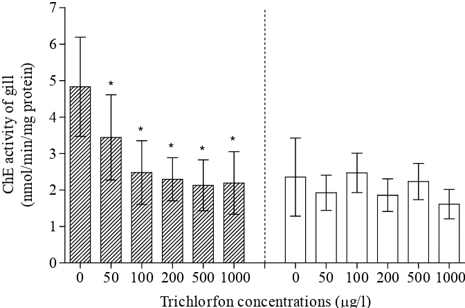
P. viridis is a requirement which has to be conducted prior to using ChE activity as biomarker. Dissected organs which were gill, foot, mantle and posterior adductor muscle (PAM) were examined for substrate specificity and inhibitors sensitivity using selective and non-selective substrates and inhibitors. The results indicated that highest level of the ChE activity was observed in gill and followed by foot, mantle and PAM. The substrate specificity approach using various alkylthiocholines indicated that the ChEs of P. viridis constitute an acetylcholinesterase (AChE) and a butyrylcholinesterase (BuChE). Likewise, the inhibitors sensitivity approach using eserine, BW284C51, and iso-OMPA sustained the substrate approach which recorded typical AChE and atypical BuChE. Hence, the study revealed that ChEs of P. viridis gill contain a typical AChE and an atypical BuChE. The results also suggested that the P. viridis’s gill is the most suitable organ for employing the ChE activity as a biomarker in marine monitoring.
phagocytic activity of green mussels from the polluted sites demonstrated a significant higher activity than that of green mussels from the pristine site, Pangkep. However, there were no significant differences of phagocytic activity between the polluted sites. This might indicate that the existing pollutants in Jakarta Bay were more neurotoxic rather than immunotoxic substances. The results showed clearly that both selected biomarkers were potential valuable tools for effect-based monitoring and pollution impacts in coastal zones of Indonesia. The hot spot biomonitoring contributes to a tailor-made toolbox focussing on risk-based coastal zone management in Indonesia.
Copyright©2008 Bogor Agricultural University
Copyright are protected by law,
It is prohibited to cite all or part of this dissertation without referring to and mentioning the source. Citation only permitted for the sake of education, research, scientific writing, report writing, critical writing or reviewing scientific problem; and citation doesn’t inflict the name and honor of Bogor Agricultural University.
SELECTED BIOMARKERS; IN VIVO AND IN SITU
APPLICATIONS
KHUSNUL YAQIN
Dissertation
Partial Fulfillment For Obtaining a Doctoral Degree
In Environment and Natural Resources Management Study Program
GRADUATE SCHOOL
BOGOR AGRICULTURAL UNIVERSITY BOGOR
The title of dissertation : Selected Biomarkers; In Vivo and In Situ
Applications
Name : Khusnul Yaqin
NIM : P062030151
Approved by :
Supervisor Committee
Prof. Dr. drh. Bibiana Widiyati Lay, M.Sc Chair
Dr. Ir. Etty Riani, M.S Dr. Ir. Zainal Alim Masud, DEA
Committee Member Committee Member
Acknowledged by
Director of Environment and Dean of Graduate School Natural Resources Management
Study Program
Prof. Dr. Ir. Surjono H. Sutjahjo, M.S Prof. Dr. Ir. Khairil A. Notodiputro, M.S
FOREWORD
I would first like to express my sincere thanks and gratitude to my supervisor committee (Prof. Dr. drh. Bibiana Widiyati Lay, M.Sc, Dr. Ir. Etty Riani, MS, Dr. Zainal Alim Masud, DEA) for their guidance in accomplishing the studies. I would like to extend my deep and warm thanks and gratitude to Prof. Peter-Diedrich Hansen for facilitating me to use the ecotoxicology laboratory in Institute for Ecological Research and Technology, Department of Ecotoxicology Technische Universitaet Berlin, Germany which made the studies possible. He also trained me to dig out scientific experiences in ecotoxicology. I wish also to thank Dr. Eckehard Unruh, Birgit Fischer, Dr. Gerd Huscheck, Martin Kern from Department of Ecotoxicology Technische Universitaet Berlin for the constructive advices and Birgit Hüssel from Alfred Wagner Institute for the organization of blue mussel, Mytilus edulis.
I wish to thank DAAD (Deutscher Akademischer Austausch Dients/German Academic Exchange Service) for supporting my works during the study time in Indonesia and Germany through the sandwich scheme scholarship. The scholarship not only assisted my works, but also gave me and my family to interact and understand the German academic atmosphere and cultural paradigm.
Bogor, 8th of September 2008
BIOGRAPHY
The author was born in Gresik 26 July 1968. He spent his academic experiences from elementary to senior high school in that city. Afterward, he migrated to Makassar for pursuing his study in Department of Fishery, Hasanuddin University from 1987 to 1992. He was registered as a lecturer in that department in 1994 until now. In 2001, he continued his study on master degree in Department of Marine Ecology, University of Aarhus, Denmark which was supported by DANIDA. In this department, he started to work on marine ecotoxicology subjects under direction of Dr. Vibeke Simonsen and Dr. Janeck Scott-Forsmand. He completed the study in 22 January 2003. Accordingly, he pursued his scientific experiences in ecotoxicology by registering on doctoral degree in Environmental Science Study Program of Graduate School of Bogor Agricultural University in 2003. His doctoral work was supported by DAAD in sandwich scheme scholarship. The scholarship set up him to synthesize tropical and temperate academic atmospheres by taking a class experience in Bogor Agricultural University for one year and laboratory works in Department of Ecotoxicology, Technische Universitaet Berlin for three years. In addition, in the Department of Ecotoxicology he was involved as a leading scientist in an international laboratory inter-calibration excersise on a new Microbial Assay Technique for Risk Assessment.
During doctoral study he has published some papers:
1. Yaqin, K. 2006. Ecotoxicological assessment of aquatic genotoxicity using the comet assay. HAYATI Journal of Biosciences, 13: 124-130. 2. Khusnul Yaqin, Bibiana Widiyati Lay, Etty Riani, Zainal Alim Masud,
TABLE OF CONTENT
FOREWORD ... ii
LIST OF TABLES ... viii
LIST OF FIGURES ... ix
LIST OF APPENDIXES ... xiii
I. GENERAL INTRODUCTION ... 1
1.1. Background ... 1
1.2. Logical Framework ... 3
1.3. Problem Formulation ... 7
1.4. Objectives of the Studies ... 11
1.5. Purpusoses of the Studies ... 12
1.6. Hypothesis ... 12
1.7. Novelties ... 13
1.7. References ... 13
II. THE USE OF SELECTED BIOMARKERS, PHAGOCYTIC
AND CHOLINESTERASE ACTIVITIES, TO DETECT THE
EFFECTS OF DIMETHOATE ON MARINE MUSSEL (
Mytilus
edulis
)………. ... 17
2.1.. Abstract ... 17
2.2.. Introduction ... 17
2.3.. Material and Methods ... 19
2.4. Results ... 22
2.5. Discussion ... 26
2.6. Reference ... 30
III. EVALUATION ON SENSITIVITY OF SELECTED
BIOMARKERS, CHOLINESTERASE ACTIVITY AND
SIPHONING RATE OF
Mytilus edulis
TO TRICHLORFON .... 35
3.1. Abstract ... 35
3.2. Introduction ... 35
3.4. Results ... 43
3.5. Discussion ... 54
3.6. References ... 64
IV. CHARACTERIZATION OF CHOLINESTERASE ACTIVITY
IN GREEN MUSSEL (
Perna viridis
) AS A POTENTIAL
BIOMARKER IN MARINE BIOMONITORING ... 68
4.1. Abstract ... 68
4.2.. Introduction ... 68
4.3. Materials and Methods ... 71
4.4. Results ... 74
4.5. Discussion ... 81
4.6. References ... 87
V. HOT SPOT BIOMONITORING OF MARINE POLLUTION
EFFECTS USING SELECTED BIOMARKERS,
CHOLINESTERASE AND PHAGOCYTIC ACTIVITY, OF
TROPICAL GREEN MUSSEL (
Perna viridis
) ... 91
5.1. Abstract ... 91
5.2. Introduction ... 91
5.3. Material and Methods ... 93
5.4. Results ... 98
5.5. Discussion ... 101
5.6. Reference ... 107
VI. GENERAL DISCUSSION ... 113
6.1. Biomarker as a Counterpart of a Classic Chemical Analysis Approach ... 113
6.2.. Mussel as Eco-sentinel Organism ... 119
6.3.. Organophosphate Pesticides Pollution and Biomarkers ... 125
6.4.. Cholinesterase Activity: An Enzymatic Biomarker ... 129
6.5.. Phagocytic Activity as Biomarker ... 135
6.6.. Siphoning Activity: Behavioural Biomarker ... 141
6.7. Conclusion ... 145
6.8. Recommendations ... 146 6.9. References ... 146
LIST OF TABLES
1. Pesticides production in Indonesia. Source of raw data: Statistics of large and medium industry, BPS (1999-2004). ... 3 2. Usage of pesticides in brackishwater and freshwater ponds (2001-2006)
(DKP 2006). ... 4 3. The various alkylthiocholine substrates and cholinesterase inhibitors. ... 73 4. Mean of cholinesterase activity of three substrates as measured in various
organs of green mussel with comparison to ASCh. ... 76 5. The influence of anti-ChE substances on ChE activity green mussel gill with
different substrates. ... 79 6. The influence of anti-ChE substances on ChE activity of green mussel foot
with different substrates. ... 80 7. The influence of anti-ChE substances on ChE activity of green mussel
posterior adductor muscle (PAM) with different substrates. ... 81 8. The influence of anti-ChE substances on ChE activity of green mussel mantle
with different substrates. ... 82 9. ChE kinetic parameters from aquatic animals. ... 84
LIST OF FIGURES
1. The role of biomarkers in ecological risk assessment (ERA) (Modified from Hansen 2007). ... 6 2. Circulating hemocyte density of M. edulis before the treatment. Data were
expressed as median (25 % and 75 % quartile, 5 % and 95 % confidence interval). ... 23 3. Phagocytosis activity of M. edulis hemocytes before the treatment. Data
were expressed as median (25 % and 75 % quartile, 5 % and 95 %
confidence interval). ... 23 4. Circulating hemocytes density of M. edulis after 14 days of dimethoate
exposure. Data were expressed as median (25 % and 75 % quartile, 5 % and 95 % confidence interval). * indicated the different number of
hemocytes from the treatments and from those observed in the control (p < 0.05). ... 24 5. Phagocytic activity of M. edulis hemocytes after 14 days of dimethoate
exposure. Data were expressed as median (25 % and 75 % quartile, 5 % and 95 % confidence interval). * indicated the different phagocytic activity of mussels among the treatments (p < 0.05). ... 24 6. ChE activity of M. edulis gill after 14 days of dimethoate exposure. Data
were expressed as median (25 % and 75 % quartile, 5 % and 95 %
confidence interval). * indicated the different enzyme activity of the treatments compare to the control (p < 0.05). ... 25 7. Effects of trichlorfon on the siphoning rate of M. edulis. The siphoning
rate of mussels exposed to trichlorfon for 96-h (striated area with ± standard deviation). The siphoning rate of post-trichlorfon exposed mussels which were incubated in clean water for 7 days (empty area with ± standard deviation). * indicate significant difference from control (0 μg/l) (P < 0.05). ... 44 8. Effects of trichlorfon on the ChE activity from hemolymph of M. edulis.
The ChE activity in hemolymph of mussels exposed to trichlorfon for 96-h (striated area with ± standard deviation). The ChE activity in hemolymph of post-trichlorfon exposed mussels which were incubated in clean water for 7 days (empty area with ± standard deviation). * indicates significant difference from control (0 μg/l) (P < 0.05). ... 45 9. Effects of trichlorfon on the ChE activity from gill of M. edulis.
The ChE activity in gill of mussels exposed to trichlorfon for 96-h (striated area with ± standard deviation). The ChE activity in gill of post-trichlorfon exposed mussels which were incubated in clean water for 7 days (empty area with ± standard deviation). * indicate significant difference from control (0 μg/l) (P < 0.05). ... 46
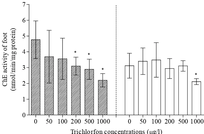
10. Effects of trichlorfon on the ChE activity from foot of M. edulis. The ChE activity in foot of mussels exposed to trichlorfon for 96-h (striated area with ± standard deviation). The ChE activity in foot of post-trichlorfon exposed mussels which incubated in clean water for 7 days (empty area with ± standard deviation). * indicate significant
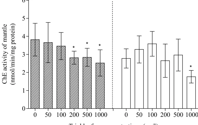
difference from control (0 μg/l) (P < 0.05). ... 47 11. Effects of trichlorfon on the ChE activity from mantle of M. edulis.
The ChE activity in mantle of mussels exposed to trichlorfon for 96-h
(striated area with ± standard deviation). The ChE activity in mantle of post-trichlorfon exposed mussels which were incubated in clean water for 7 days (empty area with ± standard deviation). * indicate significant
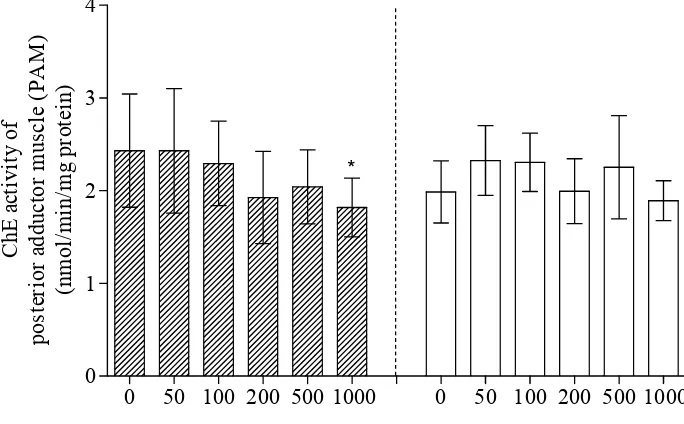
difference from control (0 μg/l) (P < 0.05). ... 48 12. Effects of trichlorfon on the ChE activity from PAM of M. edulis.
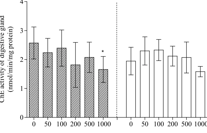
The ChE activity in PAM of mussels exposed to trichlorfon for 96-h (striated area with ± standard deviation). The ChE activity in posterior adductor muscle of post-trichlorfon exposed mussels which were incubated in clean water for 7 days (empty area with ± standard deviation). * indicates significant difference from control (0 μg/l) (P < 0.05). ... 49 13. Effects of trichlorfon on the ChE activity from the digestive gland of
M. edulis. The ChE activity in digestive gland of mussels exposed to
trichlorfon for 96-h (striated area with ± standard deviation). The ChE activity in digestive gland of post-trichlorfon exposed mussels which
incubated in clean water for 7 days (empty area with ± standard deviation). * indicates significant difference from control (0 μg/l)
P < 0.05). ... 50 14. Comparison between pre- and post-incubated mussels M. edulis after
exposed to trichlorfon concentrations in term of the ChE activity from the six organs. The ChE activity in the six organs of mussels exposed to trichlorfon for 96-h (striated area with ± standard deviation). The ChE activity in the six organs of post-trichlorfon exposed mussels which were incubated in clean ASW for 7 days (empty area with ± standard
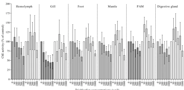
deviation). * indicate significant difference from control (0 μg/l) (P < 0.05). ... 51 15. Effects of trichlorfon on the ChE activity from three organs of M. edulis.
The ChE activity in three organs of mussels exposed to trichlorfon for 96-h (striated area with ± standard deviation). The ChE activity in three organs of post-trichlorfon exposed mussels which were incubated in clean water of original habitat of mussels for 7 days (empty area with ± standard deviation). * indicate significant different from control (0 μg/l) (P < 0.05). ... 52 16. Correlation between the siphoning rate and the ChE activity from the gill,
p < 0.001. ... 53 17. Correlation between the siphoning rate and the ChE activity from the mantle,
p < 0.001. ... 53
18. Correlation between the siphoning rate and the ChE activity from
posterior adductor muscle, p < 0.001. ... 54 19. Sampling station (ST) in Pangkajene Kepulauan (Pangkep). ... 72 20. Effect of aectylthocholine (ASCh) concentration as substrate on esterase
activity in supernatant extract 10000 g (S10) from gill P. viridis. Insert is
Hanes-Woofl-plot... 74 21. Comparison of esterase activity of various tissue of green mussel with
various substrates (3 mM). Pooled organs from sixteen green mussels were measured for the enzyme activity (nmol/min/mg of protein). ... 75 22. ChE activity of various organs of green mussel with acetylthiocholine
(ASCh) as substrate and the inhibitors (10μM); Eserine, BW 284C51 and Iso-OMPA. The enzyme activity was measured by pooling organs from sixteen green mussels (nmol/min/mg of protein). Bars indicated
standard deviation. * Significantly different from control (P <0.01). ... 77 23. ChE activity of various organs of green mussel with acetyl beta metyl
thiocholine (A-β-MSCh) as substrate and the inhibitors (10μM); Eserine, BW 284C51 and Iso-OMPA. The enzyme activity was measured by pooling organs from sixteen green mussels (nmol/min/mg of protein). Bars indicated standard deviation (SD). *Significantly different from control (P <0.01). ... 78 24. ChE activity of various organs of green mussel with Butyrylthiocholine
(BuSCh) as substrate and the inhibitors (10μM); Eserine, BW284C51 and Iso-OMPA. The enzyme activity was measured by pooling organs from sixteen green mussels (nmol/min/mg of protein). Bars indicated
standard deviation (SD). * Significantly different from control (P <0.01). ... 78 25. Indonesian Map ... 94 26. Sampling stations (ST) in Jakarta Bay. ... 94 27. Cholinesterase activity of different organs of green mussel, Perna viridis
from Pangkep Indonesia. Data were expressed as median (25 % and 75 % quartile, 5 % and 95 % confidence interval). ... 98 28. ChE activity of green mussel gill, Perna viridis from the pollution
gradient of Indonesian waters. Kamal Muara and Cilincing are expected as polluted areas, while Pangkep is expected as a clean area. Data were expressed as median (25 % and 75 % quartile, 5 % and 95 % confidence interval). * indicate the different enzyme activity among the sampling sites (p < 0.05). ... 99 29. Circular hemocytes of green mussel P. viridis collected from the selected
areas of Indonesia waters; Pangkep, Kamal Muara, and Cilincing. Data were expressed as median (25 % and 75 % quartile, 5 % and 95 %
confidence interval)... 100
30. Phagocytotic Index of green mussel P. viridis collected in the selected area of Indonesia waters; Pangkep, Kamal Muara, and Cilincing. Data were expressed as median (25 % and 75 % quartile, 5 % and 95 %
confidence interval). * indicate the different phagocytic activity of mussel hemolymph collected from polluted sites to the reference site
(Pangkep) (p < 0.05). Y-axis is logarithmic scale. ... 100
31. General structure of OP pesticide (Connell 2005). ... 126
32. Dimethoate. ... 126
33. Trichlorofon. ... 126
34. Reduction of the siphoning activity in relation to the ChE activity inhibition from mantle of blue mussels, M edulis after exposed to trichlorofon concentration for 96 h. ... 133
35. Hypothetical dose response of organophosphate pesticides (modified from Peakall 1992). ... 135
LIST OF APPENDIXES
1. Protocol of measurement of cholinesterase activity... 156 2. Protocol of phagocytic activity of mussel hemocytes for fieldwork
monitoring and laboratory exposition. ... 160 3. Correlation between the trichlorfon induced ChE activity of mussel’s
organs and the siphoning rate. ... 166 4. Backward multiple regression procedure on the trichlorfon induced ChE
activity of mussel’s organs and the siphoning rate. ... 167
I. GENERAL INTRODUCTION
1.1. Background
The term of biomarker has been obtaining intriguing attention, although it has been defined in various meaning thereby the clear definition is rather vague (Schlenk 1999). The National Academy of Science in the USA has defined the term of biomarker as a xenobiotically-induced variation in cellular or biochemical components or processes, structures, or functions that is measurable in a biological system or sample (NRC 1987). Moreover, Walker et al. (2001) has defined biomarker as any biological response to an environmental chemical at the individual level or below demonstrating a departure from the normal status. Thus, biochemical, physiological, histological, morphological and behavioral measurements are to be considered as biomarkers. Rather similar meaning of the biomarker definition has been proposed by Depledge (1993) emphasized on the matter of measurement and the purpose of biomarkers in screening test and monitoring in fields. The definition is biochemical, cellular, physiological or behavioural variations that can be measured in tissue or body fluid samples or at the level of whole organisms to provide evidence of exposure and/or effects from one or more contaminants. The last two definitions have attempted to restrict the term of biomarker that merely addressed to biological responses at individual level. The limitation of biomarker definition had been widened and modified to the level of population, community and ecosystem as illustrated by Adams (Adams 1990). Accordingly, Walker et al. (2001) had termed the responses at higher organizational levels – population, community and ecosystem – as a bioindicator. On the other hand McCarty and Munkittrick (1996) broadened the terminology of bioindicator by including biochemical, physiological, or ecological structures or processes which have correlations or causal links to biological effect measured at one or more levels of biological organization. Eventually, the current dissertation uses the working biomarker definition as what proposed by Walker et al. (2001) to pave the way for the discussions of biomarkers appropriately and to avoid overlapping with the terminology that has been used as bioindicator.
from specific to unspecific responses to delineate and record the effect of xenobiotic compounds to living organisms (Beliaeff and Burgeot 2002; Sherry 2003; Hagger et al. 2006). As early warning tool, the biomarkers are also efficacious in allowing the initiation of bioremediation strategies before irreversible deleterious damage of ecological consequence are taking place (Cajaraville et al. 2000). The specific biomarkers could be possible replacing chemical analysis of the surrounding environment due to their sensitivity, while non-specific biomarkers provide a generalized indication that a living organism may be suffering from stress induced by the presence of xenobiotic compounds (Connell et al. 1999). The present dissertation is aimed to provide evidence and explore the use of three selected biomarkers by conducting in vivo and in situ
studies. Evident-based concept of the biomarkers originated from blue mussels
Mytilus edilus were applied to green mussels, Perna viridis. This effort was addressed to give a rational basis of the use of P. viridis as eco-sentinel organism in effect-based biomonitoring campaign using biomarkers in Indonesian coastal areas since the selected biomarkers such as phagocytic and cholinesterase activities has not been studied yet in the region. Therefore, the studies that discussed in this dissertation will support Indonesian government or environmental managers to manage their marine ecosystem.
The three selected biomarkers are considered as a specific biomarker i.e. cholinesterase (ChE) activity and non-specific biomarkers such as phagocytic activity and siphoning rate in terms of in vivo and hot spot in situ applications. Nevertheless, it should be kept in mind that ChE activity can be inhibited by others contaminants other than organophosphorous and carbamate pesticides such as heavy metals (Guilhermino et al. 1998; Tabche et al 1997; Elumalai et al.
2007), PAH (Tabche et al. 1997; Akcha et al 2000; Moreira et al. 2004), and detergent (Tabche et al. 1997) so that the use of that enzyme as biomarker could be extended (Walker et al. 2001).
biological responses such growth and reproduction which may have a relevancy on ecological levels. As consequence, the use of the three different selected biomarkers either in laboratory or field scales may open an opportunity to show how toxicants interfere to biological integrity from biochemical, cellular to behavioral levels that may envisage consequences on ecological levels. Determination of ecological status of such zones which involve biomarkers from different levels of biological integrity as rational basis along with the chemical analysis approach will be meaningful efforts for supporting environment managers and governments in protecting, remediating and managing the environment concerning the anthropogenic activities and the deleterious impacts of wastages.
1.2. Logical Framework
Pesticides are chemical compounds that have broad-spectrum applications in anthropogenic activities. They are used from domestic, agricultural, sport, public health to industrial sectors (Sobiech and Henry 2003). In a developing country like Indonesia the production of these chemical compounds may increase along with increasing of economic activities since the capability of the pesticides to prevent, control, eradicate and destroy any pest animal in pre and post harvest of agricultural products, in industrial sectors and to kill disease-causing organism in public health. Table 1 shows elevation of pesticide productions in Indonesia from 1999-2004. The data may reflect not only elevation of pesticides supply, but also increase of the potential use of pesticide in anthropogenic activities in Indonesia. Table 2 shows the use of pesticides in aquaculture campaigns from 2001-2006 in Indonesia.
Table 1. Pesticides production in Indonesia. Source of raw data: Statistics of large and medium industry, BPS (1999-2004).
Year Pesticide production
Kilogram Litter
2001 1,542,455 3,919,144
2002 11,981,352 4,850,205
2003 12,208,281 10,292,928
Table 2. Usage of pesticides in brackishwater and freshwater ponds (2001-2006) (DKP 2006).
Year Pesticides used in Aquacultures (Kg)
Brackishwater pond Freshwater pond Total
2001 749,539 150,688 900,227
2002 30,542,938 1,421,447 31,964,385
2003 371,171 105,986 477,157
2004 855,307 109,216 964,523
2005 1,237,743 150,951 1,388,694
2006 370,023 677,411 1,047,434
The use of pesticides not only increases quality and quantity of food productions and other products that give benefit to humankind, but also escalates pesticide waste and pesticide-adverse effects on the environment. It is due to the fact that only small portion of pesticides (less than 5 %) can reach targeted organisms during the application, while the remaining are wasted, and contaminate the environment and its compartment (Porte and Albaiges 2002). In addition, many non-agrochemicals such as cleaning products, antibacterial soaps, lawn, garden, and swimming pools chemicals are pesticides that are washed down the drain and can become part of the waste stream which reach surface waters directly via surface runoff (Sobiech and Henry 2003). Therefore, to improve health and nutrition in developing countries Black et al. (2003) suggested using more effective pesticides such as OP pesticides and emphasizing the urgent need for adequate risk assessment of the deleterious effects of the pesticides to the environment and its compartments.
pesticides which have short biological half-lives, even though, they have long-term effects in biological system of living organism (Peakall and Walker 1994). Moreover, the chemical-based approach is extremely expensive particularly for developing countries, applicable to only a small proportion of the pollutants in the environment, provides a little biologically meaningful information, and therefore fail to notice the complexity of the studied ecosystems (Butterworth 1995). As a consequence, the chemical based analysis solely cannot answer the most critical aspect of assessment for society which is to determine how much deleterious impact of contaminants to the environment that can be prepared to tolerate and handle (Peakall and Walker 1994; Sheery 2003).
Concerning the degradable pesticides such as organophosphate pesticides, the problem of assessing deleterious environmental impacts of the pesticides which were based on chemical analysis approach is complicated by rapid degradation and bitransformation of the pesticides in aquatic environments or living organisms (Walker et al. 2001). Once the pesticides get into living organisms they will be transformed and metabolized to be metabolite compounds which differ from their parent compounds that may increase or decrease their toxicities. These complications bring about the interpretations of potential affects of the pesticides in aquatic environments and living organisms even more ambiguous (Sobiech and Henry 2003).
delineates the magnitude of the tested doses which cause the death of organisms. In fact, the tested organisms undergo biological destructive damages from molecular to behavioral levels induced by contaminants before they are dead. It means that the pre-mortality destructive damages induced by contaminants may reduce the Darwinian fitness such as metabolism process, fecundity, reproduction, and growth rate (Depledge 1993) which may be manifested in population and ecosystem levels cannot be detected by the mortality approach. Therefore, the mortality-based acute test is considered as an insufficient tool to detect and illustrate risks that will occur in the environment due to the environmental stressors in early state.
Figure 1. The role of biomarkers in ecological risk assessment (ERA) (Modified from Hansen 2007).
Pesticides
Ecosystem
Risk assessment regulation
Monitoring early recognition
Selected Biomarkers
Exposure scenarios
Risk management
Hazard characterization
Risk characterization
Hot spot biomonitoring
Laboratory scale exposure Ecological Risk Assessment (ERA)
The biomarker concept is considered as a breakthrough concept to complete the conventional approach for evaluating the environmental quality. The concept offers opportunities to picturize holistic interactions between pollutants and pollutant-induced biological damages of sentinel organisms from molecular, cellular to behavioral levels. It is acknowledged that a healthy organism exposed to increasing pollutant loads will suffer a continuum deterioration in health which shows reversible to irreversible conditions that culminate on the death of organisms (Depledge and Fossi 1994). Biomarkers have capability to recognize in which point of the continuum pollutant-exposed organisms are located so that they offer potentially an early warning system for environmental deterioration induced by pollutants (Depledge and Fossi 1994). Last but not least, biomarkers can also detect prevented adverse effecs of xenobiotic compounds on living organisms (Wu et al. 2005). Therefore, biomarkers can be used conceptually as valuable tools in detecting adverse effects of xenobiotic compounds both in laboratory and field scales (Figure 1).
1.3. Problem Formulation
Biomarkers can be used either in early step of the ERA or in the further risk characterization both in field and laboratory scales. The applicability of biomarkers in both laboratory and field scales should fulfill the requierements as rapid, sensitive, easy and cost-effective tools. ChE and phagocytic activities which are miniaturized in micro-plate application are two biomarkers that fulfill the requirements (Dizer et al. 2001; Blaise et al. 2002). Therefore, the experiment that used the two biomarkers to detect the neuro-immune disruption induced by OP pesticides using blue mussels is needed to be studied as a strategy of exposure in laboratory scale for assessing the pesticide effects.
biomarkers such as cholinesterase and phagocytic activities is the lack of ecological perpective. To solve the problem establishment of quantitative relationship between measurement of biomarker along the biological integrity is necessary (Sibley et al. 2000). Hence, in terms of OP pesticide impacts combining ChE activity that is a main target of the OP pesticide toxicity and siphoning rate as surrogate of behavioral level from blue mussels is necessary to be studied. The study allows us to recognize a transformation of ChE activity inhibition which is induced by the pesticides to the behavioral level. Accordingly, this study will facilitate the interpretation of cellular damage induced by contaminant to ecological perspective through the behavioral level.
As an early warning system, biomarkers can be inserted to early step of ERA to recognize and characterize the hazardous of environmental stressors. In the context of early recognition of the effect of existing pollutants in the environment biomarkers should be applied to assess rapidly hot spots of pollution, thereby stimulating and supporting more detailed risk assessment. To be implemented in Indonesian waters the selected biomarkers which are phagocytotic and ChE activities should be applied in indigenous mussels i.e. green mussels, P viridis.
Based on the role of biomarkers in ERA of OP pesticides four studies on the selected biomarkers from two well-accepted eco-sentinel organisms namely green mussel (Perna viridis) and blue mussel (Mytilus edulis) for tropical and temperate regions were conducted. The following are the structures of the studies.
1. In vivo test of dimethoate using ChE and phagocytic activity from blue mussel, M. edulis, as biomarkers.
2. In vivo test of trichlorfon using ChE activity and siphoning rate from blue mussel, M. edulis as biomarkers.
3. Cholinesterases (ChEs) characterization of green mussel, P. viridis, from Pangkep district South Sulawesi Indonesia. This enzyme characterization was performed using two approach i.e. substrates and inhibitors differentiations.
Dimethoate is one of organophosphate (OP) pesticides that are in fact deliberately fabricated to inhibit ChE activity of target organisms. Albeit the pesticide frequently usage in upland the presence of the pesticides in aquatic habitat from river (Abdel-Halim et al. 2006) to coastal waters (Hernandes et al. 1993) has been detected. Some OP pesticides have also been recorded to induce immune system of mobile invertebrate such as lobster (De Guise et al. 2004). Hence, the use of selected biomarkers that elucidate neuro-immune response of non-point source eco-sentinel organisms such as blue mussel, M. edulis to detect dimethoate effects is of interest. The current study was set up to deal with laboratory test to recognize a potential extendable effect of dimethoate on marine organism by using the selected biomarkers.
One of the challenges to the use of biochemical or cellular biomarkers is the lack of ecological relevance due to the ability of organisms to recover from the neurological damages after being exposed to such contaminants (McHenery et al. 1997). Hence, it is demanding to study by integrating neurological response such as ChE activity and a higher level biological integrity that has a closed reasonable relation to the induced enzyme like feeding activity. The study also attempted to record the ability of the trichlorfon-exposed mussels to recover from the neurological and behavioral failures after incubation in artificial and natural seawater.
A characterization of ChEs from preferred sentinel organisms is a prerequisite of the use of ChEs from intended organisms as biomarker either in laboratory tests or field investigations (Bocquene et al. 1990; Strum et al. 1999; Rodryguez-Fuentes and Gold-Bouchot 2004). The characterization of these enzymes provides us information at least on types of the enzymes and their parameters by which misinterpretation on data that were derived from undefined ChEs as response to contaminants can be avoided.
Geographically M. edulis is distributed widely throughout boreal and temperate waters of both northern and southern hemispheres (Soot-Ryen 1955). In contrast, P. viridis is distributed widely in the Indo-Pacific region, from Japan to New Guinea and from Persian Gulf to South Pacific Islands (Siddall 1980). Although, the two mussels have different geographical distribution, their local distributions are similar. They generally live in marine intertidal, subtidal waters though occasionally inhabit deeper water particularly where there is notable water movement (Seed 1976; Rajagopal et al. 1998). They also share common characteristics by living and growing in cluster using well-developed byssal apparatus on a variety of substrata, such as rock, wood, concrete, metal, old submerged logs, and boats (Seed 1976; Rajagopal et al. 1998).
The two mussels can be differentiated distinctly by the external color of the shell. M. edulis’s shell is covered by the conspicuous, dark yellowish brown or black, proteinaceous periostracum, while the shell color of P. viridis is bright green to dark brownish-green near the outer edge and olive-green near the attachment point. The color of the shell is influenced by several genes, the age, and the habitat of the animal. Blue mussel that inhabits in the intertidal zone has a blue-black and heavy shell, while those that lives in the sublittoral zone possesses a brown with dark brown radial markings and thin shell (Gosling 2003). Old P. viridis’s shell tends to have more brown, while younger animal has vivid green or blue-green shell (Siddall 1980). Moreover, M. edulis is characterized by the presence of an anteriror adductor muscle, while this organ is absent in P. viridis
(Gosling 2003).
As a filter feeder, both M. edulis and P. viridis feeding on phytoplankton, small zooplankton and other suspended organic materials. As a temperate animal, blue mussel has an optimum temperature and salinility for filtration rate are 5-20
oC and 15-30 ppt, respectively (Bayne et al. 1976). The optimum filtration rate of
Both in M. edulis and P. viridis sexes are separate without external sign of dimorphism (Seed 1976; Rajagopal et al. 2006). However, hermaphrodites may occur in blue mussel population. Male and female gonads are distinguishable either in M. edulis or P. viridis. Ovaries are reddish in M. edulis and bright orange in P. viridis whereas testes are cream in M. edulis and milky white in P. viridis.
The hot spot monitoring in this present study attempted to apply the two selected biomarkers i.e. ChE and phagocytic activities which are originated from
M. edulis in Indonesia waters which is populated by P. viridis. The use of P. viridis as eco-sentinel organism in stressor effects investigation campaigns in Indonesia is efficacious due to widely distribution of the animal from expected pristine to heavily polluted waters. The hot spot monitoring was conducted in three different sites. The expected pristine site is located at coastal area of Pangkajene Kepulauan (Pangkep) district, while the heavily polluted sites are located in Jakarta Bay, namely Kamal Muara and Cilincing. The three extreme environments are considered to be appropriate location models for applying the biomarkers to picturize adverse effects status of the deteriorated coastal environments.
1.4. Objectives of the Studies
The studies were aimed to elaborate the use of selected biomarkers, cholinesterase (ChE), phagoctic and siphoning activities of marine mussels to detect environmental stressors both in terms of in vivo and in situ applications. To achieve the objectives, the studies were composed as the following:
1. To detect the effects of dimethoate on ChE and phagocytic activity of M. edulis.
2. To evaluate the effects of trichlorfon on ChE activity and siphoning rate of M. edulis. The evaluation was focus on to know different sensitivity of ChE activity in different organs of M. edulis
3. To characterize ChEs enzyme of inervated organs of green mussel
P. viridis as a rational basis for the use of the ChE activity as a biomarker.
4. To study the applications of selected biomarkers, phagocytic and ChE activities, from green mussel, P. viridis in hot spot biomonitoring in Indonesian coastal waters.
1.5. Purposes of the Studies
The studies on selected biomarkers contribute to establish rational basis of screening test and pollutant response detections in biological compartments of eco-sentinel organisms particularly marine mussel. In addition, since the state of the art of the use of biomarkers is to detect the pollutant effects in living organism in early destructive conditions the hot spot study underlie biomonitoring programs to detect early deterioration of pollutants effects on living organism. Therefore, the study gives environmental manager to characterize the hazards of environmental stressor at early state which trigger more detailed ecological risk assessments.
1.6. Hypothesis
The working hypothesis of the four studies are:
1. There are adverse effects of the OP pesticide, dimethoate, on nervous and immune system of blue mussels M. edulis.
2. OP pesticide, trichlorfon induces both ChE activity and siphoning rate of
M. edulis in different sensitivity levels.
3. ChEs enzyme from different organs of M. edulis show different sensitivity to the OP Pesticide, trichlorfon.
4. There is relationship between the induced ChE activity from inervated organs of M. edulis and the siphoning rate.
5. Properties of ChEs enzyme of P. viridis differ from orthodox characteristic of vertebrate.
1.7. Novelties
The studies on the use selected biomarkers in terms of in vivo and in situ
applications proposed new findings.
1. The OP pesticides are acknowledged as anticholinesterase of living organisms from vertebrate to invertebrate. In invertebrate particularly in marine mussel many studies were conducted to recognize the effect of pesticides on ChE activity of the mussels. However, the extension effect of the pesticides on higher biological integrity such as siphoning activity that have conceptually closed relate to nervous system has been not studied yet in terms of the correlation between the ChE activity inhibition of different organs of blue mussel and the siphoning rate. One part of the studies revealed that the transformation of inhibition of ChE activity induced by the pesticide in innervated organs to inhibition of the siphoning activity of blue mussel was observed.
2. The requirement for using ChE activity as a biomarker in new eco-sentinel organism is characterization of ChEs enzyme properties of the studied animal. Prior to application of the ChE activity from P. viridis in hot spot biomonitoring, the characterization of the ChEs of the indigenous sentinel organism, P. viridis was conducted. The study suggested that ChEs of P. viridis can be classified as a typical acetylcholinesterase (AChE) and an atypical butyrilcholinesetase (BuChE).
1.7. References
Abdel-Halima, K.Y., A.K. Salama, E.N. El-khateeb, and N.M. Bakry. 2006. Organophosphorus pollutants (OPP) in aquatic environment at Damietta Governorate, Egypt: Implications for monitoring and biomarker responses. Chemosphere 63:1491–1498.
Adams, S.M. 1990. Status and use of biological indicators for evaluating the effects of stress on fish. Am Fisher Soc Symp 8: 1-8.
Akcha, F., C. Izuel, P. Venier, H. Budzinski, T. Burgeot, and J.-F. Narbonne. 2000. Enzymatic biomarker measurement and study of DNA adduct formation in benzo[a]pyrene-contaminated mussels, Mytilus galloprovincialis. Aqua. Toxicol. 49: 269-287.
Almada-Villela, P.C., J.D. Davenport, and LL.D. Gruffydd. 1982. The effects of temperature on the shell growth of young Mytilus edulis L. J. Exp. Mar. Biol. Ecol. 59: 275-288.
Bayne, B.L., R.J. Thompson, J. Widdows. 1976. Physiology: I. In B.L. Bayne [Editor]. Marine mussels: their ecology and physiology. Cambridge University Press. London: pp. 121-206.
Beliaeff, B., and Th. Burgeot. 2002. Integrated biomarker response: a useful tool for ecological Risk assessment. Environ. Toxicol. Chem. 21:1316–1322. Black, R.E., S.S. Morris, and J. Bryce. 2003. Where and why are 10 million
children dying every year? Lancet 361: 2226–2234.
Blaise, C., S. Trottier, F. Gagne, C. Lallement, and P.-D. Hansen. 2002. Immunocompetence of bivalve hemocytes as evaluated by a miniaturized phagocytosis assay. Environ. Toxicol. 17: 160-169.
Bocquene, G., F. Gaglani, and P. Truquet. 1990. Characterization and assay conditions for use of AChE activity from several marine species in pollution monitoring. Mar. Environ. Res 30:75–89.
BPS (Badan Pusat Statistik). 1999-2004. Statistik industri besar dan sedang bagian III. Badan Pusat Statistik Jakarta. Indonesia.
Butterworth, F.M. 1995. Introduction to biomonitors and biomarkers as indicators of environmental change. In F.M. Butterworth, L.D. Corkum, and J.G. Rincon, [Editors]. Biomonitor and biomarkers as indicators of environmental change: A handbook. Plenum Press. New York: pp 1-8. Cajaraville, M.P., M.J. Bebianno, J. Blasco, C. Porte, C. Sarasquete, and A.
Viarengo. 2000. The use of biomarkers to assess the impact of pollution in coastal environments of the Iberian Peninsula: a practical approach. Sci. Total. Environ 247: 295-311.
Connell, D., P. Lam, B. Richardson, and R. Wu. 1999. Introduction to ecotoxicology. Blackwell Science Ltd. Oxford.
De Guise, S., J. Maratea, and C. Perkins. 2004. Malathion immunotoxicity in the American lobster (Homarus americanus) upon experimental expousre. Aquat. Toxicol. 66:419-425.
Depledge, M.H. 1993. The rational basis for the use of biomarkers as ecotoxicological tools. In C. Leonzio [Editor]. Nondestructive biomarkers in vertebrates. CRC. London: pp. 261–285.
Dizer, H., H.C.S de Assis, and P.-D. Hansen. 2001. Cholinesterase activity as bioindicator for monitoring marine pollution in the Baltic Sea and the Mediterranean Sea. In P. Garrigues, H. Barth, C.H. Walker, and J.-F. Narbone [Editors]. Biomarker in Marine Organisms: A Practical Approach. Elsevier Science. Amsterdams: pp 331-342.
DKP (Departemen Kelautan dan Perikanan). 2006. Statistik kelautan dan perikanan. Departemen Kelutan dan Perikanan. Jakarta.
Elumalai, M., C. Antunes, L. Guilhermino. 2007. Enzymatic biomarkers in the crab Carcinus maenas from the Minho River estuary (NW Portugal) exposed to zinc and mercury. Chemosphere 66: 1249-1255.
Gosling, E. 2003. Bivalve Molluscs; Bilogy, Ecology and Culture. Fishing News Books. Oxford.
Hagger, J.A., M.B. Jones, D.R.P. Leonard,` R. Owen, and T.S. Galloway. 2006. Biomarkers and integrated environmental risk Assessment: Are there more questions than answers? Integr. Environ. Assess. Manag. 2:312–329. Hansen, P.-D. 2007. Risk assessment of emerging contaminants in aquatic
systems. Trends. Anal. Chem. 26:1095-1099.
Hernandes, F., R. Serrano, and J. Beltran. 1993. Monitoring of pesticide residues in surface waters from the Comunidad Valenciana. Research agreement between Conselleria de Agricultura Pesca (Generalitat Valenciana) and the Universitat Jaume I. Internal Report.
Landis, W.G., and M.H. Yu. 1999. Introduction to Environmental Toxicology; Impacts of Chemical Upon Ecological System. Boca Raton. Florida. McCarty, L.S., and K.R. Munkittrick. 1996. Enviromental biomarkers in aquatic
toxicology: friction, fantasy, or functional? Hum. Ecol. Risk. Assess. 2:268 – 274.
McHenery, J.G., G.E. Linley-Adams, D.C. Moore, and G.K. Rodger. 1997. Experimental and field study of effects of dichlorvos exposure on acetylcholinesterase activity in the gills of the mussel, Mytilus edulis L. Aquat. Toxicol. 38:125-143.
Moreira, S.M., M.M. Santos, R Ribeiro, and L. Guilhermino. 2004. The ‘Coral Bulker’ Oil Spil on the North Coast of Portucal: Spatial and temporal biomarker responses in Mytilus galloprovincialis. Ecotoxicology13:619– 630.
NRC (National Research Council). 1987. Biological markers in environmental health research. Environ. Health. Perspect. 74:3-9.
Peakall, D.B., and C.H. Walker 1994. The role of biomarkers in environmental assessment (3). Vertebrates. Ecotoxicology3:173-179.
Porte, C., and J. Albaiges. 2002. Residues of pesticides in aquatic organisms. Revue. Méd. Vét. 153: 345-350.
Rajagopal, S. 1991. Biofouling problems in the condenser cooling circuit of a coastal power station with special reference to green mussel, Perna viridis
(L.). Ph.D. Thesis, University of Madras, Madras, India.
Rajagopal, S., V.P. Venugopalan, G.van der Velde, and H.A. Jenner. 1998. Settlement and growth of the green mussel Perna viridis (L.) in coastalwaters: influence of water velocity. Aqua. Ecol. 32: 313-322.
Rajagopal, S., V.P. Venugopalan, G.van der Velde, and H.A. Jenner. 2006. Greening of the coasts: a review of the Perna viridis success story. Aqua. Ecol. 273-297.
Rodryguez-Fuentes, G., and G. Gold-Bouchot. 2004. Characterization of cholinesterase activity from different tissues of Nile tilapia (Oreochromis niloticus). Mar. Environ. Res. 58:505 -509.
Schlenk, D. 1999. Necessity of defining biomarkers for use in ecological risk assessments. Mar. Poll. Bull. 39: 48-53.
Seed, R. 1976. Ecology. In B.L. Bayne [Editor]. Marine mussels:their ecology and physiology. Cambridge University Press. Cambridge: pp.13-66 Sherry, J.P. 2003. The role of biomarkers in the health assessment of aquatic
Sibley, P.K., M.J. Chappel, T.K. George, K.R. Solomon, and K. Liber. 2000. Integrating effects of stressors across levels of biological organization :example using organophosphorus insecticide mixtures in field-level exposures. J. Aquat. Ecosyst. Stress. Recov. 7: 117-130.
Siddall, S.E. 1980. A clarification of the genus Perna (Mytilidae). Bull. Mar. Sci. 30:858-870.
Sobiech, S.A., and M.G. Henry. 2003. The difficulty in the determining the effects of pesticides on aquatic communities. In T.P. Simon [Editor]. Biological response signatures: indicator patern using aquatic communities. CRC Press. Boca Raton: pp 125-134.
Soot-Ryen, T. 1955. A report on the family Mytilidae (Pelecypoda). Allan Hancock Pacif. Exped. 20:1-174.
Tabche, L.M., B.R. Mora, C.G. Faz, I.G. Castelan, M.M. Ortiz, V.U. Gonzalez, and M.O. Flores. 1997. Toxic efect of sodium dodecylbenzenesulfonate, lead, petroleum, and their mixtures on the activity of acetylcholinesterase of Moina macropa in vitro. Environ. Toxicol. Water. Qual. 12; 21 - 215. Walker, C.H., S.P. Hopkin, R.M. Sibly, and D.B. Peakall. 2001. Principles of
ecotoxicology. Second edition. Taylor and Francis. London.
II. THE USE OF SELECTED BIOMARKERS, PHAGOCYTIC
AND CHOLINESTERASE ACTIVITIES, TO DETECT THE
EFFECTS OF DIMETHOATE ON MARINE MUSSEL (Mytilus
edulis)
2.1.. AbstractEffects of organophosphorous pesticide, dimethoate on blue mussels,
Mytilus edulis using selected biomarkers have been studied. Mussels were exposed to serial dilutions of dimethoate, 7.88, 15.75, 31.35 and 63.00 µg/l including positive and negative controls for 14 days. The suppression effects of dimethoate on phagocytosis activity were significant (p < 0.05) occurring at two concentrations of dimethoate (7.88 and 15.75 µg/l), but stimulation effects significantly (p < 0.05) emerged at the following concentrations (31.35 and 63.00 µg/l). The declining tendency of the ChE activity (23 % lower than the control) appeared when mussels were exposed to 7.88 and 15.75 µg/l dimethoate. Moreover, the significant inhibition of the ChE activity (p < 0.05) occurred at 31.35 µg/l dimethoate exposure. This study suggested that the phagocytosis and the ChE activity are useful biomarkers for assessing the affects of organophosporous pesticide, dimethoate on neuro-immune system of blue mussels, M. edulis.
Key words: Dimethoate, Cholinesterase, Phagocytosis, Blue Mussels 2.2.. Introduction
needed to synthesis de novo this enzyme is longer than the time of dissociation of the OP-AChE complex (Gaglani and Bocquene 2000; Hyne and Maher 2003). Likewise, the wastes of routine wide-spectrum of OP applications may cause adverse effects on non-target organisms significantly, which are raging from terrestrial to aquatic organisms (Fulton and Key 2001).
OP compounds not only inhibit cholinesterase (ChE) activity, but also interfere the immune system of organisms (Banerjee et al. 1998; Galloway and Handy 2003). These insecticides are reactive and labile that can directly damage cell membranes, protein and DNA (Videira et al. 2001; Pena-Llopis 2005). They can also reduce vertebrate ability to make either humoral or cytotic T lymphocyte responses (Voccia et al. 1999). OP insecticides were used to control mosquitoes in coastal areas were detected in laboratory induced phagacytosis activity of lobster, resulting in decreasing of lobster immune capability against virus (De Guise et al. 2004). Moreover, Anees (1978) showed that OP pesticides like dimethoate were able to reduce erythrocyte densities and hemoglobin and color index of freshwater fish (Channa punctatus) which indicate that the pesticide brought about an effect similar to the production of anemia. Hatching rate of characid fish (Prochilodus lineatus) eggs and the hatched larvae mobility were disrupted by low concentrations of pesticide containing 40% of dimethoate (Campagna et al. 2006).
2005) exposed to sublethal concentrations in the chronic test. Besides, Perret et al. (1996) reported that dimethoate caused the inhibition of ChE activity of freshwater zebra mussel (Dresissena polymorpha Pallas).
Biomarkers and marine mussels have been employed as useful tools for risk assessment of chemical compounds that are discharged in marine ecosystem (Cajaraville et al. 2000; Livingstone et al. 2000; Dizer et al. 2001b) as these mussels have a strong capacity for bioconcentration of xenobiotic (Amiard et al. 2000). In fact, M. edulis has been well studied as a sentinel organism to assess the effects of some OP pesticide pollutants using ChE activity assay (Galloway et al. 2002; Rickwood and Galloway. 2004; Brown et al. 2004) and to detect the potential immune suppression of some heavy metals and others pollutant in marine ecosystem using phagocytic activity (Pipe et al. 1999; Galloway and Depledge 2000). Notwithstanding, there is a scarcity of scientific data of dimethoate effects on M. edulis neuro-immune system to provide a basic knowledge of risk assessment of this pesticide in marine ecosystem. Hence, the studies to assess the effects of dimethoate on neuro-immune response of M. edulis
using ChE and phagocytic activity assay as biomarkers are of interest. The objective of this current study was to test the chronic effects of dimethoate on neuro-immune system of marine mussel, M. edulis using ChE and phagocytic activity assay.
2.3.. Material and Methods
Chemicals and Animal Preparation
The chemicals used in this study were purchased from Sigma (Germany), unless otherwise stated.
In Vivo Test
The in vivo study was conducted for 14 days by changing ASW every 3 days at room temperature of 5 ± 1 oC. Adjustment of the AWS pH (pH 7) was performed prior to the medium replacement to ensure the stability of the used pesticide. Eight mussels were placed into 4 l of ASW and dosed with dissolved dimethoate in methanol to final concentrations of 0.00, 7.88, 15.75, 31.50 and 63.00 µg/l, including positive control. The setup of the serial dilutions of dimethoate was referred to the concentration of which revealed an inhibition effect on ChE activity of aquatic vertebrate, Poecilia reticulata (Frasco and Guilhermino 2002). The serial nominal concentrations covered also a realistic occurrence of the pesticide in seawaters (Hernandes et al. 1993). Furthermore, a renewal of the contaminant was performed along with the renewal of the media. Mussels were fed per day by using 1 ml of commercial algae Kroonaqa® Aquatim that consist of Nannochloropsis acculata, Isochrysis galbana, and Tetraselmis suecica. The experiment was carried out in duplicate.
Cholinesterase Assay
The enzyme activity was measured following the modified Ellman method (Ellman et al. 1961), for a 96-well plate and microplate reading (Herbert et al. 1995; Dizer et al. 2002). Mussels were dissected out and gill tissue (0.32 ± 0.039 g) was homogenized in a Dounce homogenizer with 2 ml of potassium phosphate buffer (0.1 M/pH 8.0). The homogenate was centrifuged for 10 minutes at 10 000 x g and the supernatant was harvested and stored at –80 oC before analysis of ChE activity and protein content. The supernatant was diluted in 1:2 of potassium phosphate buffer (0.1 M/pH 8.0) following the enzyme measurement.
carried out for each individual of M. edulis, and the average activity was calculated.
A protein content measurement was carried out by diluting the gill extract 1:10 with distilled water. It was measured previously by placing 10 µl of the diluted extract and 10 µl of serial dilutions of γ-globuline protein standard into separate well sections of the microplate. A blank was made by placing 10 µl of distilled water into a blank section of the microplate. After the addition of 5% Bradford-reagent solution (200 µl) into the microplate wells, the samples were left in room temperature for 20 minutes to allow color development. Furthermore, the absorbance was read at 620 nm using the spectrophotometer (Spectra Thermo TECAN). Finally, AChE activity is expressed as nmoles of product developed per minute per mg of protein (nmol/min/mg protein).
Phagocytosis Assay
fluorescence was read at excitation of 485 nm and an emission of 535 nm using a fluoro meter for microplate (Dynatech, Fluorolite 1000).
A protein measurement was carried out using hemocytes only. Prior to the measurement, hemocytes were lysed with 50 µl of 0.1 N NaOH. After incubating the lysed hemocytes for 10 minutes in a shaking chamber, 10 µl of lysed hemocytes and protein standard were added to 96-microplate wells. Accordingly, 200 µl of 5% Bradford-reagent solution was added into the plate and incubated for 10 minutes to allow color development. The fluorescence of protein was measured at 620 nm using the spectrophotometer (Spectra Thermo TECAN). Finally, phagocytic activity was expressed as Relative Fluorescence Units (RFU) and calculated as a Phagocytic Index: RFU/mg hemocyte protein.
Statistical Analysis
Since both the phagocytic and the ChE activity data did not follow normal distribution, non-parametric test i.e. Kruskall-Wallis was used to differentiate the effect of administered dimethoate on the phagocyotic and the ChE activity. Dunn’s Multiple Comparison was used to recognize the differences among the treatments (Newman 1995).
2.4. Results
Phagocytic Activity
0 7.88 15.75 31.50 63.00 0.0
500000.0 1000000.0 1500000.0 2000000.0 2500000.0
Dimethoate concentrations (µg/l)
H
emo
cy
te
s d
en
sit
ie
s (c
el
l/
mL
)
0 7.88 15.75 31.50 63.00
2000 2500 3000 3500 4000
Dimethoate concentrations (µg/l)
Pha
g
oc
yt
os
is
i
nd
ex (
R
FU
/m
g
p
rot
ei
n
[image:44.612.140.462.79.305.2])
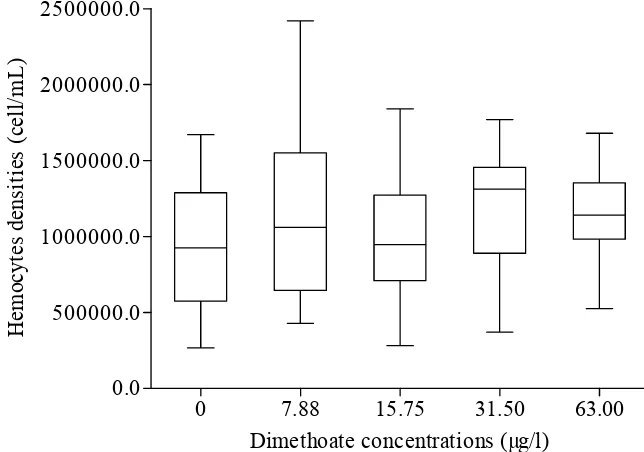
[image:44.612.110.451.383.627.2]Figure 2. Circulating hemocyte density of M. edulis before the treatment. Data were expressed as median (25 % and 75 % quartile, 5 % and 95 % confidence interval).
0 7.88 15.75 31.50 63.00 0 500000 1000000 1500000 2000000 2500000 3000000 * *
Dimethoate concentrations (µg/l)
H em o cy te s d en sit ie s (C el ls /m L )
0 7.88 15.75 31.50 63.00
0 1000 2000 3000 4000 5000 6000 7000 8000 9000 10000 11000 12000 * *
Dimethoate concentrations (µg/l)
[image:45.612.141.440.86.299.2]Ph ag ocyt o si s i n d ex ( R FU /m g p ro te in )
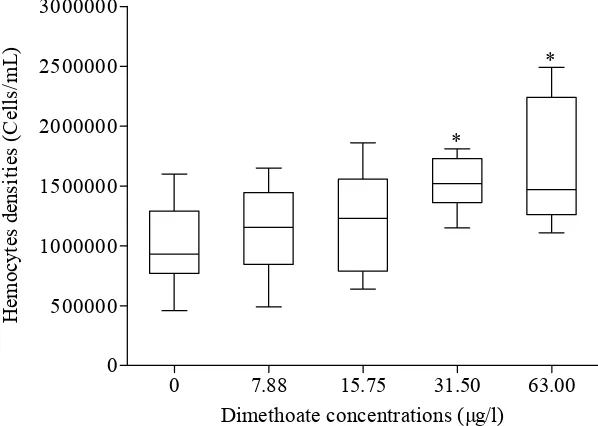
Figure 4. Circulating hemocytes density of M. edulis after 14 days of dimethoate exposure. Data were expressed as median (25 % and 75 % quartile, 5 % and 95 % confidence interval). * indicated the different number of hemocytes from the treatments and from those observed in the control (p < 0.05).
[image:45.612.149.431.403.618.2]0 7.88 15.75 31.50 63.00 0 10 20 30 40 50 60 *
Dimethoate concentrations (µg/l)
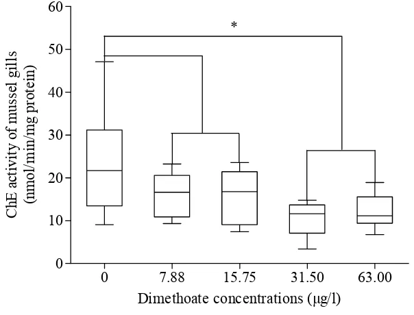
C h E ac ti v it y o f m u ss el gi ll s (n mo l/ min /m g p ro te in )
The dosed dimethoate to the mussels resulted in decreasing of phagocytic activity significantly at the concentrations of 7.88 µg/l and 15.75 µg/l of dimethoate (Figure 5). On the other side, the stimulation of the activity significantly occurred at 31.50 µg/l of dimethoate (p < 0.05) compared to previous levels and persisted significantly at the same level at 63.00 µg/l of dimethoate (Figure 5).
Cholinesterase Activity
[image:46.612.141.443.419.649.2]ChE assay was performed on the mussel gills at the end of the experiment. The results showed that dimethoate caused a significant effect on ChE activity at concentrations of 31.50 and 63.00 µg/l (Figure 6). Although, there were apparent reductions of the ChE activity about 23 % of the control from the mussels that were exposed to dimthoate at both concentrations of 7.88 and 15.75 µg/l, but due to high variability between individuals, these were not significant. On the other hand, significant suppression of the ChE activity (p < 0.05) occurred at concentrations of 31.50 and 63.00 µg/l compare to the control. Moreover, the statistical analysis showed that the different suppression of the ChE activity between the two treatments was not evident (Figure 6)
2.5. Discussion Phagocytic Activity
This study was unable to elucidate clearly dose-dependent phagocytic activity of M. edulis hemocytes following 14 days dimethoate exposure. Nevertheless, the circulating hemocytes density at concentrations just above the control (7.88 and 15.75 µg/l) demonstrated a tendency of elevation, yet the statistical analysis justified an undifferentiated numbers of hemocytes between them. This indicated that dimethoate at low level did not clearly alter the circulating hemocytes density. However, hemocytes density was significantly stimulated at 31.50 µg/l of dimethoate, but the following dimethoate treatment (63.00 µg/l) did not cause an elevation of circulating hemocytes density compared to previous treatment (31.50 µg/l). The alteration of hemocytes density as response to stressors such as chemical compounds is still debatable (Sokolova et al. 2004) even a tendency of stimulation under stress condition was a common response (Pipe et al. 1999). Some researchers have reported that hemocytes density of bivalve elevated as results of environmental stressors exposure (Coles
et al. 1994a; Coles et at. 1994b; Pipe et al. 1999; Dizer et al. 2001a; St-Jean et al. 2002), whereas others have shown that the stressors declined the hemocytes density (Dizer et al. 2001b; Suresh and Mohandas 1990; Auffret et al. 2002). Undefined response of hemocytes density to environmental stressors may implied that the number of circulating hemocytes do not fundamentally reflect the total size of the hemocytes population in mussels body which may alter over short time as result of dynamic association/dissociation between hemocytes and bivalve tissues (Ford et al. 1993). This current study was in accordance with the common te