Distance between parapatellar portal and intra-articular space for
needle positioning in knee osteoarthritis
John Butarbutar,1 Yeremia Tatang,1 Hori Hariyanto,2 Edu Tehupeory,3 Ludwig A. Pontoh4
1 Department of Surgery, Siloam Hospital, Pelita Harapan University, Jakarta, Indonesia 2 Department of Anesthesiology, Siloam Hospital, Pelita Harapan University, Jakarta, Indonesia 3 Department of Rheumatology, Faculty of Medicine, Hasanuddin University, Makassar, Indonesia
4 Department of Orthopaedy and Traumatology, Universitas Indonesia, Fatmawati Hospital, Jakarta, Indonesia
Abstrak
Latar belakang: Injeksi intra-artikular merupakan prosedur terapi osteoartritis (OA) yang memerlukan tingkat akurasi tinggi. Penelitian ini bertujuan menentukan rerata jarak antara kulit dan rongga intra-artikular yang dapat digunakan sebagai panduan untuk memilih panjang jarum ketika melakukan injeksi intra-artikular pada pasien osteoartritis lutut. Metode: Dua puluh satu MRI lutut diambil dari 16 pasien wanita dengan osteoartritis lutut. Panjang jarum yang dibutuhkan untuk mencapai ruang artikular didapatkan melalui rekonstruksi gambaran garis lurus dari kulit ke ruang intra-artikular. Uji t berpasangan dipakai untuk melihat perbedaan mean hasil pengukuran bagian kiri dengan bagian kanan, dengan tingkat kemaknaan nilai p < 0,05.
Hasil: Hasil menunjukkan tempat masuk dari parapatela pada medial dan lateral lebih kranial dari garis mid-patellar. Pada portal medial, jarak terdekat antara kulit dan cairan intra-artikular adalah 27,81 mm ± 7,58 mm. Jarak rerata titik portal adalah 4,46 ± 2,16 mm kranial dari garis mid-patellar, dan 14,20 ± 4,45 mm posterior dari permukaan medial patela. Pada portal lateral, jarak terdekat dari kulit dan cairan intra-artikular adalah 16,84 ± 6,79 mm. Jarak rata-rata titik injeksi adalah 11,10 ± 5,95 mm kranial dari garis mid-patellar, dan 8,91 ± 3,83 mm posterior dari permukaan lateral patela.
Kesimpulan: Studi MRI lutut menunjukkan bahwa rerata jarak antara kulit dan rongga intra-artikular lutut pada portal medial 27,81 ± 7,58 mm sedangkan pada portal lateral 16,84 ± 6,79 mm. Portal tersebut terletak kranial dari garis tengah patela dimana portal lateral lebih kranial dibandingkan medial, sehingga panjang jarum dan lokasi injeksi harus disesuaikan untuk meningkatkan akurasi injeksi intra-artikular. (Med J Indones. 2013;22:83-7)
Abstract
Background: Intra-articular injection is a common therapeutic procedure in osteoarthritis (OA) that need high accuracy. This study was aimed to measure the distance between parapatellar skin and intra-articular space as a guidance to choose the length of needle needed to perform intra-articular injection.
Methods: Twenty one knees MRI were taken from 16 females with knee osteoarthritis. The length of the needle needed
to reach intra-articular space was reconstructed by drawing straight line from skin to intra-articular luid. Paired t-test was using to analyze the mean difference of measurement of left side compare with right side with signiicant indicator
if p-value < 0.05.
Results: The entry point on both medial and lateral parapatellar were more cranial than transverse mid-patellar line. On medial portal, the closest distance from skin to intra-articular space is 27.81 ± 7.58 mm. Mean point of entry is 4.46 ± 2.16 mm cranial to mid-patellar line, and 14.20 ± 4.45 mm posterior to the prominence of medial border of patella. On lateral portal, the closest distance from skin to intra-articular space is 16.84 ± 6.79 mm. Mean point of entry is 11.10 ± 5.94 mm cranial to mid-patellar line, and 8.91 ± 3.83 mm posterior to the prominence of lateral border of patella.
Conclusion: MRI knee osteoarthritis study showed that the mean distance between skin and intra-articular joint space of medial portal is 27.81 ± 7.58 mm, and lateral portal is 16.84 ± 6.79 mm. The portals on both sides is cranial to midpoint of patella, lateral appears more proximal than medial. This should be put into consideration in choosing needle length and portal projection to increase intra-articular injection accuracy. (Med J Indones. 2013;22:83-7)
Keywords: Needle length, osteoarthritis, parapatellar skin portal
Correspondence email to: [email protected]
Intra-articular injection in osteoarthritis of the knee is common procedure nowadays. It can be done in outpatient setting, relatively simple procedure with
the beneit of delivering drugs directly inside the joint.
Many studies on pain control and disease modifying on the knees are using intra-articular injection as the method and variable portal of injection has been proposed, including superolateral, superomedial,
anteromedial, anterolateral, lateral mid-patella, and medial mid-patella portal.
Some studies note the superiority of anterolateral (71-85%) and lateral mid-patella (76-93%) in successfully enter the joint space.1,2 Anteromedial (73-75%) and
medial mid-patellar (56%) show lower accuracy rate.1,3
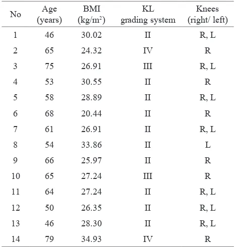
No Age
Individual data of age, BMI and KL score of study subjects
Patients distribution according to KL grade classiication
KL: Kelgren Lawrence; BMI: body mass index KL: Kelgren Lawrence; BMI: body mass index
Mean ± SD n (%)
Age (years) 60.71 ± 10.10
KL grade II 16 (76.2)
those studies of intra-articular drugs. Intra-synovial and fat pad injections are also reported to be related with pain, bleeding into the joint, bruising, and nerve injury.4-6
To overcome the problem, some studies reports increase accuracy by using additional device and technique such as
contrast with luoroscopy and mini air technique which are
either too complicated or expensive for daily practice.7-9
Injection on osteoarthritic knee could be different compare to normal knee due to changes in tibiofemoral and patella alignment, skin and soft tissue thickness in elderly and obese patients, and changes in synovium and fat pad patterns.10 This study is aimed to measure the distance
between parapatellar skin and intra-articular space using MRI inpatients with knee osteoarthritis.
METHODS
Subjects
Sixteen females with knee osteoarthritis according to American College of Rheumatology were recruited from our ongoing study on Relationship Between Tibial
Torsion and Degradation Process of Osteoarthritic Knee
Joint in Menopausal Women. The research protocol has been approved by Ethical Committee of Universitas Hasanuddin. MRI scan with their knees in extension
was done to 21 knees. Subjects with inlammatory joint
disease were excluded.
MRI examination
This study was performed with 3 tesla MRI (Philips)
using torso coil. A specially designed foot holder is used to immobilize the knee in upright position. Axial fat suppressed spin echo (FSE) proton density with a slice thickness 3 mm were obtained.
First, the skin entry point was determined, which
represent the closest distance to intra-articular luid.
The length of the needle needed to reach intra-articular space was reconstructed by drawing straight line from skin to intra-articular space. We recorded minimum (min) and maximum distance (max) from skin to
intra-articular luid, the distance of the entry point below
patellar bone prominence (post) and the entry point distance from mid-patellar line (sup). All distances were measured in millimeters (mm).
Data analysis
Data were analyzed by using paired t-test to look the mean difference of left side measurement compared with right side. The distance from both lateral and medial portal entry was measured and comparison between left
and right, and correlation with Kellgren Lawrence (KL) grading and Body Mass Index (BMI) were analyzed. Kellgren Lawrence grading system was used to classify the narrowing of joint space: grade I –doubtful narrowing of joint space and possible osteophyticlipping; grade II
–deinite-osteophytes, deinite narrowing of joint space; grade III –moderate-multiple osteophytes, deinite
narrowing of joints space, some sclerosis and possible deformity of bone contour; grade IV –large osteophytes, marked narrowing of joint space, severe sclerosis and
deinite deformity of bone contour.
RESULTS
On medial portal, the closest distance from skin to intra-articular space was 27.81 ± 7.58 mm. The longest distance, from skin to intra-articular space before the line hit medial patellar facet was 45.15 ± 9.81 mm. Mean point of entry was 4.46 ± 2.16 mm cranial to mid-patellar line, and 14.20 ± 4.45 mm posterior to
the prominence of medial border of patella. On lateral portal, the closest distance from skin to intra-articular space was 16.84 ± 6.79 mm. The longest distance from skin to intra-articular space was 30.46 ± 10.71 mm. Mean point of entry was 11.10 ± 5.94 mm cranial to mid-patellar line, and 8.91 ± 3.83 mm posterior to the prominence of lateral border of patella.
Data analysis shows that there were signiicant difference
of portal measurement between right and left knees only in distance between point of entry and patellar prominence (parapatellar medial, p = 0.011; parapatellar
lateral, p = 0.031).There was no signiicant difference in
cranial and intra-articular depth between both sides.
Statistic shows that there were no correlation between BMI with medial parapatellar intra-articular space distances ([min], p = 0.297; [max], p = 0.381), cranial distance between entry point and transverse mid-patella (p = 0.176), and distance between entry pint and patellar prominence (p = 0.553). There were also no correlation between BMI and lateral parapatellar intra-articular space distances ([min], p < 0.381; [max], p < 0.381), cranial distance between entry point and transverse mid-patella (p < 0.466), and distance between entry point and patellar prominence post (p < 0.393).
n Minimum Maximum Mean SD
MP (min) Table 3. Measurement results of parapatellar distances from the
skin to intra-articular luid
MP: medial parapatellar; LP: lateral parapatellar; (min): minimum distance from parapatellar skin to intra-articular luid; (max): maximum distance from parapatellar skin to intra-articular luid;
(sup): distance between entry point and transverse mid-patellar line; (post): distance between entry point to patellar bony promi-nence. All distances were measured in millimeters (mm)
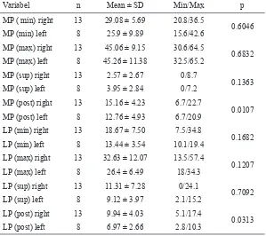
Table 4.Paired t-test comparisons between left and right knee parapatellar distances
MP: medial parapatellar; LP: lateral parapatellar; (min): minimum distance from parapatellar skin to intra-articular luid; (max): maximum distance from parapatel
-lar skin to intra-articu-lar luid; (sup): distance between entry point and transverse
mid-patellar line; (post): distance between entry point to patellar bony prominence. All distances were measured in millimeters (mm)
Variabel n Mean ± SD Min/Max p
MP ( min) right 13 29.08 ± 5.69 20.8/36.5
0.6046
MP (min) left 8 25.9 ± 9.89 15.6/42.6
MP (max) right 13 45.06 ± 9.15 30.6/64.5
0.6832
MP (max) left 8 45.26 ± 11.38 32.5/65.2
MP (sup) right 13 2.57 ± 2.67 0/8.7
0.1363
MP (sup) left 8 3.95 ± 2.84 0/7.2
MP (post) right 13 15.16 ± 4.23 6.7/22.7
0.0107
MP (post) left 8 12.76 ± 4.93 6.7/20.9
LP (min) right 13 18.67 ± 7.50 7.5/34.8
0.1682
LP (min) left 8 13.44 ± 3.54 10.1/19.4
LP (max) right 13 32.63 ± 12.07 13.5/57.4
0.1207
LP (max) left 8 26.4 ± 6.49 18/34.3
LP (sup) right 13 11.31 ± 7.28 0/24.1
0.7092
LP (sup) left 8 9.12 ± 3.97 2.1/15.2
LP (post) right 13 9.94 ± 4.03 5.1/17.4
0.0313
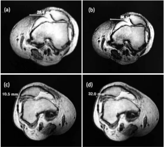
Figure 1. MRI imaging on medial and lateral portals: Minimal (a) and maxi-mal (b) distance between parapatelar lateral skin portal and joint space; minimal (c) and maximal (d) distance between parapatellar medial skin portal and joint space
There were no correlation found between KL grade and distances entry point and medial intra-articular space ([min], p = 0.368; [max], p < 0.368), cranial distance between entry point and transverse mid-patella (p < 0.240), distance between entry point and patellar prominence (p < 0.267). There were also no correlation found between KL grade and distances entry point and lateral intra-articular space ([min], p = 0.267; [max], p < 0.368), cranial distance between entry point and transverse mid-patella (p = 0.176), distance between entry point and patellar prominence (p < 0.658).
DISCUSSION
Knee joint was thought to be the easiest joint to inject
since it is located supericially and has relatively
large intra-articular space. However several studies showed different and sometimes low accuracy of knee intra-articular injection that also depend on different injection portal used by investigators.1,3,6
Most of knee intra-articular injection nowadays were
done on osteoarthritic condition with no inlammation, where minimal intra-articular luid combine with
bony deformity and malalignment could possibly lower the accuracy of putting the needle into intra-articular space. That’s why it is important to improve our understanding on knee injection portal in osteoarthritic knee.
Good portal would be one which could provide short distance between skin and intra-articular, big joint space, far from sensitive structures, less dependent on
joint luid luctuation and knee deformity, and easily deine using surrounding landmark.
MRI was used in this study since it is capable to
depict intra-articular luid which represent dynamic
intra-articular potential space and also able to do measurement of its distance to surrounding soft tissue structures.
Bony prominence is used in this study as landmark of measurement, because in our opinion, it is more readily
deine in clinical setting compare to upper and lower
pole of patella that are located inside quadriceps tendon and patellar ligament.
According to our result, point of entry on both medial and lateral parapatellar should be more cranial than
midpoint of patella, because of lack joint luid on the
level of transverse line of mid-patella and frequent potential obstruction from patellofemoral osteophytes. Accordingly, the term lateral and medial mid-patellar portal for knee injection are not appropriate and could be misleading.
The direction of the lateral facet patellar cartilage surface is almost horizontal and the direction medial facet patella is about 45 degrees tilt. Caution should be taken in directing angle of needle during insertion to minimize risk of hitting joint cartilage.
Clinician tends to use needle with smallest diameter possible for patients comfort. Based on our results, 1.5 inch needle is appropriate for medial portal and 1 inch needle for lateral portal. However, the closest distance from skin to patellar articular surface is 35.34 mm (45.15 mm - SD 9.81) in medial portal and 19.7 mm (30.46 mm - SD 10.71) in lateral portal. Therefore, the clinician should insert the needle deep enough only to penetrate joint capsule. Inserting the whole length of the needle has possible risk of damaging cartilage.
In this study, we found signiicant difference between
right and left knee on distance between entry point and patellar prominence border. It could be results of different patellar tilt between both sides. Identifying parapatellar soft spot posterior to patella will help to identify this
variation. No signiicant inluence of BMI and KL grade
to parapatellar portal point of entry showed independent
character of this portal and was a beneit for clinician.
Several consideration should be taken due to study’s weakness. Small sample population with only one gender (female) may not represent general knee OA
population. Patellar manipulation during injection
procedure to open the joint space and stretch the capsule is also cannot be reconstructed in this study because it is so variable depending on physician preference, although we believe that our result could still be applicable as a baseline for physician to predict the consequences of his manipulation to patella in directing the needle.
In conclusion, despite a fact as the most common intra-articular procedure, knee injection studies still result in
variable accuracy with lack description of portal site and needle direction. Our MRI study shows that joint space
luid on both sides was cranial to midpoint of patella,
lateral side appears more cranial than medial side, and needle projection should be directed accordingly to increase intra-articular injection accuracy.
REFERENCES
1. Jackson DW, Evans NA, Thomas BM. Accuracy of needle placement into the intra-articular space of the knee. J Bone Joint Surg Am. 2002; 84-A(9):1522-7.
2. Lavelle M, Lavelle ED, Lavelle L. Intra-articular injections. Anesthesiol Clin. 2007;25(4):853-62.
3. Esenyel C, Demirhan M, Esenyel M, et al. Comparison of four different intra-articular injection sites in the knee: a cadaver study. Knee Surg Sports Traumatol Arthrosc. 2007;15(5):573-7.
4. Lussier A, Cividino AA, McFarlane CA, Olszynski WP, Potashner WJ, De Medicis R. Viscosupplementation
with hylan for the treatment of osteoarthritis: findings from clinical practice in Canada. J Rheumatol. 1996;23(9):1579-85.
5. Sharma L, Song J, Felson DT, Cahue S, Shamiyeh E, Dunlop DD. The role of knee alignment in disease progression and functional decline in knee osteoarthritis. JAMA. 2001;286(2):188-95.
6. Jones A, Regan M, Ledingham J, Pattrick M, Manhire A,
Doherty M. Importance of placement intra-articular steroid injections. BMJ. 1993;307:1329-30.
7. Bliddal H. Placement of intra-articular injections veriied by mini-air arthrography. Ann Rheum Dis.
1999;58(10):641-3.
8. Waddell D, Estey D, Bricker DC, Marsala A.
Viscosupplementation under luoroscopic control. Am J
Med Sports. 2001;3:237-41.
9. Masala S, Fiori R, Bartolucci DA, et al. Diagnostic and therapeutic joint injections. Semin Intervent Radiol. 2010; 27(2):160-71.
10. Gaffney K, Ledingham J, Perry J D. Intra-articular
triamcinolone hexacetonide in knee osteoarthritis:
factors inluencing the clinical response. Anneheum Dis.