Inluence of 17β-estradiol treatment on the expression of NF-κB in
complete hydatidiform mole culture
Tatit Nurseta, Yahya Irwanto, Rahmi Utami
Department of Obstetric and Gynecology, Faculty of Medicine, Universitas Brawijaya, Malang, Indonesia
Abstrak
Latar belakang: Nuclear factor kappa B (NF-κB) berperan dalam proses onkogenesis. Aktiitas NF-κB pada kultur sel trofoblas mola hidatidosa komplit (MHK) dengan pemberian 17β-estradiol (E2) belum diketahui dengan jelas. Baru-baru ini, ditemukan adanya cross-talk antara reseptor estrogen (ER) dan NF-κB untuk mempromosikan kelangsungan hidup sel kanker. Pada penelitian ini, kami meneliti pengaruh pemberian estradiol (E2) terhadap ekspresi NF-κB pada kultur sel trofoblas MHK.
Metode: Penelitian eksperimental ini menilai ekspresi NF-κB pada kultur sel trofoblas MHK dengan pemberian E2 10,100, 300, 600, dan 1000 pg/mL dan tanpa E2. Ekspresi dinilai menggunakan pengecatan imunohistokimia, kemudian, sel trofoblas MHK difoto menggunakan mikroskop dengan pembesaran 400x. Selanjutnya, program Adobe Photoshop CS2 digunakan menilai kepekatan NF-κB pada inti sel. Semakin rendah nilai kepekatan sel dalam satuan RGBbv, maka semakin tinggi ekspresi NF-κB pada sel tersebut. Uji ANOVA digunakan untuk membandingkan ekspresi NF-kB. Hasil: Kepekatan sel yang menunjukkan kadar NF-κB pada kelompok kontrol didapatkan 114,839 ± 9,022. Pada pemberian E2 didapatkan berturut-turut: E2 10 pg/mL: 106,30 ± 13,95; E2 100 pg/mL: 82,47 ± 4,72; E2 300 pg/mL: 82,24 ± 2,67; E2 600 pg/mL: 69,05 ± 6,47; E2 1000 pg/mL: 68,49 ± 2,37. Terdapat penurunan progresif nilai kepekatan sel yang mengekspresi NF-κB pada kelompok yang mendapat E2 yang menunjukkan terjadinya peningkatan ekspresi NF-κB. Perbedaan bermakna dengan kelompok kontrol terjadi pada dosis E2 100, 300, 600, dan 1000 pg/mL.
Kesimpulan: Pemberian 17β-estradiol dosis bertingkat berpengaruh meningkatkan ekspresi NF-kB pada kultur sel trofoblas MHK dibanding kontrol. (Med J Indones. 2013;22:197-201. doi: 10.13181/mji.v22i4.599)
Abstract
Background: Genetic evidence has established a role of nuclear factor kappa B (NF-κB) signaling in oncogenesis. However, activity of NF-κB in complete hydatidiform mole (CHM) cell culture under 17β-estradiol (E2) treatment is not yet known. Recently, a positive cross-talk between estrogen receptor (ER) and NF-κB to promote survival and progress of cancer cells to a more aggressive phenotype was established. In the present study, we examined the inluence of E2 treatment on the NF-κB expression in CHM’s culture.
Methods: This experimental study measured the expression of NF-κB in CHM culture treated with E2: 10, 100, 300, 600, and 1000 pg/mL and without E2. Imunohistochemistry staining was used to assess the expression of NF-κB. Microphotographs were taken using 400x magniication. Adobe photoshop CS2 was used to assess the NF-κB expression in cell nucleus. The lower the color intensity of cell RGBbv, is the higher the expression of NF-κB in cells. ANOVA test was performed to compare the expression of NF-κB.
Results: NF-κB expression as indicated by color intensity in control group was 114.84 ± 9.02. NF-κB expression in E2 treatment groups were respectively: E2 10 pg/mL: 106.30 ± 13.95; E2 100 pg/mL: 82.47 ± 4.72; E2 300 pg/mL: 82.24 ± 2.67; E2 600 pg/mL: 69.05 ± 6.47; E2 1000 pg/mL: 68.49 ± 2.37. There was progressive decline in color intensity of cells with E2 treatment indicating the increase expression of NF-κB. Signiicant differences with the control group occurred in doses of E2 100, 300, 600, dan 1000 pg/mL.
Conclusion: Treatment of CHM trophoblast culture with escalating doses of E2 was associated with the increase of NF-κB expression in a dose dependent manner. (Med J Indones. 2013;22:197-201. doi: 10.13181/mji.v22i4.599)
Keywords: 17-β Estradiol, Hydatidiform mole, NF-κB
receptor signal transduction pathway that leads to increase in proliferation and inhibition of apoptosis of endometrial stromal cells.3
Nuclear factor kappa B (NF-κB) is known to have a function in the regulation of immune and inlammatory responses, but recently growing scientiic evidence also supports its role in oncogenesis. On tumor cells, a variety of molecular changes can cause the activation of NF-κB. This activation leads to deregulation of gene expression under the control of NF-κB. Changes The process of carcinogenesis from hydatidiform mole
into gestational trophoblastic tumor is still unclear and the limited available information can not clearly deine the process. Several factors are known to play a role in the process of carcinogenesis including DNA ploidy, phospholipid expression, oncogenes, tumor suppressor genes, nutrition and hormonal status.1,2 Normal and
in certain gene expression may participate in cancer growth and progression. Therefore, there might be a relationship between NF-κB and carcinogenesis.4
NF-κB is a transcription factor that is known to play an important role in controlling immune and inlammatory responses. There are presently ive members of the mammalian NF-κB/Rel family: NF-κB/p50, NF-κB/p52, c-Rel, RelA/p65 and RelB. The p50 and 65 subunits, is normally retained in the cytoplasm through interactions with inhibitor proteins IκBα and IκBβ. Inductive stimuli (TNFα, IL-1, bacterial endotoxin, etc.) lead to the phosphorylation and degradation of IκB, allowing NF-κB to enter the nucleus and regulate gene expression.5,6
Recently, estrogen receptor (ER) was found to act together with NF-κB in a positive manner to synergistically enhance certain gene transcription. There is a positive cross-talk between ER and NF-κB to promote the survival of cancer cells and progress to a more aggressive phenotype.7 However, another inding showed that 17β-estradiol (E2) could inhibit NF-κB activity and therefore enhanced the process of apoptosis. It was reported that in tamoxifen-resistant MCF-7 tumors, E2 caused down regulation of p65, a subunit of NF-κB, which was correlated with anti-proliferative and pro-apoptotic events. From these studies, it can be assumed that E2 might induce apoptosis and cause regression of tamoxifen-resistant MCF-7 tumors, and in addition, act through suppression of NF-κB as a factor of pro-survival/ anti-apoptotic.8 In another research, a novel positive interaction was shown between the ER and NF-κB dependent signalling pathways in an endometrial epithelial cell line derived from healthy endometrium. NF-κB activation modulates ER activity by an ER and NF-κB dependent mechanism and can enhance ER-mediated endogenous gene expression in an in vitro human endometrial epithelial cell model.9 In women, physiological levels of E2 during normal menstrual cycle is 10 pg/mL and may increase to 35,000 pg/ mL during pregnancy.10 This study was conducted
in order to get scientiic information on the effect of E2 on the expression of NF-κB in cultured CHM trophoblast cells. In this study we used different doses of E2, to determine at which dose it increased the expression of NF-κB in the culture of complete hydatidiform mole (CHM) trophoblast cells. The starting dose was 10 pg/mL, which is the normal level during menstruation.
METHODS
This was an experimental study conducted between March and April 2012. Ethical clearance was
obtained from Health Research Ethics Committee Saiful Anwar General Hospital no. 132/KEPK-FKUB/05/2012. Inclusion criteria were samples obtained from patient with complete mole hydatidiform, with a buble of mole in the sample. Exclusion criteria were negative β hCG test, and failure to obtain trophoblast cell growth, after culturing the samples.
Sample (1 cm3 CHM tissue) was taken by suction
curettage from patients after they signed the informed consent form. The trophoblast tissue with buble of mole was washed by normal saline (NaCl 0.9%), then put in cord solution (medium for trophoblast tissue sampling), which contains Hank’s balance salt solution (HBSS [Sigma H 1641]), gentamycine (Gentamerck), sodium hydrogen bicarbonate (SHB [Sigma], phenol red (Sigma P5530), HEPES solution (Sigma), and deionized water (WFI Otsuka). Processing of tissue for culture was performed not more than 12 hours after curettage.
Isolation and culture of CHM trophoblast cells
The bubbles of mole were washed by NaCl until free from erythrocytes, and were further washed 2 times with Dulbecco’s phosphate buffered saline (DPBS) added with a inal concentration of 100 U/mL penicillin and 100 μg/mL streptomycin. After the addition of antibiotics, this solution [DPBS] should be kept at 2-8°C and used within one week. Finally, the bubbles were washed with serum free media.
To isolate the cells, the bubbles were incubated in a solution of Collagenase (type 1) 7 mg/10 cc for 2 hours, and centrifuged 2,000 rpm for 7 minutes. The supernatant was removed, and the pellet was resuspended in serum free media. The centrifugation was repeated and the resulting pellet was resuspended in 4 mL culture medium, which contain newborn calf serum (NCS, [Sigma N4637]) and 10% FBS, then incubated in 5% CO2 incubator, in 37°C. Further, the culture was observed daily under a microscope to detect cell growth. Identiication of trophoblast cells were done by hCG β examination.
NF-κB expression study
We divided the CHM trophoblast cultures into 6 groups i.e. those which were treated with various doses of E2 (beta estradiol powder [Sigma]) i.e. 10, 100, 300, 600, 1000 pg/mL and a control group (without E2).
IHC photographs were analysed by counting and measuring red-green-blue brightness value (RGBbv) hystogram of 200 cell nuclei using adobe photoshop CS2 to collect mean data of color thickness. Cell with abundant expression of NF-κB, showed thickened color in the nuclei. The thicker the color was, the smaller was the value in the adobe photoshop CS2 hystogram.
Data analysis
ANOVA test was performed to compare NF-κB expression in CHM trophoblast cells that were treated with variable doses of E2 and control.
RESULTS
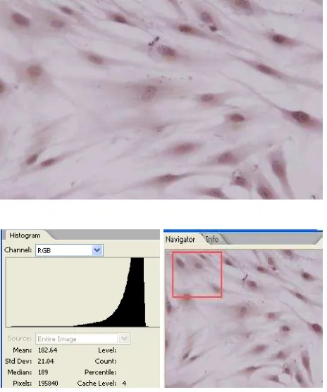
Expression of NF-κB in CHM trophoblast cells without E2 treatment (control) can be seen in igure 1. Expression of NF-κB in CHM trophoblast cells with 1000 pg/mL of E2 treatment can be seen in igure 2. Expression of NF-κB in CHM trophoblast cells with various doses E2 treatment can be seen in table 1.
Table 1 shows mean data with standard deviation of cell color thickness, which expressed NF-κB in RGBbv.
The table shows that treatment of E2 have different inluence on NF-κB expression in CHM’s cells compared with control. Cell with abundant expression of NF-κB, showed thickened color in the nuclei. The thicker the color was, the smaller was the value in the adobe photoshop CS2 hystogram. ANOVA test showed signiicant differences (p < 0.007) in the expression of NF-κB in CHM trophoblast cells with various doses of E2 treatment and control.
DISCUSSION
Estrogen is a steroid hormone family that is synthesized in the ovarian and extra ovarian tissues. Figure 1.
Figure 2.
Table 1. Mean data of cell color thickness, which expressed NF-κB in RGBbv according to varying doses of E2 and control
Expression of NF-κB in CHM trophoblast cells without E2 treatment (control) with Adobe Photo -shop CS2. Nucleus was marked with a circle and analyzed with RGB histogram. Navigator was used to avoid double counting
Expression of NF-κB in CHM trophoblast cells with 1000 pg of E2 treatment
Treatment Expression NF-κB
Means ± SD (RGBbv)
Control 114.84 ± 9.02
E2 10 pg 106.30 ± 13.95
E2 100 pg 82.47 ± 4.72
E2 300 pg 82.24 ± 2.67
E2 600 pg 69.05 ± 6.47
17β-estradiol is the primary estrogen from the ovaries. Natural estrogens are steroids hormones, while some synthetic ones are non-steroidal. Natural estrogen consists of 17β-estradiol (E2), estrone (E1) and estriol (E3). 17β-estradiol is the most potent type of estrogen, followed by E1 and E3. Estrogen receptor is an important factor in the mechanism of action of estrogen and can be found in some tissues, such as central nervous system, cardiovascular system, immune system, urogenital tract, gastrointestinal tract, kidney, lung, mammary gland, uterus and bone. Both estrogen receptors (α and β) play a different role on tissues. ER α plays an important role in mammary gland and uterus, whereas in other tissues ER β is more important.3
Signaling pathways mediated by ER is thought to cause the growth of normal tissues, preneoplasm and neoplasms. There are 3 lines of estrogen transduction signaling in carcinogenesis, including genomic, non-genomic and mitochondrial pathways. Estrogen inluence on NF-κB activity is most likely through mitochondrial pathways.11 In this case, the bound estrogen receptor in the plasma membrane and/or mitochondrial respiratory complexes will form ROS that results in kinase activation and activation of the IKKβ subunit that is responsible for the phosphorylation of serine residue in IκB. After phosphorylation, IκB protein ubiquitin-dependent degradation by the proteasome happens, and NF-κB is translocated to the nucleus, so that NF-κB becomes separated from the cytoplasm, and becomes active as a transcription factor, causes proliferation, anti-apoptosis, and eventually causing a cell to become malignant.12 Those indings corroborate the results
of this study, which showed signiicant differences (p < 0.007) between the expressions of NF-κB in CHM trophoblast cell cultures, which were given E2 with varying doses compared to control.
Our results are in line with a study on breast cancer cells with and without the anti-estrogen tamoxifen to test the extent of cross-talk between ER and NF-κB, where a microarray study was done on MCF-7 breast cancer cells that were administered with E2, and tumor necrosis factor alpha (TNFα). The study found positive cross-talks with three patterns, namely: (a) TNFα increased the activity of about 30% of E2 regulated genes, (b) E2 increased the activity of about 15% of the TNFα regulated genes (c) E2 + TNFα causes additional upregulation of about 60 genes. Moreover, these results showed the prosurvival role of ER, and NF-κB that was involved in protecting breast cancer cells to apoptosis. On the whole, there is positive cross-talk between ER and NF-κB, which act together to
promote the survival of cancer cells and to progress to a more aggressive phenotype.7 Further researches are needed to determine whether the positive cross talk between E2 and NF-κB in cultured CHM trophoblast cells occurs through receptor or non-receptor pathway by giving tamoxifen as estrogen receptor competitors. In addition, oral contraception can be provided to women after curretage of mola hydatidiform, because in preparations there are progesterone as anti estrogen.
The limitations of this study is that the measurement of the expression of NF-κB was done on microphotograph from ordinary microscope using Adobe Photoshop CS2, which was not able to assess whether the expression of NF-κB is located entirely in the nucleus or in the cytoplasm, as assessment using confocal microscopy that can obtain more accurate results.
In conclusion, there were signiicant differences in the expression of NF-κB in CHM trophoblast cell culture. Treatment of CHM trophoblast cell culture with escalating doses of E2 was associated with increased expression of NF-κB in a dose-dependent manner.
REFERENCES
1. Martaadisoebrata D. Pola Epidemiologi Penyakit Trofoblas dalam Kaitannya dengan Prognosis dan Cara Penanggulangannya. Bandung: RSHS/FKUP - Divisi Obstetri Ginekologi; 2002. Indonesian.
2. Sastradinata I.Problematik Penyakit Trofoblas di Indonesia. Palembang: Universitas Sriwijaya - Divisi Obstetri dan Ginekologi; 2009. Indonesian
3. Yager JD, Davidson NE. Estrogen carcinogenesis in breast cancer. N Engl J Med. 2006;354(3):270-82.
4. Dolcet X, Llobet D, Pallares J, Mathias-Guiu X. NF-κB in development and progression of human Cancer. Virchows Arch. 2005;446(5):475-82.
5. Cogswel PC, Guttridge DC, Funkhouser W, Baldwin Jr A. Selective activation of NF-kappa B subunits in human breast cancer: potential roles for NF-kappa B2/p52 and for Bcl-3. Oncogene. 2000;19(9):1123-31.
6. Feldman I, Feldman GM, Mobarak C, Dunkelberg JC, Leslie KK. Identiication of proteins within the nuclear factor-kappa B transcriptional complex including estrogen receptor-alpha. Am J Obstet Gynecol. 2007;196(4);394:e1-13.
7. Frasor J, Weaver A, Pradhan M, Dai Y, Miller LD, Lin CY, et al. Positive cross-talk between estrogen receptor and NF-Kappa B in breast cancer. Cancer Res. 2009;69(23):8918-25.
9. King AE, Collins F, Klonisch T, Sallenave JM, Critchley HO, Saunders PT. An additive interaction between the NFkappaB and estrogen receptor signalling pathways in human endometrial epithelial cells. Hum Reprod. 2010;25(2):510-8. 10. Nalbandian G, Kovats S. Estrogen, Immunity &
Autoimmune Disease. Curr Med Chem–Immun, Endoc & Metab Agents Journal. 2005;5(1):85-91.
11. Kumar S, Lata K, Mukhopadhyay S, Mukherjee TK. Role of estrogen receptors in pro-oxidative and anti-oxidative actions of estrogens: A perspective. Biochim Biophys Acta. 2010;1800(10):1127-35.