Cytotoxic effect of γ-sitosterol from Kejibeling (
Strobilanthes
crispus
) and its mechanism of action towards
c-myc
gene
expression and apoptotic pathway
Abstrak
Latar belakang: Penelitian ini bertujuan mengetahui efek
sitotoksisitas γ-sitosterol yang diisolasi dari Kejibeling (Strobilanthes crispus), suatu bahan tanaman obat, pada beberapa kultur sel kanker. Mekanisme dari efek yang dilihat kemudian diteliti melalui ekspresi gen penyebab kanker c-myc dan jalur apoptosis.
Metode: Penelitian in vitro ini menggunakan kultur sel
kanker kolon manusia (Caco-2), sel kanker hepar manusia (HepG2), sel kanker payudara tergantung hormon (MCF-7) dan kultur sel normal hepar manusia (Chang Liver). Efek sitotoksik diukur melalui uji MTT melalui penentuan kadar yang membunuh 50% sel (IC50). Mekanisme sitotoksik dipelajari dengan menentukan pengaruh penambahan γ-sitosterol pada ekspresi gen c-myc menggunakan metode reverse transciptase-polymerase chain reaction (RT-PCR). Pengaruh γ-sitosterol melalui jalur apoptosis dipelajari dengan menggunakan asai terminal deoxynucleotidyl transferase dUTP nick end labeling (TUNEL).
Hasil: Hasil penelitian menunjukkan bahwa γ-sitosterol
bersifat sitotoksik pada sel Caco-2, HepG2 dan MCF-7 dengan nilai IC50 masing-masing adalah 8,3; 21,8; dan 28,8 μg/mL. Tidak didapati nilai IC50 dari senyawa ini terhadap kultur sel Chang Liver. Senyawa ini terbukti menginduksi proses apoptosis pada kultur sel kanker kolon manusia dan sel kanker hepar manusia serta mampu menekan ekspresi gen c-myc di kedua sel.
Kesimpulan: γ-sitosterol bersifat sitotoksik terhadap kultur
sel kanker kolon dan hepar manusia dan efeknya dimediasi oleh penekanan ekspresi gen c-myc. Senyawa ini juga menginduksi proses apoptosis pada kedua sel.
Abstract
Background: This study aimed to analyze the cytotoxicity effect of γ-sitosterol isolated from “Kejibeling” (Strobilanthes crispus), a medicinal plant, on several cancer cell lines. The mechanisms of the effects were studied through the expression of cancer-caused gene, c-myc and apoptotic pathways.
Methods: This in vitro study was done using human
colon cancer cell lines (Caco-2), liver cancer cell lines (HepG2), hormone-dependent breast cancer cell lines (MCF-7) and the normal liver cell lines (Chang Liver). The cytotoxic effect was measured through MTT assay and the potential cytotoxic value was calculated by determining the toxic concentration which may kill up to 50% of the total cell used (IC50). Meanwhile, the cytotoxic mechanism was studied by determining the effect of adding γ-sitosterol to the c-myc gene expression by reverse transciptase-polymerase chain reaction (RT-PCR). The effect of γ-sitosterol through apoptotic pathway was studied by using terminal deoxynucleotidyl transferase dUTP nick end labeling (TUNEL) assay.
Results: γ-sitosterol was cytotoxic against Caco-2, HepG2, and MCF-7 with IC50-values of 8.3, 21.8, and 28.8
μg/mL, respectively. There were no IC50-values obtained from this compound against Chang Liver cell line. This compound induced apotosis on Caco-2 and HepG2 cell lines and suppressed the c-myc genes expression in both cells.
Conclusion: γ-sitosterol was cytotoxic against colon and liver cancer cell lines and the effect was mediated by down-regulation of c-myc expression and induction of the apoptotic pathways.
Keywords: apoptosis, c-myc gene expression, cytotoxic, RT-PCR, Strobilanthes crispus, TUNEL assay
pISSN: 0853-1773 • eISSN: 2252-8083 • http://dx.doi.org/10.13181/mji.v23i4.1085 • Med J Indones. 2014;23:203-8
Correspondence author: Susi Endrini, [email protected]; [email protected]
B a s i c M e d i c a l R e s e a r c h
Copyright @ 2014 Authors. This is an open access article distributed under the terms of the Creative Commons Attribution-NonCommercial-ShareAlike 4.0 International License (http://creativecommons.org/licenses/by-nc-sa/4.0/), which permits unrestricted non-commercial use, distribution, and reproduction in any medium, provided the original author and source are properly cited.
Susi Endrini,1 Asmah Rahmat,2 Patimah Ismail,3 Y.H. Tauiq-Yap4
1 Department of Biochemistry, School of Medicine, YARSI University, Jakarta, Indonesia
2 Department of Nutrition and Health Sciences, Faculty of Medicine, Universiti Putra Malaysia, 43400, Serdang, Selangor D.E, Malaysia
3 Department of Biomedicine, Faculty of Medicine and Health Sciences, Universiti Putra Malaysia, 43400, Serdang, Selangor D.E, Malaysia
Cancer is the second most common cause of death in the US, exceeded only by heart disease, accounting for nearly 1 of every 4 deaths.1 In 2014, about
585,720 Americans are expected to die from cancer or almost 1,600 people per day. In Indonesia, cancer has become the 7th cause of death based on a national
survey in 2007, accounting for 5.7% of all mortality. Data from the population-based cancer registry in Jakarta Province showed the leading cancers among females in 2005-2007 are breast cancer, cervical cancer, ovarian cancer, colorectal cancer and among males are bronchus and lung cancer, colorectal cancer, liver cancer, pharingeal cancer, and prostate cancer.2
Historically, natural products have served as a rich source of lead compounds for drug development against a wide array of biological targets, including various forms of cancer. Indonesia is rich in natural resources, especially medicinal plants. One of them is Kejibeling (Strobilanthes crispus). Strobilanthes
crispus ZII 109 (L) Bremek or Saricocalix crispus
ZII 109 (L) Bremek (Acanthaceae) plant is a native to countries from Madagascar to Indonesia,3 and
was irst quoted by Anderson. Thomas classiied the plant under Spermatophyta (Flowering plants and Gymnosperma).4 A study in Indonesia found that an
infusion of the dried leaves of S. crispus has been used as antidiabetic, diuretic, antilytic and laxative agents. A recent study indicated that water extract of
S.crispus contains compounds with very high binding afinity to protein molecules that bind the active part of reverse transcriptase. It inhibits the proliferation of retrovirus; an agent in viral disease such as acquired immune deiciency syndrome (AIDS) and Adult T-cell Leukemia.5 Previous studies reported that the
chloroform extract of S. crispus has been shown to be cytotoxic against human colon cancer cell lines (Caco-2) and human liver cancer cell lines (HepG2).6
The present work was aimed to analyze the cytotoxicity effect of the g-sitosterol isolated from Kejibeling (S. crispus) on several cancer cell lines. The mechanism of the effects was also studied through the expression of cancer-caused gene c-myc
and apoptotic pathways.
METHODS
Plant material
The leaves of S. crispus was harvested at the Faculty of Medicine and Health Sciences, UPM, Serdang,
Selangor. The herbarium voucher specimen were identiied and deposited by Mr. Ahmed Zainuddin from the Department of Botany, Faculty of Science and Technology, Universiti Kebangsaan Malaysia. The voucher number of S. crispus was AZ-6803.
Preparation of extract
The extraction method was obtained from Ali, et al7 with slight modiication. The dried leaves (5 kg) of
S.crispus was homogenized and soaked in chloroform 100% for a week. The crude extract was then iltered with Whatmann paper No. 4 and evaporated with rotary evaporator. The fractionation and the isolation of active compounds was performed using solvent extraction method with different polarity.
Culturing of cells
HepG-2, Caco-2, MCF-7 and Chang Liver cell lines were obtained from American Type Culture Collection (ATCC, USA). The medium for HepG-2 and Chang liver were Minimum Essential Medium with Earle’s salt (Gibco, USA). While Caco-2 and MCF-7 were grown in Dulbecco’s Modiied Eagle medium (Gibco, USA). The cells were cultured in their own medium supplemented with 10% of fetal calf serum, 100 IU/mL penicillin and 100 mg/mL of streptomycin (Gibco, USA) using 25-cm2 lasks (Nunc, Denmark), in a CO2 incubator (Sanyo, Japan) at 37°C.
MTT assay
The viability of cells was determined with trypan blue. Exponentially growing cells were harvested, counted with hemocytometer, and diluted with medium, yielding a concentration of 1 x 10⁵ cells
Finally, the absorbance was read with ELISA reader Cycle kit (Invitrogen, USA). The polymerase Chain Reactions were performed by 30 cycles ampliication for 1 minute at 94°C, 2 minutes at 55°C and 3 minutes at 72°C. The PCR products were analyzed by electrophoresis on 1.5% agarose gel.
The sequences of primers were as follows :
c-myc sense :
5’-CAAGAGGCGAAGACACAACGTCT-3’
c-myc antisense :
5’-AACTGTTCTCGTCGTTTCCGCAA-3’ Sequencing
The sequencing technique was done on Automatic Sequencer (USA) and the chromatograms were analysed with Chromatos software and blasted to the database in gene bank.
Terminal deoxynucleotidyl transferase dUTP nick end labeling (TUNEL) assay
The TUNEL assay was carried out using Apoptosis Detection System, Fluorescein (Promega, USA). Firstly, the cells were ixed by immersing slides in freshly prepared 4% paraformaldehyde solution in PBS (pH 7.4) in a Coplin jar for 25 minutes at 4°C. The cells were then washed by immersing the slides in fresh PBS for 5 minutes at room temperature. The washing step was repeated and cells were permeabilized by immersing the slides in 0.2% Triton X-100 solution in PBS for 5 minutes.
After this, the slides were rinsed by immersing in fresh PBS for 5 minutes at room temperature. The washing step was repeated once again. The excess liquid was removed by tapping the slides. The cells were then covered with 100 µL of equilibration buffer at room temperature for 5-10 minutes. While the cells were equilibrating, the Nucleotide Mix was thawed on ice and suficient terminal deoxynucleotidyl transferase (TdT) incubation buffer for all experimental reactions were prepared (TdT incubation buffer consists of 45 mL equilibration buffer, 5 mL Nucleotide mix and 1mL TdT enzyme, for each samples). After incubation, the
50 mL TdT incubation buffers were blotted into each cells on a 5 cm2 area. The cells were covered with
Plastic Coverslips to ensure even distribution of the reagent. The slides were then incubated at 37°C for 60 minutes inside the humidiied chamber to allow the tailing reaction to occur. The chamber slides were covered with aluminum foil to protect from direct light. After incubation, the reactions were terminated by immersing the slides in 2X SSC in a Coplin jar for 15 minutes at room temperature. The samples were then washed by immersing the slides in fresh PBS for 5 minutes at room temperature. The washing steps were repeated twice to remove unincorporated luorescein-12-dUTP. The staining processes were done in a Coplin jar by immersing the slides in 40 mL of propidium iodide solution freshly diluted to 1 mg/mL in PBS for 15 minutes at room temperature in the dark. After that, the samples were washed by immersing the slides in deionized water for 5 minutes at room temperature and repeated twice for a total of 3 washings. Finally, drop of anti-fading solution was added cells and slides were mounted with coverslips, sealed with rubber cement or clear nail polish and let dry for 5-10 minutes. Sample were viewed and analysed immediately under a Confocal Laser Scanning Microscope (CLSM).
RESULTS
γ-sitosterol (Figure 1) was obtained from fraction 97-102 as white needle shaped crystal from the crude chloroform extract of S. crispus. After washing with n-hexane and recrystallisation from MeOH, this compound gave a single spot on the TLC plate. The conirmation of structure were done using infra red and mass spectrometry spectrum. The cytotoxic effect of γ-sitosterol has been determined and displayed the strongest cytotoxic effect on colon carcinoma cell lines (Caco-2), liver cancer cell line (HepG2), and hormone-dependent breast cancer cell lines (MCF-7) with IC50 -values of 8.3, 21.8, and 28.8 mg/mL, respectively (Figure 2). There were no IC50-values obtained from this compound against Chang Liver cell line.
O
H
Figure 1. Molecular structure of γ-sitosterol from S. crispus
Figure 2. The effect of γ-sitosterol from S. crispus on different human cell lines. The protocol applied was similar to the previ -ous one. IC50 of 8.3, 21.8, and 28.8 µg/mL were obtained for
Caco-2, HepG2 and MCF-7 cell lines, respectively
1 2 3 M
1031bp
900
800 700
600
500
100 400
200 300
218bp
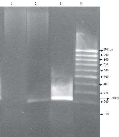
Figure 3. Effect of γ-sitosterol from S. crispus on the expression of c-myc gene in Caco-2 cell line. PCR products were analysed
on a 1.5% agarose gel. M, 100 bp DNA ladder marker; lane 1, Caco-2 treated with 30 μg/mL γ-sitosterol; lane 2, Caco-2 treated with 20 μg/mL γ-sitosterol; lane 3, Caco-2 control (untreated)
Figure 4. Effect of γ-sitosterol from S. crispus on the expression of c-myc gene in HepG2 cell line. PCR products were analysed
on a 1.5% agarose gel. M, 100 bp DNA ladder marker; lane 1, HepG2 treated with 30 μg/mL γ-sitosterol; lane 2, HepG2 treated with 20 μg/mL γ-sitosterol; lane 3, HepG2 control (untreated).
Gene expression was observed in lanes 2 and 3, but not in lane 1
DISCUSSION
The c-myc oncogene contributes to the genesis of many human cancers. Recent insights into its expression and function have led to new cancer therapeutic opportunities.8 Many plant extracts have been reported to inhibit cell proliferation through the down regulation of c-myc expression.9,10 In this study, the c-myc
expression was suppressed by γ-sitosterol which was isolated from S. crispus. To observe the effectiveness of γ-sitosterol in suppressing oncogenes, mRNAs was extracted from the treated cells. Due to instability of RNA and for the PCR purpose, mRNA was converted to cDNA before proceeding to the PCR process. The PCR has been selected as the most suitable technique to amplify the quantity of oncogenes, so that the suppression of oncogenes can be visualized clearly after a gel electrophoresis analysis. Besides, different oncogenes have their own temperature for denaturation, annealing, elongation in a number of cycles to get the best PCR products. Results showed that the effect was
M 1 2 3
1031bp 900
800
100 200 300 400 500 600
700
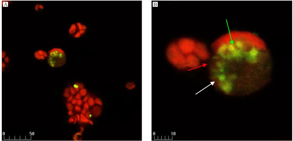
Figure 5. Confocal micrograph of Caco-2 cells treated with 30 μg/mL γ-sitosterol from S. crispus. Condensed nucleus (green arrow), prominent cell shrinkage (red arrow), and apoptotic bodies (white arrow). A: mixture of PI and FITC labelled apoptotic cell as yellow intensely. B: zoom of A for observed a single cell (apoptotic bodies). x400 magniication
dose defendent and it seemed to be correlated with the IC50- value of each treatment.
Recent studies have demonstrated that apoptosis involved in cell death induced by chemotherapeutic agents including cisplatin, cytarabine, camptothecin, amsacrine, etoposide and teniposide.11,12 There is an
accumulating evidence that the eficacy of antitumor agents is related to the intrinsic propensity of the target tumor cells to respond to these agents by apoptosis.13-15 Apoptosis, a physiological mode of cell death, is characterized by reduced cell volume, condensed chromatin in the nucleus, formation of internucleosomal DNA fragmentation and loss of membrane integrity, and generation of apoptotic bodies.16,17
The methods used to assess DNA strand breaks are based on labeling/staining the cellular DNA. The labeled/stained DNA is subsequently analysed by luorescence microscopy or confocal laser scanning microscopy for better results. Extensive DNA degradation is a characteristic event, which often occurs, in the early stages of apoptosis. Cleavage of the DNA may yield double-stranded, low molecular weight DNA fragments (mono-and oligonucleosomes) as well as single strand breaks (nicks) in high molecular weight DNA. Those DNA strand breaks can be detected by enzymatic labeling of the free 3-OH termini with modiied nucleotides. Suitable
labeling enzymes include terminal deoxynucleotidyl transferase (end labeling). TdT is able to label blunt ends of double stranded DNA break independent of a template. The end-labeling method has also been termed TUNEL (TdT-mediated X-dUTP nick end labeling).18 The TUNEL reaction is more speciic for
apoptosis and may be helpful to differentiate cellular apoptosis and necrosis.19 This study demonstrated that
γ-sitosterol from S. crispusmay induced the apoptosis in Caco-2 and HepG2 cell lines. The morphology of chromatin condensation, DNA fragmentation and several apoptotic bodies were found by confocal laser scanning microscope in the treated groups. There were no apoptotic phenomena observed in the untreated group.
In conclusion, our results veriies that γ-sitosterol from S. crispus was cytotoxic against colon and liver cancer cell lines and the effect was mediated by down-regulation of c-myc expression which induced the apoptotic pathways.
Acknowledgment
The authors thank IRPA grant 06-02-04-0050.
Conlict of interest
The authors afirm no conlict of interest in this study.
REFERENCES
1. American Cancer Society. Cancer facts and igures 2014. Atlanta : American Cancer Society; 2014.
2. Wahidin M, Noviani R, Hermawan S, Andriani V, Ardian A, Djarir H. Population-based cancer registration in Indonesia. Asian Paciic J Cancer Prev. 2012;13(4):1709-10.
3. Sunarto PA. Materia Medika Indonesia. Jakarta: Direktorat Jenderal Pengawasan Obat dan Makanan; 1977. Indonesian. 4. Brummit RK, Powell CE. Authors of plant names. Britain:
Royal Botanic Gardens; 1992. p.731.
5. Kusumoto JT, Shimada I, Kakiuchi N, Hattori M, Namba
T. Supriyatna S. Inhibitory effects of Indonesian plant
extracts on reverse transcriptase of an RNA tumour virus (I). Phytotherapy Research. 1992;6(5):241-4.
6. Susi E, Asmah R, Patimah I, Tauiq-Yap YH. Comparing
of the cytotoxicity properties and mechanism of Lawsonia inermis and Strobilanthes crispus extract against several
cancer cell lines. J Med Sci. 2007;7(7):1098-102.
7. Ali AM, Mackeen MM, Intan-Sainar I, Hamid M, Lajis NH, El-Sharkawy SH, et al. Antitumour-promoting and
antitumour activities of the crude extract from the leaves of
Juniperus chinensis. J Ethnopharmacol. 1996;53(3):165-9. 8. Chi V. Dang. Myc on the path to cancer. Cell.
2012;149(1):22-35.
9. Tuntiwechapikul W, Taka T, Songsomboon C, Kaewtunjai N, Imsumran A, Makonkawkeyoon L, et al. Ginger
extract inhibits human telomerase reverse transcriptase
and c-Myc expression in A549 lung cancer cells. J Med Food. 2010;13(6):1347-54.
10. Unger C, Popescu R, Giessrigl B, Rarova L, Herbacek I, Seelinger M, et al. An apolar extract of Critonia morifolia inhibits c-Myc, cyclin D1, Cdc25A, Cdc25B, Cdc25C and Akt and induces apoptosis. Int J Oncol. 2012;40(6):2131-9.
11. Kolfschoten GM, Hulscher TM, Schrier SM, van Houten VM, Pinedo HM, Boven E. Time-dependent changes
in factors involved in the apoptotic process in human ovarian cancer cells as a response to cisplatin. Gynecol
Oncol. 2002;84(3):404-12.
12. Solary E, Plenchette S, Sordet O, Rébé C, Ducoroy P, Filomenko R, et al. Modulation of apoptotic pathways triggered by cytotoxic agents. Therapie. 2001;56:511-8. 13. Clary A, Larrue A, Pourquier P, Robert J. Transcriptional
down-regulation of c-Myc expression in an erythroleukemic cell line, K562, and its doxorubicin-resistant variant by two
topoisomerase II inhibitors, doxorubicin and amsacrine.
Anticancer Drugs. 1998;9(3):245-54.
14. Lane DP. A death in the life of p53. Nature. 1993;362:786-7. 15. Clarke AR, Purdie CA, Harrison DJ, Morris RG, Bird
CC, Hooper ML, et al. Thymocyte apoptosis induced by p53-dependent and independent pathways. Nature. 1993;362:849-52.
16. Nicoletti I, Migliorati G, Pagliacci MC, Grignani F, Riccardi C. A rapid and simple method for measuring thymocyte apoptosis by propidium iodide staining and low cytometry. J Immunol Methods. 1991;139(2):271-9. 17. Evans DL, Dive C. Effects of cisplatin on the induction
of apoptosis in proliferating hepatoma cells and
nonproliferating immature thymocytes. Cancer Res.
1993;53(9):2133-9.
18. Bortner CD, Oldenburg NB, Cidlowski JA. The role of DNA fragmentation in apoptosis. Trends Cell Biol. 1995;5(1):21-6.
19. Gold R, Schmied M, Giegerich G, Breitschopf H, Hartung HP, Toyka KV, et al. Differentiation between