A chimera of
Urtica dioica
agglutinin and tobacco chitinase
displays both agglutination and chitinase activity
Mirjam P. Does *, Ben J.C. Cornelissen
Section for Plant Pathology,Institute for Molecular Cell Biology,BioCentrum Amsterdam,Kruislaan318,1098SM Amsterdam,The Netherlands
Received 18 February 1999; received in revised form 27 May 1999; accepted 30 June 1999
Abstract
To obtain a protein with agglutination activity as well as chitinase activity, a fusion protein was designed in whichUrtica dioica
agglutinin (UDA)-isolectin I and the catalytic domain of tobacco (Nicotiana tabacumcv. Samsun NN) chitinase I were assembled. A construct was made containing sequences encoding the signal peptide and the isolectin sequence of the precursor to UDA-isolectin I, followed by the linker and the catalytic domain of tobacco chitinase I. Due to the introduction of a stopcodon, the precursor to this chimera lacked the seven carboxyl-terminal amino acids necessary for vacuolar targeting of tobacco chitinase I. The construct was expressed in transgenic tobacco (Nicotiana tabacumcv. Samsun NN) under control of the cauliflower mosaic virus 35S promoter. Analysis of transgenic plants showed that the fusion protein UDA – Chi is targeted extracellularly. Both crude leaf extracts of transgenic tobacco and purified fusion protein showed agglutination activity on trypsin-treated rabbit erythrocytes. The molar agglutination activity of the UDA – Chi chimera was shown to be similar to that of mature UDA. The chimera has chitinase activity that differs from that of tobacco chitinase I. © 1999 Published by Elsevier Science Ireland Ltd. All rights reserved.
Keywords:Fusion protein; Stinging nettle lectin; Tobacco chitinase I; Agglutination activity; Chitinase activity
www.elsevier.com/locate/plantsci
1. Introduction
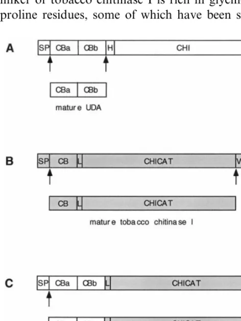
Urtica dioica agglutinin (UDA) consists of two cysteine-rich chitin-binding domains [1 – 3]. Both are homologous to hevein, a small chitin-binding protein of 43 amino acids from the rubber tree [4,5]. The presence of a carbohydrate-binding site in each chitin-binding domain enables UDA to agglutinate erythrocytes [1,6]. In most stinging nettle (U. dioicaL.) ecotypes, UDA is present as a mixture of isolectins [3,7]. Each isolectin is pro-cessed from a precursor (Fig. 1A), which com-prises an amino-terminal (N-terminal) signal peptide followed by two chitin-binding domains, a hinge region of 16 amino acids and a carboxyl-ter-minal (C-tercarboxyl-ter-minal) chitinase domain of 244 amino
acids [3,8]. A vacuolar sorting determinant has been shown to be present within the 25 C-terminal amino acids of the UDA precursor [9]. The signal peptide, the hinge region, as well as the chitinase domain are processed from the precursor to yield mature UDA (Fig. 1A). Because of the presence of the two chitin-binding domains and the homology of the chitinase domain with other plant chiti-nases, the precursor to UDA is classified as a plant class V or Chia5 chitinase [10,11]. Recently, pro-cessing of the precursor to UDA-isolectin I was studied in transgenic tobacco plants [9]. UDA purified from these tobacco plants had retained its chitin-binding activity, agglutination activity and antifungal activity against chitin-containing plant pathogenic fungi [1,9,12]. Fungal growth-inhibi-tion by UDA does not occur by lysis of the fungal cell wall. It does, however, depend on the phase of fungal growth and is temporal, suggesting that fungi adapt to the presence of UDA [9].
* Corresponding author. Tel.: +31-20-5257839; fax: + 31-20-5257934.
E-mail address:[email protected] (M.P. Does)
Mature tobacco chitinase I is comprised of one chitin-binding domain fused to a linker and a chitinase domain [13,14]. The structure of the pre-cursor to this chitinase is similar to the UDA precursor and consists of a signal peptide, the single chitin-binding domain, a linker, the catalytic domain of 244 amino acids and a C-terminal propeptide of seven amino acids (Fig. 1B). This propeptide has been shown to be necessary and sufficient for vacuolar targeting [15]. Removal of this vacuolar sorting determinant causes secretion of the mature basic chitinase into the extracellular space [15 – 17]. Depending on the isoform of the tobacco chitinases I, the linker consists of 10 (chitinase A) or 15 (chitinase B) amino acids [18]. Unlike the hinge region of the UDA precursor, the linker of tobacco chitinase I is rich in glycine and proline residues, some of which have been shown
to be 4-hydroxylated [19]. Tobacco chitinase I displays antifungal activity on several fungi that contain chitin in their cell walls. Unlike UDA, tobacco chitinase I causes lysis of the hyphal tips as a result of its hydrolase activity [20].
In this paper, we report the production of a fusion protein in transgenic tobacco (Nicotiana tabacum cv. Samsun NN), in which we have brought together the different biological activities of both UDA and tobacco chitinase I. The UDA – Chi chimera consists of the mature UDA-isolectin I sequence fused to the linker and the catalytic domain of tobacco chitinase I (chitinase B). We show that the purified fusion protein displays both agglutination and chitinase activity.
2. Materials and methods
2.1. Cloning procedures
The chimeric UDA – ChiDCT7 fusion gene con-taining the UDA-isolectin I leader sequence fol-lowed by the sequences encoding the signal peptide and UDA-isolectin I of the UDA precur-sor, the linker and the catalytic domain of tobacco chitinase I (chitinase B), was made by the follow-ing procedure.
The gene encoding the precursor to UDA-isolectin I (UdChia5.1.1) from stinging nettle eco-type Weerselo was cloned between the cauliflower mosaic virus 35S promoter and the nopalin syn-thase terminator on vector pMOG181 as described previously [3,9]. To isolate the UDA-isolectin I sequence, polymerase chain reaction (PCR) was performed on this clone using the primers 35S1 (5%-CGACACGCTTGTCTACTCC-3%) and UD9 (5%-GCAGCGGTACTGGCATTTG-3%). PCR
am-plification was performed on 100 pg of plasmid DNA in a 100 ml reaction mixture containing 2.5 U Pfu DNA polymerase (Stratagene), 200 mM of each of the dNTPs (Pharmacia) and 100 pmol of both primers, using a DNA Thermal Cycler (Perkin Elmer). The PCR fragment was obtained after an initial step of 4 min at 95°C, followed by 30 cycles of 1 min at 95°C, 2 min at 57°C and 2 min at 72°C and a final step of 8 min at 72°C. Subsequently, the fragment was digested with
BamHI.
To isolate the tobacco chitinase sequences, a clone was used containing the tobacco (N. Fig. 1. Schematic representation of how the precursors to (A)
tabacum cv. Samsun NN) basic chitinase cDNA on vector pMOG181 [16]. PCR was performed using the primers Chi5 (5%
-CCTGGTGGTCCCA-CACC-3%) and LS19 (5% -TTCCCAGT-CACGACGTTGT-3%) on 100 pg plasmid DNA as
already described. The chitinase PCR fragment was obtained after an initial step of 4 min at 95°C, followed by 35 cycles of 1 min at 95°C, 2 min at 52°C and 2 min at 72°C, and a final step of 8 min at 72°C. This fragment was digested withHindIII. The BamHI-blunt (UDA-isolectin I) and
blunt-HindIII (chitinase) PCR fragments were together cloned in the expression cassette on pMOG181. For extracellular targeting of the fusion protein, a stopcodon was introduced in the sequence of the chitinase domain. Therefore, a vacuolar targeting mutant chitinase construct (pMOG189) was used which had been created previously [16]. This con-struct encodes the basic tobacco chitinase I lacking the C-terminal vacuolar targeting signal of seven amino acids. The mutated chitinase gene was cloned from pMOG198 into pMOG181 (ChiDCT7). The stopcodon was introduced into the UDA – ChiDCT7 fusion by exchanging the
PstI/HindIII fragment from the UDA – Chi fusion by the stopcodon-containing PstI/HindIII frag-ment from ChiDCT7. The construct was se-quenced using the dideoxynucleotide chain-termination method [21]. The EcoRI/
HindIII fragment from pMOG181:UDA – ChiDCT7 was cloned into the binary vector pMOG402 [22]. Cloning procedures were per-formed as described elsewhere [23].
2.2. Tobacco transformation
Transfer of the pMOG402:UDA – ChiDCT7 construct to Agrobacterium tumefaciens and to-bacco (N. tabacum cv. Samsun NN) transforma-tion were performed as previously described [9]. Seeds of primary transformants and of their progeny were selected on plates containing kanamycin (200 mg/ml).
2.3. Analysis of transgenic plants
Total leaf extracts were isolated by grinding leaf samples in sodium acetate buffer (50 mM NaOAc, pH 5.2). Extracellular washing fluid (EWF) was isolated according to De Wit and Spikman [24], using sodium acetate buffer. Protein
concentra-tions were determined by the Bradford method [25] using bovine serum albumen as standard. Protein electrophoresis was performed using 12.5% sodium dodecyl sulfate (SDS)-polyacry-lamide gels [26] or using 20% Tricine-SDS-poly-acrylamide gels [27]. Immunological detection by
a-UDA antibodies [3] was performed as described previously [3]. For immunological detection with
a-CHI antibodies [16], antibodies were diluted 5000-fold and incubated with the blot overnight at room temperature.
2.4. Purification of the UDA–Chi fusion protein from transgenic tobacco
The UDA – Chi fusion protein was isolated from a homozygous high expressor line. Leaves were de-veined and homogenized in ice-cold extraction buffer (50 mM NaOAc, pH 5.2, 0.1% ascorbic acid) using a blender. One teaspoon of charcoal was added per 200 ml extraction buffer. The mix-ture was squeezed through four layers of gauze and kept on ice. Subsequently, the filtrate was centrifuged at 3200×g for 30 min at 4°C. Super-natant was filtered through eight layers of gauze and centrifuged at 20,000×g for 60 min at 4°C. The supernatant was again filtered through eight layers of gauze and brought to pH 4.0 using 2 N acetic acid. NaCl was added to a concentration of 0.1 M and the mixture was kept on ice for at least 1 h. After centrifugation for 60 min at 20,000×g
and 4°C, the supernatant was filtered through eight layers of gauze.
membrane (Amicon, Inc.) using a 400 ml stirred cell (Amicon, Inc.).
The concentrate was dialyzed against phosphate buffered saline (PBS) and applied to a Su-perdex™75 gel filtration column (HiLoad™16/60; Pharmacia), equilibrated with PBS. Gel filtration occurred at 0.8 ml/min. Fractions containing the fusion protein were pooled and gel filtration was performed repeatedly, until fractions with pure fusion protein were obtained. Pooled fractions were concentrated and dialyzed against 50 mM potassium phosphate buffer pH 6.0.
Protein concentration was determined using a bicinchoninic acid protein assay kit (Sigma). Pu-rity of the fusion protein was checked by 12.5% SDS-polyacrylamide gel electrophoresis and both immunological detection using a-UDA antibodies anda-CHI antibodies, and silverstaining using the Silverstain plus kit (Biorad).
2.5. Purification of tobacco chitinase I from transgenic tobacco
Homozygous seeds of transgenic tobacco ex-pressing the gene encoding tobacco chitinase I [16] were obtained from ZENECA MOGEN. Chiti-nase I was isolated from transgenic tobacco leaves according to the procedure described previously [20]. Protein concentration was determined using the bicinchoninic acid protein assay kit (Sigma). Purity was checked by 12.5% SDS-polyacrylamide gel electrophoresis and immunological detection using a-CHI antibodies, and silverstaining.
2.6. Agglutination assays
Agglutination assays using crude leaf extracts were performed on microscope slides, with squashed leaf samples to which trypsin-treated rabbit erythrocytes [1] and PBS were added.
Agglutination tests with purified UDA – Chi fu-sion protein and purified tobacco chitinase I were performed as follows: to 30ml trypsin-treated rab-bit erythrocytes, 0.25, 0.5, 1.0, 2.0, 3.0 and 4.0 mg of purified protein was added. Five times concen-trated PBS was added to a final volume of 60 ml. To inhibit possible proteolytic activity from residual trypsin or other proteolytic enzymes, tests were also performed in the presence of protease inhibitors. Therefore, 2.4 ml of a 25× stock solu-tion of a protease inhibitor cocktail (Complete™;
Boehringer Mannheim) in PBS was first added to 30 ml erythrocytes, followed by the different amounts of fusion protein. The final volume was brought to 60 ml with five times concentrated PBS.
2.7. Chitinase assay
Chitinase activity measurements were carried out using carboxylmethyl-chitin-Remazol-Bril-liant-Violet 5R (CM-chitin-RBV; LOEWE Bio-chemica, Sauerlach, Germany) as a substrate [28] as described previously [29].
3. Results
3.1. UDA–ChiDCT7 precursor construct
Construct UDA – ChiDCT7 encoding the pre-cursor to the mature chimera UDA – Chi (Fig. 1C) was made by fusion of sequences coding for the signal peptide and the mature isolectin of the precursor to UDA-isolectin I [3] (Fig. 1A) to the coding sequence for the linker and the catalytic domain of tobacco chitinase I [16] (Fig. 1B). Due to the introduction of a stopcodon, the precursor construct UDA – ChiDCT7 lacks the C-terminal propeptide of seven amino acids (DCT7), which has been shown to be necessary for the targeting of tobacco chitinase I to the vacuoles [15]. Because of this deletion, we expected the fusion protein to be targeted extracellularly, like the tobacco chiti-nase [16]. Compared with the tobacco chitichiti-nase I, the mature fusion protein UDA – Chi contains an additional chitin-binding domain (Fig. 1). Mature tobacco chitinase I consists of 294 amino acids, while the mature fusion protein consists of 340 amino acids.
Construct UDA – ChiDCT7 was placed in an expression cassette, between the cauliflower mo-saic virus 35S promoter and the nopalin synthase transcription terminator, and transformed into to-bacco using Agrobacterium.
3.2. Production and extracellular targeting of the fusion protein UDA–Chi in transgenic tobacco
Fig. 2. Western analysis of EWF from F1 progeny using (A) a-UDA antibodies and (B) a-CHI antibodies. Proteins were
electrophorized on 20% Tris – Tricine gels. (A) and (B) Lanes 1 – 3, 5 – 6, 9 – 17, 19 – 20 and 23 – 28, EWF from different F1progeny;
lanes 4 and 18, nontransformed control; (C) lanes 7 and 22, purified stinging nettle UDA; lanes 8 and 21, purified tobacco chitinase I. The UDA – Chi fusion, UDA and tobacco chitinase I are indicated by horizontal arrows. CHI-I, tobacco chitinase I.
synthetic peptide consisting of 15 sequential amino acids of the mature UDA-isolectin I sequence [3]. In an agglutination assay, crude extracts of sev-eral high expressor lines agglutinated trypsin-treated rabbit erythrocytes (data not shown), indicating that an agglutinating protein was present in these extracts. A crude extract of a nontransformed tobacco did not agglutinate rab-bit erythrocytes.
Seeds of several primary transformants were germinated and EWF of kanamycin resistant F1
progeny was analysed by Western analysis using
a-UDA antibodies (Fig. 2A) and a-CHI antibod-ies (Fig. 2B). As is shown in Fig. 2A, a-UDA antibodies recognized purified UDA (lanes 7 and 22) and the fusion protein UDA – Chi in EWF from the F1 plants (lanes 1 – 3, 5 – 6, 9 – 17, 19 – 20,
23 – 28), but they did not cross-react with purified tobacco chitinase I (lanes 8 and 21). Upon longer exposure, a weak signal at the same level as that of UDA was found in some high expressor lines (Fig. 2A, lanes 10, 11, 12 and 24), suggesting that a small amount of the fusion protein was being processed in transgenic tobacco. When the same blot was probed witha-CHI antibodies, the fusion protein was recognized as well (Fig. 2B, lanes 1 – 3,
5 – 6, 9 – 17, 19 – 20, 23 – 28). Some cross-reacting bands are present. The two bands of approxi-mately 26 and 28 kD most likely represent endoge-nous class II chitinases [20], known to be induced by stress. The UDA – Chi chimera is clearly larger than tobacco chitinase I (Fig. 2B, lanes 8 and 21), due to the presence of an extra chitin-binding domain of about 4.5 kD. The chimera was not present in the EWF from control plants (Fig. 2A,B, lanes 4 and 18). The precursor UDA – ChiDCT7 to the mature fusion protein UDA – Chi lacks the vacuolar targeting signal. The presence of the fusion protein in the EWF indicates extra-cellular targeting of UDA – Chi, as expected.
3.3. Agglutination and chitinase acti6ities of purified fusion protein UDA–Chi and tobacco chitinase I
Fusion protein UDA – Chi was purified to ho-mogeneity from total extract of a homozygous high expressor F2line. Fig. 3A shows the
Fig. 3. Western analysis of purified fusion protein UDA – Chi using (A)a-UDA antibodies and (B)a-CHI antibodies. Lanes 1, 100 ng UDA – Chi; lanes 2, 50 ng UDA – Chi; lanes 3, 5 ng UDA – Chi; lanes 4, 20 ng purified tobacco chitinase I.
Samsun NN) plants, expressing the protein at high level [16]. Purity was verified by Western analysis using a-CHI antibodies (Fig. 3) and silverstaining of the protein gel (not shown).
Agglutination activity was tested for both proteins. The tobacco chitinase I did not aggluti-nate trypsin-treated rabbit erythrocytes (data not shown). In contrast, UDA – Chi displayed a low but significant agglutination activity at a concen-tration of 8.3 mg/ml (0.5 mg/60 ml). Increased ag-glutination activities were shown for higher concentrations of fusion protein (Fig. 5). To ex-clude the possibility that UDA – Chi was being processed by residual trypsin or proteases from the erythrocytes, proteinase inhibitors were added to the agglutination mixtures. No differences were detected between the assays with or without the protease inhibitors (data not shown).
Purified UDA-isolectin I from transgenic to-bacco agglutinated trypsin-treated rabbit erythro-cytes as effectively at a concentration of 2.5 mg/ml [9] as UDA – Chi at a concentration of 8.3 mg/ml. Since the molecular mass of the UDA – Chi fusion protein is 36,546 Da and the molecular mass of UDA-isolectin I is 9408 Da, 8.3 mg fusion protein corresponds to approximately the same number of UDA-containing molecules as 2.5 mg UDA-isolectin I. Therefore we conclude that the molar agglutination activities of UDA-isolectin I and the UDA – Chi fusion are similar.
Chitinase activities of purified UDA – Chi, to-bacco chitinase I and purified UDA-isolectin I were measured using the dye-labelled substrate CM-chitin-RBV (Table 1). Using this substrate, the chitinase activity of the fusion protein ap-peared to be 3.6-fold lower than that of tobacco chitinase I. UDA-isolectin I did not display chiti-nase activity. Fusion of UDA-isolectin I to the catalytic domain of tobacco chitinase I therefore used for detection with a-CHI antibodies (Fig.
3B). No contamination by endogenous chitinase was detected using these antibodies. Upon longer exposure times of this blot, 5 ng fusion protein became clearly visible, while no endogenous chiti-nase was detected in the sample of 100 ng fusion protein. Purity of the fusion protein was also checked by silverstaining, shown in Fig. 4.
Tobacco chitinase I was purified from ho-mozygous transgenic tobacco (N. tabacum cv.
Fig. 4. Silverstained protein gel with 250 ng purified fusion protein UDA – Chi.
Table 1
Chitinase activities of UDA-isolectin I, tobacco chitinase I and fusion UDA–Chi, purified from transgenic tobacco
Chitinase activitya(ODu/mg)
0 UDA-isolectin I
3.690.3 Tobacco chitinase I
Fusion UDA–Chi 1.090.2
aCM-chitin-RBV was used as the substrate. Chitinase
ac-tivity is represented as units of optical density (ODu) at 550 nm permg protein.
by a flexible linker does not affect agglutination activity. This suggests that the folding and expo-sure of sugar-binding sites [6] in both chitin-bind-ing domains of the fusion protein and mature UDA are identical.
Using CM-chitin-RBV in a colorimetric assay, the enzymatic activity of the fusion protein was lower than that of the tobacco chitinase I (1.09
0.2 ODu/mg vs 3.690.3 ODu/mg). This does not necessarily mean that the actual chitinase activity was lower. It has been shown that different classes of tobacco chitinases display distinct hydrolysing and lysozyme activities, depending on the sub-strate [31]. Melchers et al. [29] have shown differ-ences in activity between tobacco class I chitinase and class II chitinase on two substrates, CM-chitin-RBV and tritiated-chitin. Although the en-zymatic activity of class I chitinase was 10,000 times higher on tritiated chitin, the activity on CM-chitin-RBV was 30 times lower than that of the class II chitinase. Therefore, the enzymatic activity of the UDA – Chi chimera on tritiated-chitin could be higher than that of tobacco chiti-nase I. We therefore conclude that the chitichiti-nase activity of the fusion protein differs from that of tobacco chitinase I.
Previously, it has been shown that the enzymatic activity of the catalytic domain of tobacco chiti-nase I on tritiated-chitin is modified by addition of the chitin-binding domain [32]. Using CM-chitin-RBV, tobacco chitinase I with and without a chitin-binding domain showed similar activities at pH 5.2 [33]. Although the presence of one chitin-binding domain did not affect the enzyme activity of the catalytic domain of tobacco chitinase I on CM-chitin-RBV substrate, the fusion protein was 3.6-fold less active compared with the tobacco chitinase I. Actually, the UDA – Chi fusion can be seen as a tobacco chitinase I with an extra chitin-binding domain added to it. Due to this addition, the interaction of the chitin-binding domains and the chitinase domain during hydrolysis might have changed.
UDA and tobacco chitinase I both display anti-fungal activity on chitin-containing plant patho-genic fungi. However, the range of susceptible chitin-containing fungi and the amount of each protein needed to obtain 50% growth inhibition differ for both these chitin-binding proteins [34,35]. For example, UDA is known to inhibit the growth of Botrytis cinerea [9,12], whereas tobacco seemed to have changed the chitinase activity of
the molecule on CM-chitin-RBV.
4. Discussion
A chimeric protein UDA – Chi, consisting of the UDA-isolectin I sequence fused to the linker and catalytic domain of tobacco chitinase I, has been produced in transgenic tobacco.
In high expressor lines, a protein with a similar molecular weight to that of UDA was detected by Western analysis using a-UDA antibodies. This protein could be a processing product of the fu-sion protein. Recently, two truncated class I chiti-nases have been identified in NaCl-adapted tobacco cells [30]. Both chitinases lacked the chitin-binding domain, but still contained a partial linker. These linkers resembled those of both to-bacco chitinase I isoforms A and B. It was sug-gested that the truncated proteins were most probably products of new genes. However, it can-not be excluded they were products of class I chitinases that had been processed between the first two glycine residues of the linker. Hence, the UDA-like protein we have detected might be the product of a similar processing event in the linker of the UDA – Chi chimera.
chitinase does not affect this fungus [34]. For inhibition of Colletotrichum lindemuthianum, very high concentrations of tobacco chitinase were needed [35], whereas much lower concentrations of UDA were sufficient [9,12,35]. On the contrary, growth of Trichoderma hamatumwas inhibited by low concentrations of chitinase and high concen-trations of UDA [34,35]. UDA and tobacco chiti-nase I have been shown to act synergistically onT.
hamatum [12]. Since the fusion protein binds to chitin, and shows agglutination and chitinase ac-tivity, it may display antifungal activity as well. One may speculate that the UDA – Chi fusion has a growth-inhibiting effect on each fungus that is susceptible for either chitinase or UDA, and there-fore has potential to be used as an antifungal on a broad range of chitin-containing fungi.
Acknowledgements
We thank ZENECA MOGEN for use of their expression vectors, a-CHI antibodies and trans-genic tobacco seeds expressing tobacco chitinase I. We also thank Jaap Fontijn for cultivating our plants in the greenhouse and Simon van Mechelen for photographic work.
References
[1] W.J. Peumans, M. De Ley, W.F. Broekaert, An unusual lectin from stinging nettle (Urtica dioica) rhizomes, FEBS Lett. 177 (1984) 99 – 103.
[2] J.J. Beintema, W.J. Peumans, The primary structure of stinging nettle (Urtica dioica) agglutinin: a two-domain member of the hevein family, FEBS Lett. 299 (1992) 131 – 134.
[3] M.P. Does, D.K. Ng, H.L. Dekker, W.J. Peumans, P.M. Houterman, E.J.M. Van Damme, B.J.C. Cornelissen, Characterization of Urtica dioica agglutinin isolectins and the encoding gene family, Plant Mol. Biol. 39 (1999) 335 – 347.
[4] B.L. Archer, The proteins of He6ea brasiliensis latex: isolation and characterization of crystalline hevein, Biochem. J. 75 (1960) 236 – 240.
[5] K. Walujono, R.A. Scholma, J.J. Beintema, A. Mariono, A.M. Hahn, Amino acid sequence of hevein, Proceedings of the International Rubber Conference, Kuala Lumpur, Vol. 2, Rubber Research Institute, Malaysia, 1975, pp. 518 – 531.
[6] K. Hom, M. Gochin, W.J. Peumans, N. Shine, Ligand-induced perturbations inUrtica dioicaagglutinin, FEBS Lett. 361 (1995) 157 – 161.
[7] E.J.M. Van Damme, W.J. Peumans, Isolectin composi-tion of individual clones of Urtica dioica: evidence for phenotypic differences, Physiol. Plant. 71 (1987) 328 – 334.
[8] D.R. Lerner, N.V. Raikhel, The gene for stinging nettle lectin (Urtica dioicaagglutinin) encodes both a lectin and a chitinase, J. Biol.Chem. 267 (1992) 11085 – 11091. [9] M.P. Does, P.M. Houterman, H.L. Dekker, B.J.C.
Cor-nelissen, Processing, targeting and antifungal activity of
Urtica dioica agglutinin in transgenic tobacco, Plant Physiol. 120 (1999) 421 – 431.
[10] F. Meins Jr, B. Fritig, H.J.M. Linthorst, J.D. Mikkelsen, J.-M. Neuhaus, J. Ryals, Plant chitinase genes, Plant Mol. Biol. Reptr. 12 (1994) S22 – S28.
[11] J.-M. Neuhaus, B. Fritig, H.J.M. Linthorst, F. Meins Jr, J.D. Mikkelsen, J. Ryals, A revised nomenclature for chitinase genes, Plant Mol. Biol. Reptr. 14 (1996) 102 – 104.
[12] W.F. Broekaert, J. Van Parijs, F. Leyns, H. Joos, W.J. Peumans, A chitin-binding lectin from stinging nettle rhizomes with antifungal properties, Science 245 (1989) 1100 – 1102.
[13] H.J.M. Linthorst, L.C. van Loon, C.M.A. van Rossum, A. Mayer, J.F. Bol, J.S.C. van Roekel, E.J.S. Meulen-hoff, B.J.C. Cornelissen, Analysis of acidic and basic chitinases from tobacco and petunia and their constitu-tive expression in transgenic tobacco, Mol. Plant Mi-crobe Interact. 3 (1990) 252 – 258.
[14] H. Shinshi, J.-M. Neuhaus, J. Ryals, F. Meins Jr, Struc-ture of a tobacco endochitinase gene: evidence that dif-ferent chitinase genes can arise by transposition of sequences encoding a cysteine-rich domain, Plant Mol. Biol. 14 (1990) 357 – 368.
[15] J.-M. Neuhaus, L. Sticher, F. Meins Jr, T. Boller, A short C-terminal sequence is necessary and sufficient for the targeting of chitinases to the plant vacuole, Proc. Natl. Acad. Sci. U.S.A. 88 (1991) 10362 – 10366. [16] L.S. Melchers, M.B. Sela-Buurlage, S.A. Vloemans, C.P.
Woloshuk, J.S.C. van Roekel, J. Pen, P.J.M. van den Elzen, B.J.C. Cornelissen, Extracellular targeting of the vacuolar tobacco proteins AP24, chitinase andb -1,3-glu-canase in transgenic plants, Plant Mol. Biol. 21 (1993) 583 – 593.
[17] L. Sticher, J. Hofsteenge, J.-M. Neuhaus, T. Boller, F. Meins Jr, Posttranslational processing of a new class of hydroxyproline-containing proteins, Plant Physiol. 101 (1993) 1239 – 1247.
[18] M. van Buuren, J.-M. Neuhaus, H. Shinshi, J. Ryals, F. Meins Jr, The structure and regulation of homeologous tobacco endochitinase genes of Nicotiana syl6estrisand N.tomentosiformis origin, Mol. Gen. Genet. 232 (1992) 460 – 469.
[19] L. Sticher, J. Hofsteenge, A. Milani, J.-M. Neuhaus, F. Meins Jr, Vacuolar chitinases of tobacco: a new class of hydroxyproline-containing proteins, Science 257 (1992) 655 – 657.
[20] M.B. Sela-Buurlage, A.S. Ponstein, S.A. Bres-Vloemans, L.S. Melchers, P.J.M. van den Elzen, B.J.C. Cornelissen, Only specific tobacco (Nicotiana tabacum) chitinases and
[21] F. Sanger, S. Nicklen, A.R. Coulson, DNA sequencing with chain-termination inhibitors, Proc. Natl. Acad. Sci. U.S.A. 74 (1977) 5463 – 5467.
[22] E. Jongedijk, H. Tigelaar, J.S.C. van Roekel, S.A. Bres-Vloemans, I. Dekker, P.J.M. van den Elzen, B.J.C. Cor-nelissen, L.S. Melchers, Synergistic activity of chitinases andb-1,3-glucanases enhances fungal resistance in trans-genic tomato plants, Euphytica 85 (1995) 173 – 180. [23] J. Sambrook, E.F. Fritsch, T. Maniatis, Molecular
Cloning: A Laboratory Manual, Second ed., Cold Spring Harbor Laboratory Press, Cold Spring Harbor, NY, 1989.
[24] P.J.G.M. de Wit, G. Spikman, Evidence for the occur-rence of race and cultivar-specific elicitors of necrosis in intercellular fluids of compatible interactions of
Cladosporium ful6umand tomato, Physiol. Plant. Pathol.
21 (1982) 1 – 11.
[25] M.M. Bradford, A rapid and sensitive method for the quantitation of microgram quantities of proteins utilizing the principle of protein-dye binding, Anal. Biochem. 72 (1976) 248 – 254.
[26] U.K. Laemmli, Cleavage of structural proteins during the assembly of the head of the bacteriophage T4, Na-ture 227 (1970) 680 – 685.
[27] H. Scha¨gger, G. von Jagow, Tricine-sodium dodecyl sulfate-polyacrylamide gel electrophoresis for the separa-tion of proteins in the range from 1 to 100 kDa, Anal. Biochem. 166 (1987) 368 – 379.
[28] S.J. Wirth, G.A. Wolf, Dye-labelled substrates for the assay and detection of chitinase and lysozyme activity, J. Microbiol. Methods 12 (1990) 197 – 205.
[29] L.S. Melchers, M. Apotheker-de Groot, J.A. van der Knaap, A.S. Ponstein, M.B. Sela-Buurlage, J.F. Bol, B.J.C. Cornelissen, P.J.M. van den Elzen, H.J.M. Linthorst, A new class of tobacco chitinases homologous to bacterial exo-chitinases displays antifungal activity, Plant J. 5 (1994) 469 – 480.
[30] D.-J. Yun, M. Paino D’Urzo, L. Abad, S. Takeda, R. Salzman, Z. Chen, H. Lee, P.M. Hasegawa, R.A. Bres-san, Novel osmotically induced antifungal chitinases and bacterial expression of an active recombinant isoform, Plant Physiol. 111 (1996) 1219 – 1225.
[31] F. Brunner, A. Stintzi, B. Fritig, M. Legrand, Substrate specificities of tobacco chitinases, Plant J. 14 (1998) 225 – 234.
[32] B. Iseli, T. Boller, J.-M. Neuhaus, The N-terminal cys-teine-rich domain of tobacco class I chitinase is essential for chitin binding but not for catalytic or antifungal activity, Plant Physiol. 103 (1993) 221 – 226.
[33] M.B. Sela-Buurlage, In6itrosensitivity and tolerance of Fusarium solanitowards chitinases andb-1,3-glucanases, Ph.D. thesis, Landbouwuniversiteit Wageningen, Wa-geningen, The Netherlands, 1996.
[34] J. Van Parijs, W.F. Broekaert, I.J. Goldstein, W.J. Peu-mans, Hevein: an antifungal protein from rubber-tree (He6ea brasiliensis) latex, Planta 183 (1991) 258 – 264. [35] W.F. Broekaert, W. Marie¨n, F.R.G. Terras, M.F.C De
Bolle, P. Proost, J. Van Damme, L. Dillen, M. Claeys, S.B. Rees, J. Vanderleyden, B.P.A. Cammue, Antimicro-bial peptides from Amaranthus caudatus seeds with se-quence homology to the cysteine/glycine-rich domain of chitin-binding proteins, Biochemistry 31 (1992) 4308 – 4314.