BIOTRANSFORMATION AND CYTOTOXIC ACTIVITY OF GUAIACOL DIMER
Galuh Widiyarti
*, Jamilah Abbas, and Yulia Anita
Research Center for Chemistry (RCC-Chem), Indonesian Institute of Sciences (LIPI), Kawasan PUSPIPTEK Serpong, Tangerang Selatan, Banten 15314 Indonesia
Received October, 31 2013; Accepted April 2, 2014
ABSTRACT
Guaiacol, a phenolic compound is known as an anticancer. Dimerization of guaiacol has been done by biotransformation using peroxidase enzyme as biocatalyst. This enzyme was isolated from Indonesian plant, kailan (Brassica oleraceae var. alboglabra). Analysis of dimerization product was carried out by TLC, IR, LC-MS, and NMR. Whilst analysis of in-vitro cytotoxic activity was carried out by MTT method against breast cancer T47D and MCF7 cells. The result showed that the dimerization reaction gave O-para dehydroguaiacol. The in-vitro cytotoxic activity analysis showed that O-para dehydroguaiacol compound has potency as anti-breast cancer.
Keywords:guaiacol; peroxidase; dimerization; anti-breast cancer
ABSTRAK
Guaiakol merupakan senyawa fenolik yang telah diketahui sebagai antikanker. Telah dilakukan dimerisasi guaiakol secara biotransformasi dengan menggunakan enzim peroksidase sebagai biokatalis. Peroksidase diisolasi dari tanaman lokal Indonesia, kailan (Brassica oleraceae var. alboglabra). Analisis produk dimer dilakukan dengan menggunakan KLT, IR, LC-MS, dan NMR, sedangkan analisis aktivitas sitotoksik terhadap sel kanker payudara T47D dan MCF7 dilakukan secara in-vitro dengan metode MTT. Pada studi ini, dimerisasi guaiakol menghasilkan senyawa O-para dehidroguaiakol. Analisis aktivitas sitotoksik menunjukkan bahwa, senyawa O-para dehidroguaiakol mempunyai potensi sebagai anti-kanker payudara.
Kata Kunci:guaiakol; peroksidase; dimerisasi; anti-kanker payudara
INTRODUCTION
Cancer is a top ten killer diseases in the world, the 7th after heart, stroke, pneumonia, tuberculosis, diabetes, and HIV/AIDS diseases. Yayasan Kanker Indonesia (YKI) stated that number of breast cancer patients in Indonesia until 2010 is 2nd after cervical cancer, but now, by increasing of the breast cancer patients about 60%, breast cancer is 1st, while cervical cancer is 2nd. World Heath Organization (WHO) stated that breast cancer as most common cancer in women and the leading cause of cancer death in women, so categorized as International Classification of Diseases (ICD) [1].
During the last few decades, chemopreventive and chemotherapeutic compounds against various types of cancer have been isolated from a number of plants. Medicinal plants have been always a useful source for the research of new biologically active compounds [2]. Some phytochemicals from plants and fruits that is known as a chemopreventif agent than can suppress tumorogenesis are phenolic compounds such as catechin, eugenol, and gingerol [3-5].
Guaiacol, a phenolic compound possesses potent free radical scavenger, anti-inflammatory, anticancer activities. It is material for drug of antiseptic, local anesthetic and expectorant cough medicine [6]. Guaiacol is a bioactive compound in the form colorless aromatic oil derived from Guaiacum officinale (Lignum sanctum) or Guaiacum sanctum (Lignum vitae), including in family of Zygopolyllaceae. As phenolic compound, guaiacol is susceptible to enzymatic and nonenzymatic oxidation giving rise to a variety of derivative product. The phenolic oxidative coupling reaction is a key step for the biosynthesis of natural product [7-8].
Peroxidase (PO) is a group of oxidoreductase that catalyzes oxidation reaction of numerous compounds by peroxide as an oxidant [9-10]. PO has higher redox potentials and thus is stronger oxidizing agents. PO have been successfully employed to catalyze oxidative coupling of phenol and aromatic amines to produce the dimer derivatives of naphthol, eugenol, and catechin with satisfactory yields, and applied for the removal of phenols from industrial wastewater [11].
so disadvantage in biocatalytic applications. The higher thermal and environmental stability PO is PO from plant such as horseradish or soybean, thus this is more attractive biocatalysts [12-13]. Commercially, production of horseradish peroxidase (HRP) is done by extraction process of horseradish root that grown in temperate countries relatively cool, as agricultural weeds and difficult to obtain [9,14-15]. Therefore, it is necessary to develop applications of HRP from Indonesian native plants. Local plants that are similar to horseradish family are kalian (Brassica oleraceae var. alboglabra), sawi putih (Brassica rapa var. Pekinensis), and sawi hijau (Brassica juncea) including in family of Brassicaceae. The previous study has shown that the highest activity of PO was isolated from kailan at pH 6 [16]. Therefore we used kailan as source of PO in this study.
The aim of this study is synthesis guaiacol dimer, by biotransformation, using PO as a biocatalyst. Identification of guaiacol dimer compound was carried out by TLC, spectrophotometer FT-IR, LC-MS, and NMR. Guaiacol is known as an anticancer and it is necessary to study in-vitro cytotoxic activity of guaiacol dimer on inhibition human cancer, especially human breast cancer T47D and MCF7 cells using Mosmann’s method with dimethylthiazol diphenyltetrazolium bromide (MTT) coloring.
EXPERIMENTAL SECTION
Materials
Materials used were Guaiacol (Nacalai) as starting material, phosphate buffer, 5% H2O2, 5% HCl,n-hexane, ethyl acetate, butanol, distilled water, and analytical grade chemicals were used for mass spectra, structure, and in-vitro cytotoxic activity analysis. Kailan (Brassica oleraceae var. alboglabra) was obtained from traditional market in Serpong, Tangerang Selatan.
Instrumentation
Instrumentation used in this study was dimerization process unit, evaporation unit, and one set of diguaiacol identification unit. Thin Layer Chromatography (TLC) was carried out using precoated silica gel plates (Merck Kieselgel 60F 254, 0.25 mm). IR spectra were measured by Fourier Transform Infra Red (FT-IR) Spectrophotometer Shimadzu prestige 21. Mass spectra (MS) was obtained with Liquid Chromatography-Mass Spectroscopy (LC-MS) Mariner Biospectrometry spectrometer using Electrospray Ionization (ESI) System and positive ion mode.1H-NMR (500 MHz) and13C-NMR (125 MHz) spectra were recorded on a Nuclear Magnetic Resonance (NMR) JEOL spectrometer, while the 2D-NMR experiments were conducted using the standard
JEOL software for COSY and DEPT for molecular structure analysis.
Procedure
Preparation of phosphate buffer
A total of 13.9 g NaH2PO4.H2O (Sodium
dihydrogenphosphat-monohydrat) was dissolved in 1 L of aquadest (as solvent A) and 35.85 g Na2HPO4.2H2O
(di-Natriumhydrogenphosphat dihydrat) in 1 L of aquadest (as solvent B). The mixture of solution A (1 L) and B (500 mL) gave phosphate buffer pH 6.
Preparation of enzyme from Brassica oleraceae var alboglabra
Leaves of Brassica oleraceae var. alboglabra (773 g) were washed and cut into small size, then mixed with buffer pH 6 (775 mL) using a blender, and flittered. The crude enzyme was then stored in the refrigerator until it is used [11].
Synthesis of guaiacol dimer
50 mL of peroxidase enzyme was added to 6 mL of guaiacol (4-hydroxy-5-methoxybenzene), and 3 mL of 5% H2O2 was added and stirred for 3 min at room temperature (27 °C). 3 mL of 5% HCl was then added to stop the reaction. The mixture was then extracted with EtOAc/n-BuOH 9:1. The combined extract was concentrated at 45 °C under vacuum to yield brown residues containing of a mixture dimerization products [6].
Purification of dimerization products
Dimerization products were purified by column chromatography (silica gel Merck 64271) eluting with n-hexane, a gradient of EtOAc to 100%, followed by EtOAc/MeOH 1:1. The purified of dimerization products were identified by spectroscopic methods (LC-MS, FT-IR, NMR) [17].
Analysis methods
Column chromatography was carried out on silica gel Merck (70-230 mesh and 230-400 mesh). Thin layer chromatography (TLC) was performed on precoated silica gel plates, and spots were visualized under UV light (254 and 365 nm) irradiation and by spraying with 10% sulphuric acid solution followed by heating at 110 °C. IR spectra were measured on a FT-IR Shimadzu prestige 21. 1H-NMR (500 MHz) and 13
In-vitro Cytotoxic Activity Analysis
The inhibitory effect of guaiacol dimer product on human breast cancer T47D and MCF7 cells were assessed using MTT method (Mosmann’s method) with 3-(4,5-dimethylthiazol-2-yl)-2,5-diphenyltetrazolium bromide (MTT) coloring. The development of cells culture performed by growing of cells culture in RPMI 1640-serum Phosphate Bovine Serume (PBS) medium and incubated in a humidified atmosphere of 37 °C and 5% of CO2 for 24 h. Serum medium was replaced with new serum medium and incubated in a humidified atmosphere of 37 °C and 5% of CO2 again in order to obtain sufficient cells number for testing. After a sufficient number of cells were obtained, cell medium was removed, and washed, and then RPMI medium added. The cells were transferred into tubes and centrifuged at 1200 rpm for 5 min. Supernatant was discarded and the precipitate was added to the RPMI 1640 medium containing 10% PBS. The cells density was calculated by hemocymeter and cells number was counted and then made dilution by adding with RPMI-serum medium to obtain cell number of 2 X 10⁴cells/mL cell suspense. Cells with densities 1-2 X 10⁴cells/well in 96 well-plates were cultivated in a humidified atmosphere at 37 °C and 5% of CO2for 24 h. Afterwards the cell cultures were replaced, washed with PBS, and added with 100 µL fresh culture medium containing guaiacol dimer sample at concentration of 100, 50, 25, 10, 5, 2.5, and 1 µg/mL. Furthermore, the plates were incubated at 37 °C in CO2 5% for 48 h. At the end of the treatment, medium was replaced and the cells were washed with PBS and added with 100 µL of a new fresh medium containing MTT 5 mg/mL. Plates were incubated in a humidified atmosphere at 37 °C in 5% CO2 for 4 h to bioreduction of the MTT dye into purple formazan crystals. After 4 h, the medium containing MTT was discarded, washed with PBS, added with 200 µL isopropanoat solution, and incubated at room temperature for 12 h to complete the solubilization of the formazan crystals. The bioreduction of MTT was assessed by measuring the absorbance of each well at 550 nm by ELISA reader. The proliferation inhibition was expressed as a percentage of the absorbance control cells minus abcorbance treatment cells divided by abcorbance control cells [18].
RESULT AND DISCUSSION
Isolation of HRP
We have isolated HRP from Indonesian plant, kailan (Brassica oleraceae var alboglabra). The specific activity of crude enzyme was determined by Bergmeyer and Lowry methods [19-20]. The amount of crude HRP from 773 g of leaves of Brassica oleraceae var
alboglabrathat was mixed with 775 mL of buffer pH 6 was 1350 mL with specific activity of 14.577 U/mg, The specific activity of this crude HRP was lower than the specific activity of commercially HRP (65 U/mg).
Analysis of Guaiacol Dimer
Peroxidase enzymes are usually used as biocatalyst for dimerization of phenolic compounds, such as naphthol, eugenol, and catechin [11]. We employed kailan’s peroxidase (crude HRP) to catalyze phenolic compound of guaiacol for dimerization process. The dimer is a chemical compound that is composed of two identical or similar molecules and bound together. Dimerization of guaiacol is the process of combining the two monomers of guaiacol in the presence of crude HRP as biocatalyst. Guaiacol is a phenolic compound so peroxidase catalyzes the oxidative coupling of this phenolic compound using hydrogen peroxide (H2O2) as the oxidizing agent and hydrogen acceptor for guaiacol as substrate. The reaction is a three-step cyclic reaction by with the enzyme is first oxidized by H2O2, and then reduced in two sequential one-electron transfer steps from reducing substrate. The coupling of guaiacol always occurs in ortho or para of phenolic hydroxyl group position, so dimerization due to o-o, o-p, or p-p C-C coupling can be expected.
This dimerization reaction product was reddish brown oil. It reacted with FeCl3 reagent indicating the presence of a phenolic group. TLC analysis showed that synthesis product contained guaiacol dimer which was indicated by a spot with Rf value of 0.2 of guaiacol dimer, lower than Rf of guaiacol (0.63) as starting material. The guaiacol dimer compound was purified by column chromatography and eluting with n-hexane/EtOAc 3:7, so obtained a oily brown crystal (98.3 mg, yield 1.47%).
The mass spectrum showed a (M+H)+1at m/z 247 which corresponded to molecular formula of C14H14O4 and molecular weight of guaiacol dimer is 246 g/mol. The IR spectra of guaiacol dimer exhibited free
hydroxyl (νmax = 3455.9 cm -1
), aromatic ring (C=C aromatic) (1595-1446) absorption, C-O-C
(νmax= 1257 cm -1
), C-H aromatic (νmax2880 - 3200 cm -1
), while IR spectra of guaiacol exhibited free hydroxyl
(νmax = 3502.7 cm -1
), aromatic ring (C=C aromatic)
(1454.4-1599.1) absorption, C-O-C (νmax = 1219 cm -1
),
C-H aromatic (νmax 2841.8 – 3007.3 cm -1
). The IR spectra of guaiacol dimer showed that the new compound is coupling oxidative of guaiacol.
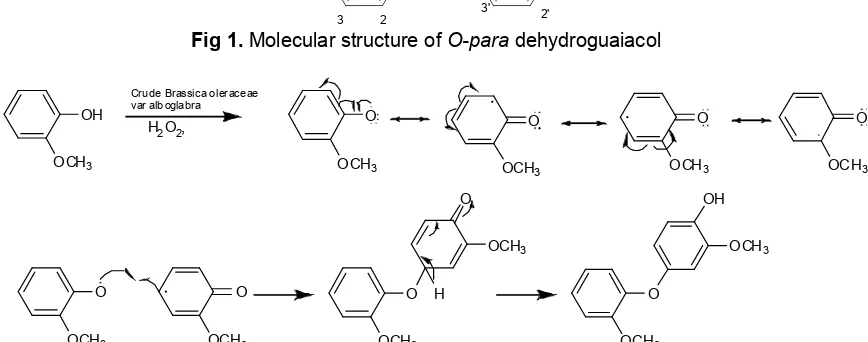
O Fig 1.Molecular structure ofO-paradehydroguaiacol
Fig 2. Proposed mechanism for the oxidative radical dimerization of guaiacol catalyzed by Crude of Brassica oleraceae var alboglabra.
quaternary carbons, including a hydroxyl (δ =
141.6 ppm). Furthermore,1H and13C-NMR spectra also displayed the presence of three sets of signals. The first
set, a four-proton doublet of doublet at δH 6.46/δC = 110.6 (1Hdd J = 2.6 and 8.4 Hz), δH6.87/δC= 119.1 (1H dd J 1.3 and 7.8 Hz), δH6.89/δC = 121.1 (1H dd J 1.3
and 7.8) and δH 6.69/δC = 112.6 (1H dd J 1.3 and 7.8 Hz). The second set of signals, consisting of two
proton doublet at δH6.65 /δC = 103.0 (1H d J 2.6) and
δH 6.83/δC=114.5 (1Hd J8.4 H), and also the three set of signals, consisting of one proton. multiplet at
δH 7.06 /δC = 123.9 (1H, m J 2.6 and 7.3 Hz), and also
there are six proton singlet at δH3.88/ δC= 56.1 (3H, s)
and δH3.83/δC= 56.1 (3H, s), established the presence of a two methoxy substituents.
A combination of the COSY and HMQC experiments permitted the assignments of all of the protonated carbons. It remained to establish the position of the substituents on the dimers of guaiacol skeleton. In
the HMBC spectrum, the hydroxyl group (δH= 5.38) was
correlated to the quarternary carbons at δC = 141.6 (C-4), 114.5 (C-3) and 147.2 (C-1). This finding clearly indicated that the hydroxyl group was located at C-4.
The other resonance at δC = 141.6 also gave cross peaks with three of olefinic protons of the guaiacol (at
δ = 6.83; 6.46 and 6.65). Methoxy group (δH= 3.83) was
correlated to the quarternary carbons at δC= 150.7 (C-5) and 147.2 (C-1). The HMBC spectrum also exhibited
methoxy group (δH = 3.88) which correlated to the
quarternary carbons at δC= 150.5 (C-5’). Proton at 7.06 correlated to the carbon 150.5 (C-5’), 121.1 (C-6’), 119.1 (C-2’). Proton at 6.89 correlated to the carbon 150.5 (C-5’), 112.6 (C-1’), 123.9 (C-3’) and
146.8 (C-4’). 2-Methoxy phenol moiety was connected to the C-4’ as oxigented- para couplet correlation. Proton at 6.99 and 6.87 correlated to the carbon 150.5 (C-5’), 121.1 (C-6’), 123.9(C-3’) and 146.8 (C-4’). The doublet signal at 6.65 and doublet of doublet signal at 6.46 (J 2.6 and 8.4) showed the HMBC correlation
between H-6 (δH 6.65) and C-4’ (δC146.8). The COSY spectrum of guaiacol dimer also showed the
connection of a doublet of doublet signal at δ 6.87 and multiplet signal at δ 7.06 (J 2.6 and 7.3 Hz), other
connection could be showed a doublet signal δ 6.99 and the doublet of doublet signal δ 6.87 and 6.89 (J1.3 and 7.8).
Based on the above spectroscopic analysis, the guaiacol dimer compound was characterized as dimer of 4-hydroxy-5-methoxybenzene with other name O-p-dehydroguaiacol (dehydrodiguaiacol) (Fig. 1). The product showed O-paracoupled guaiacol, leading to O-p-dehydroguaiacol. The mechanism of biotransformation of guaiacol catalyzed by crude enzyme of Brassica oleraceae var alboglabra peroxidase to produce O-p-dehydroguaiacol is shown in Fig. 2.
In-vitroCytotoxic Activity
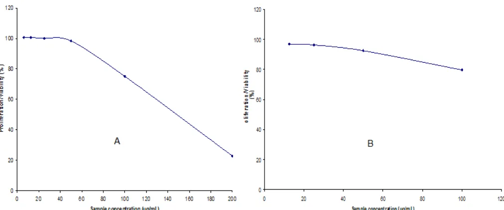
Fig 3.The result of in-vitrocytotoxic activity test of dehydrodiguaiacol against human breast cancer T47D (A) and MCF7 (B) cells
Fig 4.The result ofin-vitrocytotoxic activity test of doxorubicin as standard against human breast cancer T47D (A) and MCF7 (B) cells
showed that dehydrodiguaiacol more active than guaiacol as monomer (starting material). Dehydrodiguaiacol could inhibit the growth of human breast cancer, T47D and MCF7 cells about 77.2 and 22.9%, respectively, while guaiacol as monomer could inhibit the growth of human breast cancer, T47D and MCF7 cells only about 51.4 and 9.5%, respectively. The cytotoxicity of sample was expressed with IC50 value. The IC50 express 50% death induced by the concentration and is a measure of the effectiveness of a compound in inhibiting the growth of cancer cell. The cytotoxic activity of dehydrodiguaiacol against breast cancer T47D and MCF7 cells by MTT method yielded IC50145.9 and 215.2g/mL, respectively (Fig. 3). Whilst
the concentration of doxorubicin that inhibit 50% of growth of human breast cancer T47D and MCF7 cells (IC50) was 1.1 and 4.2 g/mL, respectively (Fig. 4). Based on the IC50 values, dehydrodiguaiacol has activity much lower than doxorubicin.
estrogen receptor, thus dehydrodiguaiacol has potency as anti-breast cancer [21].
CONCLUSION
Crude of kailan (Brassica oleraceae var alboglabra) peroxidase could be used as biocatalyst for synthesis of dimerization-oxidative coupling of guaiacol. The biotransformation of guaiacol produce O-para dehydroguaiacol (dehydrodiguaiacol) compound. Dehydrodiguaiacol product has potency as anticancer therapeutic agent for human breast cancer, against breast cancer T47D and MCF7 cells.
ACKNOWLEDGEMENT
The authors are grateful to Indonesian Institute of Sciences (LIPI) for funding this research through Competitive Research Grant 2012. We also thanks to Prof. Dr. Muhammad Hanafi for discussing this research, Mrs. Puspa Dewi for help in LC-MS analysis and Mrs. Sofa Fajriah for NMR analysis.
REFERENCES
1. Anonymous, 2011, http://sehatherba.com/artikel-kanker/pengertian-kanker-cancer.html, accessed on January 17, 2012.
2. Cragg, G.M., and Newman, D.J., 2005, Ethnopharmacol., 100 (1-2), 72–79.
3. Stuart, E.C., Scandlyn, M.J., and Rosengren, R.J., 2006,Life Sci., 79 (25), 2329–2336.
4. Park, K.D., Lee S.G., Kim, S.U., Kim, S.H., Sun, W.S., Cho, S.J., and Jeong, D.H., 2004, Bioorg. Med. Chem. Lett., 14 (20), 5189–5192.
5. Thambi, D., and Bharat, B.A., 2004, Cancer Lett., 215, 129–140.
6. Niemetz, R., Gross, G.G., 2003, Phytochemistry, 64 (7), 1197–1201.
7. Bortolomeazzi, R., Verardo, G., Liessi, A., and Callea, A., 2010,Food Chem., 118 (2), 256–265. 8. Actis-Goretta, L., Romanczyk, L.J., Rodriguez,
C.A., Kwik-Uribe, C., and Keen, C.L., 2008,J. Nutr. Biochem., 19 (2), 797–808.
9. Mohammed, S.A., Abulnaja, K.O., Ads. A.S., Khan, J.A., and Kumosani, T.A., 2011, Food Chem., 128 (3), 725–730.
10. Hamid, M., and Rehman, K., 2009, Food Chem., 115 (4), 1177–1186.
11. Tzeng, S-C., and Liu, Y-C., 2004, J. Mol. Catal. B: Enzym., 32 (1-2), 7–13.
12. O’Brien, P.J., 2000, Chem. Biol. Interact., 129 (1-2), 113–139.
13. van de Velde, F., van Rantwijk, F., and Sheldon, R.A., 2001,Trends Biotechnol., 19 (2), 73–80. 14. Burton, S.G., 2003, Trends Biotechnol., 21 (12),
543–549.
15. de Araujo, B.S., de Oliveira, J.O., Machado, S.S., and Pletsch, M., 2004, Plant Sci., 167 (5), 1151–1157.
16. Widiyarti, G., Filailla, E., Mulyani, H., and Anita, Y, 2012,Proc. Int. Conf. Biotechnol., 321–326.
17. Cichewicz, R.H., Clifford, L.J., Lassen, P.R., Cao, X., Freedman, T.B., Nafie, L.A., Deschamps, J.D., Kenyon, V.A., Flanary, J.R., Holman, T.R., and Crews, P., 2005, Bioorg. Med. Chem., 13 (19), 5600–5612.
18. Mosmann, T., 1983,J. Immunol. Methods, 65 (1-2), 55–63.
19. Bergmeyer, H.U., 1994, Method of enzymatic analysis I, 3rded., Weinheim, New York, 571. 20. Pomory, C.M., 2008, Anal. Biochem., 378 (2),
216–217.