Indonesian Journal of Biotechnology
VOLUME 22(1), 2017, 6–12 | RESEARCH ARTICLE
Biofilm forma on analysis and molecular iden fica on of
copper-resistant bacteria isolated from PT Freeport Indonesia’s tailings
Maria Massora1,∗, Erni Martani2, Eko Sugiharto3, Roberth Sarwom4, and Tumpal Sinaga4
1Biotechnology Study Program, Graduate School of Universitas Gadjah Mada, Jalan Teknika Utara, Depok, Sleman, Yogyakarta 55281,
Indonesia
2Department of Microbiology, Faculty of Agriculture, Universitas Gadjah Mada, Jalan Flora, Yogyakarta 55281, Indonesia
3Department of Chemistry, Faculty of Mathema cs and Science, Universitas Gadjah Mada, Jalan Sekip Utara, Yogyakarta 55281, Indonesia 4Environmental Department, PT Freeport Indonesia, Jalan Mandala Raya No. 1 OB-2, Timika 99920, Indonesia
∗Corresponding author:[email protected]
ABSTRACTCopper is an essen al macronutrient for living organisms. Nevertheless, at high concentra ons, it is toxic to most forms of life, including microorganisms. In this research, we examined the biofilm forma on ability and iden fied the molecular characteris cs of copper-resistant bacteria isolated from PT Freeport Indonesia’s tailings. Four bacteria isolates from PT Freeport Indonesia’s tailings were used in this study. Qualita ve analysis of biofilm forma on by copper-resistant bacteria was performed using the Scanning Electron Microscopy (SEM) method and Micro ter Plate Biofilm Assay. The results showed that the C53 isolate could be categorized as a strong biofilm former, and three other isolates (C38, C40, and C43) as medium biofilm formers. The iden ty of the selected isolates was based on 16S rRNA gene sequence analysis: C38 isolate had a 99% similarity toBacillus cereusstrain HM85, C43 isolate had a 99% similarity toBacillus sub lisstrain EN16, C40 isolate had a 99% similarity toLycinibacillus fusiformisstrain MB52, and C53 isolate had a 98% similarity toPseudomonas aeuruginosastrain GGRJ21. The capability of the C53 isolate to form strong biofilm can be exploited in bioremedia on processes aiming to remove copper from tailings.
KEYWORDS16S rRNA; biofilm; copper resistant bacteria; minimum inhibitory concentra on; scanning electron microscopy
1. Introduc on
Microorganisms play an important role in the environmen-tal fate of heavy meenvironmen-tal toxic with a multiplicity of mecha-nisms causing transformation between insoluble and sol-uble forms. Although some heavy metals are essential trace elements, at high concentrations, they are toxic to all branches of life, including microbes, by forming complex compounds within the cell (Roy and Ganguly 2015).
Living organisms require copper as an essential mi-cronutrient (Osredkar and Suskar 2011; Espírito Santo et al. 2014). However, at high concentrations, it is very toxic to most forms of life other than microorganisms ( An-dreazza et al. 2011;Espírito Santo et al. 2014). Mining activities in modern societies, extensive industrial use of copper, and its widespread use of pesticide in crop produc-tion are the major sources of copper polluproduc-tion in soils and water (Andreazza et al. 2011). Soil bacteria are responsi-ble for diverse ecological processes, such as biochemical cycling of the elements, plant growth, decomposition of organic matter, maintenance of soil structure, and detox-ification (Chen et al. 2008; Desai et al. 2008). Copper
accumulation could induce harmful effect to soil bacteria damaging the biological process and the soil quality (Chen et al. 2008;Freitas et al. 2009;He et al. 2009).
2. Materials and methods
2.1. Bacterial cultures and growth medium
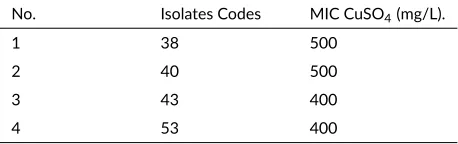
The bacterial cultures used in this research were coded as C38, C40, C43, and C53. They were isolated from several sampling locations of PTFI’s tailings in Timika, Papua (Massora et al. 2016). They were resistant to copper and had a minimum inhibitory concentration (MIC) of 400 mg/L to 500 mg/L copper-sulfate. MIC of each bacterial isolates is shown in Table1. These bacteria were grown in Luria-Bertani (LB) broth containing the following (per liter): tryptone 10 g, yeast extract 5 g, NaCl 10 g, glucose 0.1 g. LB medium was sterilized by autoclaving at 121°C for 20 min. After sterilization, Copper sulfate was added and the pH was adjusted to 7.0 (Andreazza et al. 2011).
2.2. Qualita ve analysis of biofilm forma on
The biofilm formation was analyzed using Scanning Elec-tron Microscopy (SEM) in LPPT Universitas Gadjah Mada. The isolates of copper-resistant bacteria (106
CFU/mL) were inoculated in Luria-Bertani broth and in-cubated overnight at room temperature on a shaker at 170 rpm. The coverslip was placed at the bottom of a Petri dish. Five (5) mL cultures of copper-resistant bacteria were placed on the coverslip surface until completely sub-merged and was incubated for 24 h at 37°C temperature. After incubation, the coverslip was added with 2% glu-taraldehyde and stored at 4°C overnight. The sample in coverslip was prepared according to specimen preparation for SEM analysis (Ratnayake et al. 2012).
2.3. Quan ta ve analysis of biofilm forma on
The quantitative analysis of biofilm formation ability by copper-resistant bacteria was modified fromMathur et al.
(2006) and Merritt et al.(2011). The isolate of copper-resistant bacteria (106CFU/mL) was inoculated into
Luria-Bertani and incubated overnight at room temperature with a shaker at 170 rpm. After incubation, the isolate was added each 100μL into four wells of a microtiter plate. Mi-crotiter plates were incubated at room temperature for 24 and 48 h. Then, the isolates were removed and the plates were washed with aquadest. They were added with 125μL of 0.1% crystal violet solution for each well and stored at room temperature for 10 minutes. The plates were washed with aquadest after crystal violet solution was removed, and dried in inverse position for 1-2 weeks. When the
TABLE 1MIC of copper-resistant bacterial isolates
No. Isolates Codes MIC CuSO4(mg/L).
1 38 500
2 40 500
3 43 400
4 53 400
Source: Massora et al. 2016
plate dried, biofilm was diluted in 200 μL of PBS, then it was stored for 10-15 minutes at room temperature. For quantitative analysis of biofilm formation, 125 μL of the sample was transferred to a new microtiter plate and was measured with micro Elisa auto reader at 492 nm. Biofilm formation ability was classified according toMøretrø et al.
(2003). The sample was classified as weak biofilm former if A492< 0,20; medium former if 0.20 ≤ A492≤ 1.0; and
strong former if A492> 1.0. Based on the ability to resist
cooper and to form a biofilm, the four selected isolates were identified based on their molecular characteristics.
2.4. Molecular iden fica on of copper-resistant and biofilm forming bacteria
The isolates were identified by 16S ribosomal RNA gene sequencing as follows. Pure Culture of Copper-Resistant Bacteria was grown overnight in LB broth for isolation of genomic DNA by using Promega Wizard Genomic DNA Purification Kit (Promega, Madison, WI). 16S rDNA was amplified by using the universal bacterial 16S rDNA primers. Two primers corresponding toEscherichia coli
positions 27F (5’-AGAGTTTGA TCCTGGCTC-3’) and 1492R (5’-GGTTACCTTGTTACGACT-3’) were used for PCR amplification of the 16S ribosomal RNA (18). The PCR reaction mixture (25μL) consisted of 1 μL of DNA template, Ready to Go Mixture PCR, 22 μL of nuclease-free water, 1 μL of primer 27F (10 pmol/μL) and 1 μL of primer 1429R (10 pmol/μL). The 16S rRNA gene was amplified using 35-cycle PCR (initial denaturation), 95ºC for 5 min; subsequent denaturation, 94°C for 30 s; anneal-ing temperature, 52°C for 1 min; extension temperature, 72°C for 1 min and the final extension was carried out at 72°C for 5 min, followed by a 4°C hold. The PCR amplification products were analyzed by electrophoresis on a 1.5% agarose gel. The appearance of 1.5 kb band showed that 16S rDNA had been amplified and ready for sequencing. Sequencing was carried out at Genetic Sci-ence Malaysia. DNA SequSci-ence Similarity and Phyloge-netic Analysis GenBank BLAST (N) was used for homol-ogy searches. Molecular evolutionary and phylogenetic analyses were conducted using MEGA version 6 (Tamura et al. 2013).
3. Results and discussion
3.1. Biofilm qualita ve analysis by SEM
(a) (b) (c) (d)
FIGURE 1Biofilm forma on by C53 isolate (a); C38 isolate; (b) C40 isolate; (c) C43 isolate and (d) C53 isolate that was analyzed using Scanning electron microscopy (SEM). All isolates produce EPS matrix around their cells that enable bacterial cell aggrega on.
stresses or microbially deleterious substances than plank-tonic cells (Annous et al. 2009).
A biofilm is a group of microorganisms in which cells stick to each other on a surface. These adherent cells are frequently embedded within a self-produced matrix of EPS. EPS contributes to protect cells from hostile environ-ments and can bind significant amounts of heavy metals. The composition of EPS depends on the present microor-ganisms, temperature, and availability of nutrients. The biofilm formation by these strains is considered a natural strategy to maintain a favorable niche in stressful environ-ments with increased metals concentrations (Annous et al. 2009;Cabarkapa et al. 2013).
As reported byWorkentine et al.(2008), the biofilm may reduce metal toxicity by altering bacterial physiol-ogy to protect the sensitive chemical targets of the reactive metal species. Upon metal binding, the concentration of the free toxic ions in the cytoplasm is minimized. Biosorp-tion of toxic metals is known from cell membranes, cell walls and EPS of biofilms. For example, the EPS matrix and the contained polysaccharides were reported to bind heavy metals. Thus, the metal tolerance of the bacteria be-longing to that biofilm was enhanced (Annous et al. 2009;
Meliani and Bensoltane 2016).
3.2. Quan ta ve analysis of biofilm forma on
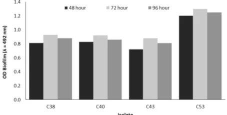
Analysis of copper resistance is not always parallel with the ability to form a biofilm. Therefore, all isolates showed high potency as copper-resistant bacteria when tested using the Microtiter plate biofilm assay (Figure2). The result showed that the biofilm formation on differ-ent isolates varied with time. After 48 h all isolates were found to produce biofilm. The readings of A492at time 48
h, for C38, C40, C43 and C53 isolates were 0.81, 0.83, 0.72, and 1.2 respectively. According to Møretrø et al. (2003), the readings of A492 at time 72 hours, C53 Iso-late can be categorized as strong biofilm former (1.3), and 3 isolates (C38, C40, and C43) as medium biofilm former (0.93, 0.92, and 0.88, respectively).
The time course for attachment varies depending on the organism and must be determined empirically, al-though when using this system, many organisms com-monly studied will form a biofilm within 48 h (Merritt
et al. 2011). In this research, isolates formed a biofilm at 48 h. The highest absorbance value of the isolates was at 72 h and the highest descendance value was at 96 h.
Bacteria that form biofilm are known to possess greater resistances to stress conditions than their plank-tonic counterparts that dispersed in the environment, in-cluding the susceptibility to sanitizers and other antimicro-bials (D’Souza et al. 2014;Tang et al. 2012). Biofilm is formed when bacterial cells attach to one another and/or adhere to a living or inert contact surface. The attached bacterial cells are enclosed in a self-produced polymeric matrix. The organisms can increase their ability to colo-nize and survive in a harsh environmental condition if they are able to form this biofilm (Monier and Lindow 2003;
Tang et al. 2012).
Biofilm growth and its adhesion are mutually depen-dent processes, increase consistency with biofilm matu-ration process. The dispersion of microorganisms from biofilm can be defined as a kind of adaptive response to the conditions present in the environment in which biofilm is developing, for instance, as the adaptive response to the starvation condition (Alipour et al. 2009). The application of the same treatment to bacterial population suspended in the liquid media does not cause activation gene. The resis-tance of the bacteria in the biofilm is most probably the consequence of the activity of several rather than a single mechanism (Cabarkapa et al. 2013). The results showed that the isolates had the potential to form biofilm and re-move copper from the contaminated environment.
3.3. Molecular iden fica on of copper-resistant bacte-ria
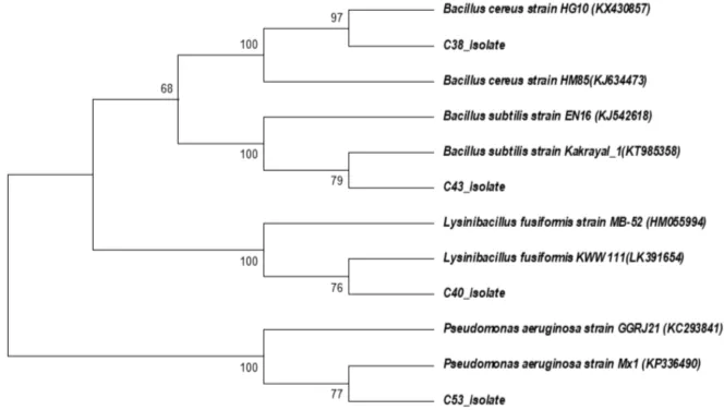
The bacterial isolates were selected based on their high concentration of copper resistance were identified by 16S rRNA gene sequence analysis. Nucleotide sequences were used for GenBank blast analysis and construction of the phylogenetic tree. The Gene of 16S rRNA was ampli-fied using universal primer set (27f and 1492r), with PCR product of 1500 bp in size.
The phylogenetic tree showed evolutionary distance among copper-resistant bacteria based on 16S rRNA gene sequence (Figure3). The phylogenetic tree showed that C38 isolate and Bacillus cereusstrain HG10 were clus-tered together with a bootstrap support of 97%, and a strong bootstrap support of 100% with Bacillus cereus strain HM85. B. cereusstrain HG10 was isolated from activated sludge andB. cereusstrain HM85 was isolated from polluted heavy metal soil.
C43 Isolate formed a clade withBacillus subtilisstrain Kakrayal_1 with a bootstrap support of 79% and a strong bootstrap support of 100% with B. subtilisstrain EN16.
Bacillus is an important bacterial genus for bioremedia-tion of heavy metals in different heavy metal contaminated areas. B.subtilisstrain Kakrayal_1 was isolated from wa-ter environmental andB. subtilisstrain EN16 was isolated from soil.
C40 Isolate formed a clade with Lycinibacillus fusiformisstrain KWW 111 with a bootstrap support of 76% and a strong bootstrap support of 100% with L. fusiformisstrain MB-52. L. fusiformisstrain KWW 111 was isolated from sedimentation basin of the water system andL. fusiformisstrain MB-52 was isolated from biofilms in chlorinated drinking water systems.
C53 Isolate formed a clade with Pseudomonas aeu-ruginosa strain Mx1 with a bootstrap support of 77% and a strong bootstrap support of 100% with P.
aeu-ruginosa strain GGRJ21. P. aeuruginosa strain Mx1 is a dichloro propionate-degrading bacteria. P. aeurugi-nosa strain GGRJ21 was isolated from soil rhizosphere. C53 isolate can be categorized as a strong biofilm for-mer. As the adaptive response to the starvation conditions,
P. aeuruginosa cells produce the alginate lyase enzyme that dissolves alginate, namely the biofilm polysaccharide (Alipour et al. 2009).
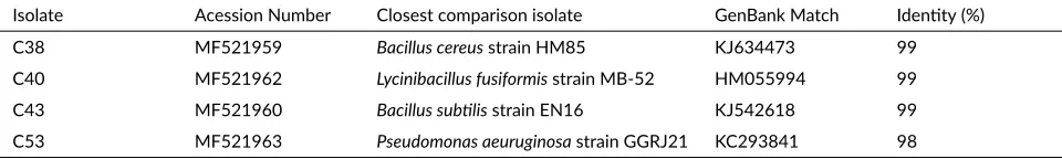
The identity of selected isolates was based on 16S rRNA gene sequence analysis (Table2). C38 isolate had 99% similarity toB. cereusstrain HM85. C43 Isolate had 99% similarity toB. subtilisstrain EN16. C40 Isolate had 99% similarity toL. fusiformisstrain MB52 and C53 iso-late had 98% similarity toP. aeuruginosastrain GGRJ21. The sequences of the isolates have been submitted to the GenBank via the submission portal and every isolate had an accession number (Table2).
In the environmental conditions, bacteria are capa-ble to develop diverse mechanisms as adaptive responses, which enable them to adapt to new environmental con-ditions such as DNA damages, temperatures, starvations, oxidative stresses, and low water activities. This type of adaptive responses has been characterized in details as the bacteria suspended in liquid media, and it is most probable that such adaptations are present in the bacterial biofilm. Catalase production in P. aeruginosa via the activation of Kat B-inducible gene in the response to the treatment with 50 mM of hydrogen peroxide is one of the simple examples of adaptive responses caused by changes of the environmental conditions (Cabarkapa et al. 2013). The genome-wide transcriptional response of copper-adapted and copper-shockedP. aeruginosacultures has revealed a core set of genes comparably regulated under both condi-tions (Teitzel et al. 2006). This core set includes different transport genes suggesting that copper tolerance is mainly achieved by copper efflux. A CueR-like regulator
TABLE 2The closest phylogene c rela ves of bacterial isolates from PT Freeport Indonesia’s tailings.
Isolate Acession Number Closest comparison isolate GenBank Match Iden ty (%) C38 MF521959 Bacillus cereusstrain HM85 KJ634473 99 C40 MF521962 Lycinibacillus fusiformisstrain MB-52 HM055994 99 C43 MF521960 Bacillus sub lisstrain EN16 KJ542618 99 C53 MF521963 Pseudomonas aeuruginosastrain GGRJ21 KC293841 98
vates transcription of several target genes, includingcueA, upon copper addition (Thaden et al. 2010; Rademacher and Masepohl 2012).
The monocistroniccsoRgene is localized directly up-stream of thecopZAoperon inB. subtilis, which encodes a copper chaperone and a copper-ATPase (Smaldone and Helmann 2007). csoR repressescopZA transcription at low copper concentrations by binding a palindromic se-quence (TACCCTAC-N4-GTATGGTA) overlapping the
copZ promoter. csoR is no longer binds the promoter and transcription are relieved at elevated copper concen-trations. In a mutant which lacks ofcsoRrepressor, the
copZAoperon is constitutively transcribed (Rademacher and Masepohl 2012).
Gram-negative and Gram-positive bacteria utilize copper-ATPases as principal defense determinants to ex-crete excess copper from the cytoplasm. Copper- resis-tanat bacteria examined so far induceATPaseexpression, and different species utilize functionally and structuraly different regulators to controlATPasegene transcription. Under copper-limiting conditions, gram-positive bacteria repress transcription , and Gram-negative bacteria acti-vateATPase gene transcription (Rademacher and Mase-pohl 2012).
C38, C40, C43, dan C53 isolates were indigenous bac-teria from tailings which contain copper. The isolates can form biofilm and also developed resistance mechanism by accumulating copper. The ability of the isolates to accu-mulate copper was higher than previously known copper-resistant bacteria. B. pumilusandBacillus sp. isolated from copper mining waste grew up in 300 mg/L of Cu(II) (Andreazza et al. 2011). These mechanisms could be uti-lized for detoxification and removal of copper or the other heavy metal from polluted environmental. Therefore, the next research is encouraged to know the mechanisms of isolates for reducing or detoxification of heavy metals.
4. Conclusions
The main Based on 16S rRNA gene sequence analysis: C38 isolate had 99% similarity toB. cereusstrain HM85. C43 isolate had 99% similarity toB. subtilisstrain EN16. C40 isolate had 99% similarity to L. fusiformis strain MB52 and C53 isolate had 98% similarity toP. aeurug-inosastrain GGRJ21. All of the isolates had biofilm form-ing ability which isolate C53 can be categorized as a strong biofilm former, and 3 other isolates (C38, C40, and C43)
as medium biofilm formers. The capability of C53 iso-late to form strong biofilm formation can be exploited in a bioremediation process which aims to remove copper from tailings.
Acknowledgments
The authors gratefully acknowledge financial support from PT Freeport Indonesia and “Hibah Penelitian Diser-tasi Doktor” of the Ministry of Research and Technology in 2017.
Authors’ contribu ons
MM, EM, ES designed the study. MM, EM, RS, TS con-ducted the field data collection. MM carried out the lab-oratory work. MM, EM, ES, RS, TS analyzed the data. MM, EM, ES wrote the manuscript. All authors have read and approved the final version of the manuscript.
Compe ng interests
The authors declare no competing interest.
References
Alipour M, Suntres ZE, Omri A. 2009. Importance of dnase and alginate lyase for enhancing free and li-posome encapsulated aminoglycoside activity against
Pseudomonas aeruginosa. J Antimicrob Chemother. 64:317–325. doi:10.1093/jac/dkp165.
Andersson S, Nilson M, Dalhammar G, Rajaro GK. 2008. Assesment of carrier materials for biofilm formation and denitrification. Vatten 64:201–207.
Andreazza R, Pieniz S, Okeke BC, Camargo FAO. 2011. Evaluation of copper resistant bacteria from vine-yard soils and mining waste for copper biosorption. Braz J Microbiol. 42:66–74. doi: 10.1590/S1517-83822011000100009.
Annous BA, Fratamico PM, Smith JL. 2009. Quorum sensing in biofilms: why bacteria behave the way they do. J Food Sci. 74:24–37. doi: 10.1111/j.1750-3841.2008.01022.x.
Cerca N, Pier GB, Vilanova M, Oliveira R, Azeredo J. 2005. Quantitative analysis of adhesion and biofilm formation on hydrophilic and hydropho-bic surfaces of clinical isolates of Staphylococ-cus epidermidis. Res Microbiol 156(4):506–514. doi:10.1016/j.resmic.2005.01.007.
Chen YQ, Ren GJ, An SQ, Sun QY, Liu CH, Shuang JL. 2008. Changes of bacterial community struc-ture in copper mine tailings after colonization of reed
(Phragmites communis). Pedosphere 18(6):731–740. doi:10.1016/S1002-0160(08)60068-5.
Desai C, Jain K, Madamwar D. 2008. Evaluation of in vitro Cr(VI) reduction potential in cy-tosolic extracts of three indigenous bacillus sp. isolated from Cr(VI) polluted industrial land-fill. Bioresource Technology 99(14):6059–6069. doi:10.1016/j.biortech.2007.12.046.
D’Souza EL, Meria QGS, Barbosa IM, Athayde AJAA. 2014. Biofilm formation byStaphylococcus aureus
from food contact surfaces in a meat-based broth and sensitivity to sanitizers. Braz J Microbiol 45:67–75. Espírito Santo C, German N, Elguindi J, Grass G, C R.
2014. Biocidal mechanisms of metallic copper sur-faces. In: G Borkow, editor. Use of biocidal surfaces for reduction of healthcare acquired infections. Cham: Springer International Publishing. p. 103–136. Freitas O, Delerue-Matos C, Boaventura R. 2009.
Optimization of Cu(II) biosorption onto Ascophyl-lum nodosum by factorial design methodology. Journal of Hazardous Materials 167(1):449–454. doi:10.1016/j.jhazmat.2009.01.001.
He Z, Gao F, Sha T, Hu Y, He C. 2009. Iso-lation and characterization of a cr(vi)-reduction ochrobactrum sp. strain cscr-3 from chromium land-fill. Journal of Hazardous Materials 163(2):869–873. doi:10.1016/j.jhazmat.2008.07.041.
Jain AN, Udayashankara TH, Lokesh K. 2014. Review on bioremediation of heavy metals with microbial iso-lates and amendments on soil residue. Int J Sci Res 3:118–123.
Khusnuryani A, Martani E, Wibawa T, Widada J. 2014. Molecular identification of phenol-degrading and biofilm-forming bacteria from wastewater and peat soil. Indones J Biotechnol. 19(2):99–110. doi:10.22146/ijbiotech.9299.
Massora M, Martani E, Sugiharto E, Sarwom R, Sinaga T. 2016. Isolation and selection of copper-resistant bac-teria from PT Freeport Indonesia’s tailings. Biotech-nology Conference IV. Yogyakarta: UGM Biotechnol-ogy Research Center.
Mathur T, Singhal S, Khan S, Upadhyay D, Fatma T, Rat-tan A. 2006. Detection of biofilm formation among the clinical isolates of staphylococci: an evaluation of three different screening methods. Indian J Med Mi-crobiol. 24(1):25–29. doi:10.4103/0255-0857.19890.
Meliani A, Bensoltane A. 2016. Biofilm-mediated heavy metals bioremediation in PGPR Pseudomonas. J Bioremediat Biodegrad. 7(5):1–9. doi: 10.4172/2155-6199.1000370.
Merritt JH, Kadouri DE, O’Toole G. 2011. Growing and analysing static biofilms, current protocols in microbi-ology. New York: John Wiley & Sons, Inc.
Monier JM, Lindow SE. 2003. Differential sur-vival of solitary and aggregated bacterial cells promotes aggregate formation on leaf surfaces. Proc Natl Acad Sci USA 100(26):15977–15982. doi:10.1073/pnas.2436560100.
Møretrø T, Hermansen L, Holck AL, Sidhu MS, Rudi K, Langsrud S. 2003. Biofilm formation and the pres-ence of the intercellular adhesion locus ica among staphylococci from food and food processing envi-ronments. Appl Environ Microbiol 69(9):5648–5655. doi:10.1128/AEM.69.9.5648-5655.2003.
Osredkar J, Suskar N. 2011. Copper and zinc, biological role and significance of copper/zinc imbalance. J Clin Toxicol. 3:1–18.
Rademacher C, Masepohl B. 2012. Copper-responsive gene regulation in bacteria. Microbiology 158(10):2451–2464. doi: 10.1099/mic.0.058487-0.
Ratnayake K, Joyce DC, Webb RI. 2012. A conve-nient sample preparation protocol for scanning electron microscope examination of xylem-occluding bacterial biofilm on cut flowers and foliage. Sci Hortic. 140(Supplement C):12–18. doi:10.1016/j.scienta.2012.03.012.
Roy S, Ganguly S. 2015. Bioremediation activity of microorganisms in soil environment contaminated by heavy metals. J Biol Chem Res. 32:323–330.
Smaldone GT, Helmann JD. 2007. Csor regu-lates the copper efflux operon copza in Bacil-lus subtilis. Microbiology 153:4123–4128. doi:10.1099/mic.0.2007/011742-0.
Tamura K, Stecher G, Peterson D, Filipski A, Kumar S. 2013. Mega6: Molecular evolutionary genetics anal-ysis version 6.0. Mol Biol Evol 30(12):2725–2729. doi:10.1093/molbev/mst197.
Tang PL, Pui CF, Wong WC, Noorlis A, Son R. 2012. Biofilm forming ability and time course study of growth of Salmonella typhi on fresh pro-duce surfaces. Int Food Res Journal J 19(1):71–76. doi:10.1128/JB.00837-06.
Teitzel GM, Geddie A, De Long SK, Kirisits MJ, Whiteley M, Parsek MR. 2006. Survival and growth in the pres-ence of elevated copper: Transcriptional profiling of copper-stressed pseudomonas aeruginosa. J Bacteriol. 188(20):7242–7256. doi:10.1128/JB.00837-06. Thaden JT, Lory S, Gardner TS. 2010.
doi:10.1128/JB.01528-09.
Umrania VV. 2006. Bioremediation of toxic heavy metals using acidothermophilic autotro-phes. Biores Technol. 97(10):1237–1242. doi:10.1016/j.biortech.2005.04.048.