Endothelial Progenitor Cells in Diabetic Vasculopathy
Anna Meiliana
1,2* and Andi Wijaya
1,2*
R E V I E W A R T I C L E
1Post Graduate Program in Clinical Biochemistry, Hasanuddin University, Makassar
2Prodia Clinical Laboratory, Jakarta
B
ACKGROUND:
The discovery of endothelial
progenitor cell (EPC) a decade ago by Asahara
et al has refuted the previous belief that
vasculogenesis only occurs during embryogenesis.
The reduced circulating concentration of EPCs is a
surrogate marker of endothelial function and has
been implicated in the pathogenesis of many vascular
diseases.
CONTENT:
Diabetes is linked to impaired vascular
function, including alterations in both endothelial
cells and EPCs. A number of studies have shown
that individuals with diabetes have decreased level
of circulating EPCs and that the severity of disease
is inversely proportional to EPC levels. In vitro,
hyperglycemia increases the rate of EPC senescence
and the angiogenic function of EPCs from patients
with either type 1 or type 2 diabetes is impaired such
that they are poorly proliferative and fail to incorporate
into forming vessel-like structures.
Given the comprehensive role of EPC alterations in
diabetes complications, modulation of the levels and/
or function of EPCs may be considered a potential
therapeutic strategy.
SUMMARY:
The available data demonstrating that
decrease or dysfunction of EPCs may have a prominent
role in the pathogenesis of all diabetes complications.
Further approaches, such as EPC administration, may
represent novel treatments for diabetic vasculopathy
Abstract
in the future. To date, many barriers remain to such
a therapeutic approach. Firstly, there is no specific
marker for EPC at present. Secondly, techniques of
EPC isolation are not standardized, preventing direct
comparison between various studies. The long-term
effects of transplanted EPCs are currently unknown.
Introduction
*Address correspondence to this author at: Prodia Clinical Laboratory, Jl. Cisangkuy No. 2 Bandung; e-mail: anna_m@ prodia.co.id, [email protected]
Diabetic Complications
The impaired neovascularization found in the
diabetic setting may be explained by the inability of
diabetic EPCs to respond to stromal-derived factor-1
(SDF-1), which plays a crucial role in EPC homing (13).
Dysfunctional EPCs in the diabetic setting are also
associated with impaired re-endothelialization (14).
Furthermore, EPC mobilization was also impaired
after ischaemic reperfusion injury in a streptozotocin
induced diabetic mouse model, a finding that was
associated with altered SDF-1 and VEGF release as well
as impaired hypoxia inducible factor-1a upregulation
(15). These in vivo animal studies demonstrated that
diabetes is associated with impaired EPC mobilization,
differentiation and ability to form tubules.
The administration of both autologous and
heterologous unfractionated BM cells, which contain
EPCs, has produced dramatic improvement in
neovascularization in various ischaemic states (16-26).
However, the results of autologous EPC transplantation
in the diabetic setting may not be as promising. As
discussed above, in patients with DM, EPC numbers
are lower and their function is impaired.
Diabetes complications represent a huge burden
for patients and health services. The fight against
each single complication has led to significant
improvements in diabetes care, especially for
microvascular complications, yet macroangiopathy
remains a major source of morbidity and mortality. A
common approach for the prevention and treatment of
diabetes complications relies on the understanding of
their complex pathophysiology (27).
While it was originally thought that a single patient
tends to develop the cluster of micro- or macrovascular
complications, recent prospective studies show that
typical markers of microvascular dysfunction, such as
microalbuminuria or retinal vascular abnormalities,
are associated with an increased risk of macrovascular
events (28,29). These and other data suggest that there
must be a unifying pathogenetic model underlying
diabetes complications. To date, the most credited
and supported model proposes that oxidative stress
originating from mitochondria activates all subcellular
damage pathways (30). However, subsequent events
diverge for each complication, and there is not a
supracellular unifying hypothesis. The discovery that
a subset of circulating immature cells contributes to
vascular homeostasis has been a major achievement in
many fields of basic science.
Endothelial progenitor cells (EPCs) are circulating
immature cells that contribute to vascular homeostasis
and compensatory angiogenesis. During the last
decade, data have become available indicating that
alterations in EPCs may have an important causative
role in the development and progression of virtually
all diabetes complications (27).
Endothelial Progenitor Cells (EPCs)
EPCs were discovered in 1997 as circulating cells with
the ability to differentiate into mature endothelium
and take part in neoangiogenesis (1). EPCs share
markers of hematopoietic (CD34 and CD133) and
endothelial (KDR, CD31, and vWf) lineages (31), are
derived from bone marrow, and can be mobilized to
the peripheral circulation in response to many stimuli
(32). Tissue ischemia, through the release of growth
factors and cytokines, mobilizes EPCs, which, once
in the peripheral circulation, specifically home on the
ischemic sites to stimulate compensatory angiogenesis
(33). Moreover, EPCs constitute a circulating pool of
cells able to form a cellular patch at sites of endothelial
injury, thus contributing directly to the homeostasis
and repair of the endothelial layer. Taken together,
these observations suggest that EPCs have a major
role in cardiovascular biology; in fact, the extent of
the circulating EPC pool is now considered a mirror
of cardiovascular health. Virtually all risk factors for
atherosclerosis have been associated with decrease
and/or dysfunction of circulating EPCs (34), while
an expanded EPC pool is associated with a decreased
cardiovascular mortality (11).
absent on mature endothelial cells and monocytic cells
(36) Thus, CD133+VEGFR2+ cells more likely reflect
immature progenitor cells, whereas CD34+VEGFR2+
may also represent shedded cells of the vessel wall. At
present, it is unclear whether CD133 only represents a
surface marker or has a functional activity involved in
regulation of neovascularization. Overall, controversy
exists with respect to the identification and the origin
of endothelial progenitor cells, which are isolated from
peripheral blood mononuclear cells by cultivation
in medium favoring endothelial differentiation. In
peripheral blood mononuclear cells, several possible
sources for endothelial cells may exist: (1) the rare
number of hematopoietic stem cells, (2) myeloid cells,
which may differentiate to endothelial cells under the
cultivation selection pressure, (3) other circulating
progenitor cells (eg, “side population” cells), and (4)
circulating mature endothelial cells, which are shed off
the vessel wall (37) and adhere to the culture dishes
(38).
A combination of CD34, kinase insert
domain-containing receptor (KDR) and CD133 are commonly
used to define EPCs at present. The use of
fluorescence-activated cell sorter (FACS) analysis to define EPCs
appears to be the most optimal approach but may be
technically challenging and the best combination of the
‘true’ markers for EPCs is currently unknown (39).
The study of EPC biology consists of two related
aspects: quantitative evaluation of the EPC pool
and functional assessment. Circulating EPCs can
be quantified directly ex vivo using flow cytometry,
which is considered the gold standard for this purpose
(40); typical surface antigens to identify EPCs are
CD34, CD133, and KDR. Functional characteristics are
explored in vitro using standardized protocols (38).
Proliferation refers to the ability of EPCs to expand
and form colonies in culture: EPCs should proliferate
in response to growth factors released locally after
vascular damage or tissue ischemia. Adhesion is a
further step required for both reendothelization and
angiogenesis; it is assessed as the ability of EPCs to
adhere to a monolayer of mature endothelium in
culture. Migration of EPCs through the extracellular
matrix is crucial for the growth of new vessels and
is generally assessed in vitro as the ability to invade
the lower side of a Boyden-like chamber. Finally, after
EPCs have adhered to the vessel wall, migrated into
the interstitium, and expanded locally, they should
spatially organize to form vascular structures; this
property can be assessed in vitro as a tube formation
assay in which EPCs are seeded with human umbilical
vein endothelial cells on a gel of extracellular matrix
proteins.
Two types of EPC had different morphology,
proliferation rates, and survival behaviors. They also
had different gene expression profiles, leading to
different function in vitro. Despite such differences
in gene expression and in vitro function, they equally
contributed to neovasculogenesis in vivo in that early
EPC secreted angiogenic cytokines, whereas late EPC
supplied a sufficient number of endothelial cells (41).
Late EPC was different from early EPC in the
expression of VE-cadherin, Flt-1, KDR, and CD45.
Late EPC produced more nitric oxide, incorporated
more readily into human umbilical vein endothelial
cells monolayer, and formed capillary tube better than
early EPC. Early EPC secreted angiogenic cytokines
(vascular endothelial growth factor, interleukin 8)
more so than late EPC during culture in vitro. Both
types of EPC showed comparable in vivo vasculogenic
capacity (41).
Distinct origins of the different types of EPCs exist
that have different functions in neovascularization.
Mixed transplantation of these cells results in
synergistic neovascularization through cytokines and
MMPs (42).
EPCs in Neovascularization
Improvement of neovascularization is a therapeutic
option to rescue tissue from critical ischemia (43).
The finding that bone marrow–derived cells can
home to sites of ischemia and express endothelial
marker proteins has challenged the use of isolated
hematopoietic stem cells or EPCs for therapeutic
vasculogenesis.
However, the number of incorporated cells with an
endothelial phenotype into ischemic tissues is generally
quite low. How can such a small number of cells
increase neovascularization? A possible explanation
might be that the efficiency of neovascularization may
not solely be attributable to the incorporation of EPCs
in newly formed vessels, but may also be influenced
by the release of proangiogenic factors in a paracrine
manner (38).
expression of growth factors such as VEGF, HGF, and
IGF-1. Moreover, adherent monocytic cells, which were
cultivated under similar conditions, but do not express
endothelial marker proteins, also release VEGF, HGF,
and G-CSF (44). The release of growth factors in turn
may influence the classical process of angiogenesis,
namely the proliferation and migration as well as
survival of mature endothelial cells (45). However,
EPCs additionally incorporated into the newly formed
vessel structures and showed endothelial marker
protein expression in vivo.
EPCs in Endothelial Regeneration
In the past, the regeneration of injured endothelium
has been attributed to the migration and proliferation
of neighboring endothelial cells. More recent studies,
however, indicate that additional repair mechanisms
may exist to replace denuded or injured arteries.
Bone
marrow
transplantation
experiments
revealed that bone marrow–derived cells can contribute
to reendothelialization of grafts and denuded
arteries (46-48). However, in a model of transplant
arteriosclerosis, bone marrow–derived cells appear
to contribute only to a minor extent to endothelial
regeneration by circulating cells (49). These data
again indicate that there might be at least two distinct
populations of circulating cells that principally are
capable to contribute to reendothelialization, namely
mobilized cells from bone marrow and non-bone
marrow–derived cells. The latter ones may arise from
circulating progenitor cells released by non-bone
marrow sources (eg, tissue resident stem cells) or
represent vessel wall–derived endothelial cells (49,
46-48).
A rapid regeneration of the endothelial monolayer
may prevent restenosis development by endothelial
synthesis of antiproliferative mediators such as
nitric oxide. Indeed, enhanced incorporation of
-galactosidase–positive, bone marrow–derived cells
was associated with an accelerated reendothelialization
and reduction of restenosis (46,47). Similar results
were reported by Griese et al, who demonstrated
that infused peripheral blood monocyte–derived EPC
home to bioprosthetic grafts and to balloon-injured
a
h
t
i
w
d
e
t
a
i
c
o
s
s
a
g
n
i
e
b
r
e
t
t
a
l
e
h
t
,
s
e
i
r
e
t
r
a
d
i
t
o
r
a
c
significant reduction in neointima deposition (50).
Likewise, infusion of bone marrow–derived CD34-/
CD14+ mononuclear cells, which are not representing
the population of the “classical hemangioblast,”
contributed to endothelial regeneration (51). The
regenerated endothelium was functionally active
as shown by the release of NO (51), which is
supposed to be one of the major vasculoprotective
mechanisms. Consistently, neointima development
was significantly reduced after cell infusion (51).
Whereas the regeneration of the endothelium by EPCs
protects lesion formation, bone marrow–derived
stem/progenitor cells may also contribute to plaque
EPCs Mobilization Differentiation and Homing
angiogenesis, thereby potentially facilitating plaque
instability (52). However, in a recent study, no influence
of bone marrow cell infusion on plaque composition
was detected in nonischemic mice (53). An increase
in plaque size was only detected in the presence of
ischemia, suggesting that ischemia-induced release of
growth factors predominantly accounts for this effect
(53).
Overall, these studies implicate that regardless
of the origin of circulating endothelial progenitor
cells, this pool of circulating endothelial cells may
exert an important function as an endogenous repair
mechanism to maintain the integrity of the endothelial
monolayer by replacing denuded parts of the artery
(Figure 1). One can speculate that these cells may
also regenerate the low grade endothelial damage
by ongoing induction of endothelial cell apoptosis
induced by risk factors for coronary artery disease
(54).
Moreover, various risk factors for coronary artery
disease, such as diabetes, hypercholesterolemia,
hypertension, and smoking, affect the number and
functional activity of EPCs in healthy volunteers (10)
and in patients with coronary artery disease (55).
In addition, factors that reduce cardiovascular
risk such as statins (56,46,47,57) or exercise (58)
elevate EPC levels, which contribute to enhanced
endothelial repair. The balance of atheroprotective
and proatherosclerotic factors, thus, may influence
EPC levels and subsequently reendothelialization
capacity.
The release of EPCs from the bone marrow is
regulated by a variety of growth factors, enzymes,
ligands, and surface receptors. Activation of
matrix metalloproteinase-9, which promotes the
transformation of membrane-bound Kit ligand to
a soluble Kit ligand and the subsequent move of
cKit-positive stem and progenitor cells, including
a common hematopoietic and angioblast precursor
(hemangioblast), to the vascular zone of the bone
marrow microenvironment, are initial steps in this
complex process (59). The signals, which initiate the
diversion of the hemangioblast to either hematopoietic
precursor cells or EPCs, are largely unknown at present
but may include angiogenic growth factors from the
periphery.
To date, no clear definition exists as to when
an endothelial progenitor cell turns into a mature,
fully differentiated endothelial cell in vivo. One
possibility could be the loss of CD133 and a parallel
or subsequent expression of von Willebrand factor in
conjunction with the appearance of other endothelial
characteristics. The starting point of this differentiation
process may by the migration of EPCs from the bone
marrow to the systemic circulation. After homing, ie,
after adhesion and insertion into the monolayer of
surrounding mature vascular ECs, this process may
be completed (60).
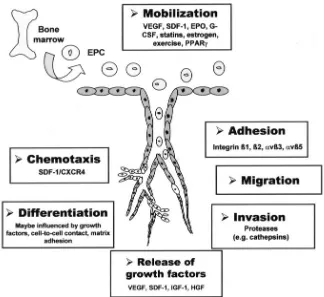
Recruitment and incorporation of EPCs requires
a coordinated sequence of multistep adhesive and
signaling events including chemoattraction, adhesion,
and transmigration, and finally the differentiation to
endothelial cells (Figure 2) (38).
The local bone marrow microenvironment, the
,
Figure 2. Mechanism of EPC homing and differentiation. Reprinted by permission from Wolters Kluwer Health, Circ. Res, Copyright (2004).
for VEGF-receptor-1+ stem cells, whereas in later
stages PlGF functions are mediated by MMP-9 (68).
Thus, increasing the local concentration of MMP-9
in the bone marrow cleaves membrane bound Kit
ligand (mKitL) and, finally, releases soluble Kit ligand
(KitL; also known as stem cell factor) (59). After all,
this process transfers endothelial and hematopoietic
progenitor cells from the quiescent to the proliferative
niche. However, the question of whether G-CSF–
induced stem cell mobilization depends on MMP-9
is still a matter of debate (59,69,70). This controversy
might be explained by the fact that MMP-9 plays a
pivotal role in growth factor-induced hematopoietic
progenitor mobilization in wild-type animals, whereas
compensatory upregulation of enzymes with a similar
activity profile to MMP-9 might mask the impact of
MMP-9 deficiency in the knockout model. As discussed,
eNOS is essential to maintain adequate progenitor cell
mobilization in response to distinct stimuli, including
VEGF, statins, exercise, and estrogen, in the regulation
of stem and progenitor cell mobilization (58,62,71,72).
The defective mobilization was caused by the lack of
eNOS (Nos3) provided by the bone marrow stromal
microenvironment.
Therefore, eNOS deficiency in the bone marrow
microenvironment impaired the mobilization of
stem and progenitor cells from the bone marrow. In
contrast, intravenous injection of stem and progenitor
cells circumvented the defective mobilization from the
bone marrow and improved the neovascularization
after induction of hind limb ischemia. Therefore,
eNOS-derived NO is a physiological regulator of stem
and progenitor cell mobilization in the bone marrow
stromal microenvironment.
Given that patients with coronary artery disease
showed a diminished NO bioavailability in peripheral
endothelial cells, one may speculate that this also
translates into the bone marrow. EPC numbers are
lower in patients with coronary artery disease or
diabetes (55,74) and are correlated with NO-dependent
vasorelaxation measured in the forearm (10).
in embryonic and adult target tissue leads to an
expression of chemotactic factors inducing migration
of angioblasts and EPCs toward the region to be
vascularized by vasculogenesis.
SDF-1 gene expression is regulated by the
transcription factor hypoxia-inducible factor-1 in
endothelial cells, resulting in selective in vivo
expression of SDF-1 in ischemic tissue in direct
proportion to reduced oxygen tension. HIF-1-induced
SDF-1 expression increases the adhesion, migration
and homing of circulating CXCR4-positive progenitor
cells to ischemic tissue.
These data show that the recruitment of
CXCR4-positive progenitor cells to regenerating tissues is
mediated by hypoxic gradients via HIF-1-induced
expression of SDF-1 (80).
In brief, the directed migration of angioblasts is
regulated by a combination of different soluble and
interaction molecules that build overlapping attractive
and repulsive gradients of soluble factors forming the
migratory route for the endothelial precursor cells. It
can be suggested that the correct positioning of the
endothelial cells after they start to migrate toward the
target tissue is dependent on a fine balance of signals.
Soluble factor from regions that should be vascularized
diffuse to the endothelial precursors, attract them and
keep them in their migratory phenotype state until
they reach their final position (81).
Vascular endothelial growth factor caused
the phosphorylation of AMPK, acetyl-coenzymeA
(CoA) carboxylase (ACC), and eNOS in human cord
blood-derived EPCs. The expression of EC markers,
including VE-cadherin and intercellular adhesion
molecule1 (ICAM-1), was also increased but blocked
by Compound C, an AMPK inhibitor. AICAR, an
AMPK agonist, increased the phosphorylation of ACC
and eNOS and the expression of EC markers in a time-
and dose-dependent manner, which reinforces the
positive effect of AMPK on EPC differentiation.
The activation of eNOS by AMPK during EPC
differentiation provides a novel mechanism for
the pleiotropic effects of statins in benefiting the
cardiovascular system (82).
There is accumulating evidence that platelets
mediate the effect of hematopoietic cytokines to recruit
bone marrow–derived cells to the vasculogenic niche.
Thrombopoietic cytokines thrombopoietin (TPO)
and stem cell factor (SCF) are significantly elevated
shortly after ischemic tissue injury, and transgenic
mice deficient in TPO or the TPO receptor (c-MPL)
demonstrate significantly impaired hind – limb
revascularization and inhibited angiogenic tumor
growth (83).
Aggregation of activated platelets was necessary
for the recruitment of bone marrow–derived c-Kit+
ScaI+ Lin- progenitor cells and CD34+ cells to sites of
endothelial disruption (84). Hematopoietic cells were
unable to adhere directly to the exposed extracellular
matrix, but were
tethered to platelet surface P-selectin via P-selectin
glycoprotein ligand (PSGL)-1 binding. Platelet release
of SDF-1 provided an ongoing retention signal for
bone marrow–derived cells at these sites (84). The
mechanism by which platelets release SDF-1, VEGF
and other growth factors in a site-specific fashion is
likely to involve glycoprotein (GP) IIb– dependent
platelet aggregation (84), inward receptor signaling,
and MMP-9–mediated SDF-1 release (83). In addition
to platelet-derived SDF-1, exposed smooth muscle
cells also produce SDF-1 after arterial injury, and
platelet activation via CXCR4 signaling leads to
P-selection upregulation, further promoting arrest of
bone marrow–derived cells to activated endothelium
(85).
Taken together, these data suggest that aggregation
and activation of platelets at sites of exposed
subendothelium and vasculogenesis play a major role
in the recruitment, differentiation, and incorporation
of bone marrow–derived progenitor cells.
These insights have profound clinical implications;
inhibition of platelet-deployed growth factors or their
receptors may be an effective strategy to block tumor
growth, whereas activation of these pathways may
be used to accelerate revascularization and tissue
regeneration (86).
Adverse effects of EPC mobilization have
been described as contribution of EPCs to tumor
neovascularization in some tumor models (89).
Moreover, circulating progenitor cells have been
implicated in the neovascularization of arteriosclerotic
lesions of allografts and in further atherosclerotic plaque
progression in an ischemic setting (52,53). However,
transfusion of EPCs enhanced re-endothelialization
and reduced neointima formation after vascular injury
(47). One may speculate that the endothelial repair
capacity might override the potential harmful effects
of plaque neovascularization. Thus, future studies
have to determine the overall influence of EPC levels
on atherosclerotic disease progression and prognosis
(32).
Cardiovascular Risk Factors and EPCs
Small clinical studies have shown that the number
of circulating EPCs inversely correlates with risk
factors for atherosclerosis (10,55). Circulating CD34/
KDR-positive progenitor cells are reduced to ~50%
in patients with CAD compared with control groups.
In addition, EPCs isolated from patients with CAD
displayed an impaired migratory response, which was
inversely correlated with the number of cardiovascular
risk factors (55).
In patients with arterial hypertension, systolic
blood pressure negatively correlates with the number
of circulating CD133+ and CD34+/KDR+ EPCs,
whereas the clonogenic potential (number of colony
forming units–ECs) is not impaired by arterial
hypertension (55). Experimental data demonstrate
that angiotensin II, a potent mediator of detrimental
effects in arterial hypertension, can accelerate the
onset of EPC senescence by gp91 phox – mediated
increase in oxidative stress, leading to an impaired
proliferation activity of EPCs. Angiotensin II–induced
EPC senescence was inhibited by treatment with the
angiotensin II type 1 receptor blocker valsartan (90).
Recent studies have underlined the detrimental
effects of types 1 and 2 diabetes on EPC function (74,91).
Tepper et al demonstrated that in type 2 diabetes
proliferation capacity of EPCs was reduced, adhesion
capacity on activated human ECs was impaired.
Hypercholesterolemia was associated with
reduced EPC numbers in 20 age-matched patients
with hypercholesterolemia (8). Proliferative capacity,
migratory activity, and in vitro vasculogenesis were
negatively influenced by hypercholesterolemia. The
underlying mechanisms are probably an increased
rate of EPC senescence/apoptosis, as demonstrated
after incubation of EPCs with oxidized LDL (92).
A role for HDL in promoting the repair of injured
endothelium by stimulating the recruitment of
endothelial progenitor cells (EPCs) into the endothelial
layer (93).
Smoking has been identified as an important
risk factor for reduced EPC numbers in one of the
first studies on cardiovascular risk factors by Vasa et
al (55). However, Wang et al recently demonstrated
that the role of nicotine is more complex than
initially expected (94). In an experimental study, they
demonstrated that low concentrations of nicotine
(10-8-10-12 mol/L) increased EPC number and activity,
whereas higher (toxic) concentrations (>10-6 mol/L)
were associated with cytotoxicity. In humans, Kondo
et al demonstrated that chronic smokers (n=15) exhibit
reduced EPC levels that can be restored after smoking
cessation within 4 weeks (9).
In humans, a significant increase in progenitor
cell numbers was observed in patients who resumed a
standardized physical activity during a rehabilitation
program (58), in patients with CAD (95), and in healthy
individuals exercising for ≥30 minutes (96).
CRP, at concentrations known to predict
adverse vascular outcomes, directly inhibits EPC
differentiation, survival, and function, key components
of angiogenesis and the response to chronic ischemia.
This occurs in part via an effect of CRP to reduce EPC
eNOS expression. The PPAR agonist rosiglitazone
inhibits the negative effects of CRP on EPC biology.
The ability of CRP to inhibit EPC differentiation and
survival may represent an important mechanism that
further links inflammation to cardiovascular disease
(97).
He et al demonstrate that human EPCs possess
a unique property to withstand oxidative injury and
that elevated expression of manganese superoxide
dismutase (MnSOD, a mitochondria-located SOD) is
a critical intrinsic mechanism protecting EPCs against
oxidative stress (100).
The finding that GPx-1 expression is essential for
EPC functions may also have clinical implications,
given that patients with chronic heart failure (101) and
with type 2 diabetes (102) showed a downregulation
of GPx-1. This in turn may contribute to the reduced
EPC numbers and functions in patients with
coronary artery disease and severe heart failure
(55,103). However, other antioxidative enzymes such
as superoxide dismutases and catalases are also
downregulated in these patients (101,102). Thus, it
seems mandatory to understand the specific role of the
various antioxidative enzymes for EPC functions. In
the future, a specific interference with the expression
and activity of antioxidative enzymes in progenitor
cells from patients might be a therapeutic strategy to
improve their regenerative potential (104).
Schroeter et al (105) have now demonstrated
that infusion of leptin-stimulated human EPCs
reduces
neointima
formation
and
enhanced
reendothelialization through upregulation of v 5-
and 4-integrin–dependent adhesion to platelets in
ferric chloride–induced vascular injury. With this
elegant approach, the effect of leptin on EPC-mediated
endothelial recovery can be readily discerned from
leptin-enhanced thrombosis. This not only extends the
mechanisms by which platelet-derived chemokines,
P-selectin, and 2 integrins can support the arrest of
EPCs and CD34+ progenitor cells at sites of arterial
injury (85,106) but also adds a new dimension to the
functional profile of leptin.
PPAR- activation with agonist (GW501516 or
L-165041) increased the proliferation of human EPCs
and protected them from hypoxia-induced apoptosis.
In addition, PPAR- activation enhanced EPC
functions, such as transendothelial migration, and
tube formation. These actions by PPAR- activation
in EPCs were dependent on the phosphatidylinositol
3-kinase/Akt pathway (107).
What are the mechanisms by which risk factors
mediate their negative effect on progenitor cells?
It appears that the signaling pathways mediating
progenitor cell impairment are similar to the previously
identified regulators of endothelial cell function and
atherosclerosis and include a dysregulation of nitric
oxide (NO) and reactive oxygen species (ROS). A
diabetic environment or high glucose exposure in
vitro is associated with reduced nitric oxide (NO)
bioavailability in cultured EPCs, and plasma levels of
endogenous NO – synthase inhibitors (asymmetrical
dimethyl-L-arginine) are associated with clinically
reduced EPC numbers (108). In experimental studies,
endothelial NO synthase (eNOS)-derived NO was
shown to be essential for basal, VEGF-, and SDF-1–
induced migration of EPCs or bone marrow–derived
progenitor cells, and eNOS-/- progenitor cells showed
a reduced homing capacity in ischemia models in vivo
(109,110). The underlying mechanisms mediating the
NO effects, eg, on chemokines or integrin expression or
signaling, need to be further defined. Despite various
studies supporting an important role of eNOS for
progenitor cell mobilization and function (58,71,109),
under certain conditions, eNOS uncoupling may lead
to an increased ROS production. To what extent the
redox balance in stem cells (as opposed to cultured
EPCs or crude bone marrow homogenates used in the
study(111) favors such uncoupling processes is unclear
and deserves further studies.
In healthy individuals and patients with coronary
artery disease, age is associated with a reduced
number and function of cultured EPCs, circulating
CD34+KDR+ cells, or CD133+ cells and of granulocyte
macrophage colony forming units (GM-CFUs) in the
bone marrow (11,55,112).
On a molecular level aging is linked to a reduction
of telomere length. The proliferative history of a cell
is written on telomeres: telomere erosion reflects the
number of past divisions experienced by a cell and
its proliferative potential (113). In addition, telomere
erosion may contribute to telomere shortening.
When long telomeres protect chromosomal ends,
cells can undergo repeated cell divisions. Conversely,
telomere shortening beyond a critical length leads to
genomic instability, DNA damage, p53 activation, and
ultimately cell cycle arrest (114).
EPCs
Alteration in Diabetes
Both cytometric and culture methods have extensively
demonstrated that type 1 and type 2 diabetic patients
have less circulating EPCs than matched healthy
subjects. Moreover, diabetic EPCs display functional
impairment, such as reduced proliferation, adhesion,
migration, and incorporation, into tubular structures
(74,91,116). The mechanisms underlying EPC reduction
in diabetes include weak bone marrow mobilization,
decreased proliferation, and shortened survival in
peripheral blood (127).
Poor collateral formation in diabetes may be
attributed to weaker bone marrow stimulation from
the ischemic tissue. Fadini et al (27) have recently
confirmed this hypothesis, showing that bone marrow
mobilization of EPCs after ischemia-reperfusion injury
is defective in diabetic rats. Inability to mobilize EPCs
was associated with downregulation of HIF-1 and
weakened release of marrow – stimulating factors, such
compensatory angiogenesis (15). Another study has
shown that progenitor cell mobilization restored
blood flow in diabetic mice (117). It is conceivable
that HIF-1 deregulation in diabetes depends on an
overproduction of reactive oxygen species (ROS).
Although molecular mechanisms that regulate
EPC release in peripheral blood are complex and not
fully understood, a role for the phosphatidylinositol
(PI) 3-kinase/protein kinase-B and endothelial nitric
oxide (NO) synthase pathways has been shown
(72,118). As diabetes is characterized by altered
activation of PI 3-kinase/Akt pathways and by
reduced NO bioavailability (119), dysfunction of
these subcellular pathways may be involved in the
defective mobilization of EPCs from bone marrow.
Hyperglycemia may be the common feature that
affects survival and function of EPCs because similar
alterations have been demonstrated in both type 1
and type 2 diabetes. In vivo, hyperglycemia induces
oxidative stress by increasing the production of ROS
and alters leukocyte and endothelial function (30).
Recently, Krankel et al. (120) have convincingly
demonstrated that high glucose impairs proliferation,
survival, and function of cultured EPCs, with
concomitant-decreased NO production and matrix
metalloproteinase-9 activity. Furthermore, activation
of mitogen-activated protein kinases has been revealed
as a potential mechanism of EPC dysfunction induced
by high glucose (121). A definite demonstration is
that correction of hyperglycemia by insulin therapy
(15,122) can indeed restore the normal EPC pool.
Fadini et al (27) shown that patients with
the metabolic syndrome have decreased levels of
CD34+KDR+ EPCs compared with patients without
the syndrome (116). Circulating CD34+ cells are
synergically decreased by clustering components of
the metabolic syndrome, and their levels negatively
correlate with the homeostasis model assessment
value, a measure of insulin resistance (123). In fact,
insulin resistance, the typical hallmark of metabolic
syndrome, is characterized by a defective activation of
the PI 3-kinase/Akt pathway and decreased endothelial
NO synthase activity, which are considered essential
for EPC mobilization and function. Oxidative stress
plays a crucial role in the pathogenesis of diabetes
complications (30), as well as in the entire atherogenic
process. Therefore, stress-induced apoptosis may be
one mechanism of EPC reduction in diabetes. The
literature provides ample evidence that EPCs might
decrease because of increased apoptosis and that EPCs
are stress sensitive (124).
In summary, reduction in circulating EPCs
in diabetic patients may recognize at least three
pathophysiological explanations: impaired bone
marrow mobilization, defective proliferation, and
enhanced apoptosis. Remarkably, in accordance
with Brownlee’s unifying hypothesis (30), oxidative
stress appears as a major determinant of all of these
mechanisms. Interestingly, two very recent studies
have demonstrated that the natural transketolase
activator, benfotiamine, which is theoretically able to
prevent the subcellular damage pathways triggered
by oxidative stress, restored EPC-mediated healing of
ischemic diabetic limbs in mice (125) and prevented
hyperglycemia – mediated EPC dysfunction via
modulation of the Akt pathway (126).
EPCs and the Diabetic Paradox
marrow–derived cells are mobilized and recruited
at sites of retinal neovascularization in response to
VEGF and SDF-1 (127,128). Therefore, not only local
endothelial cells, but also EPCs may be involved in the
development of proliferative retinopathy. This seems
counterintuitive, as diabetes complications that may
affect the same patient, such as diabetic retinopathy
and PAD, can have opposing EPC alterations, the
one being associated with increased and the other
with decreased EPC levels. Parallely, a single diabetic
patient can presentat the same time with complications
of excessive angiogenesis (proliferative diabetic
retinopathy) and of poor angiogenesis (symptomatic
PAD) (27).
An unexplained paradox puzzles diabetologists:
diabetic patients must face both poor vessel growth in
ischemic heart and limbs and increased angiogenesis in
retinal complications (129,130). Endothelial progenitor
cells (EPCs) are marrow-derived cells involved in
adult neovascularization and endothelial homeostasis
(1,131). It has been postulated that low EPCs in
peripheral blood may have a role in cardiovascular
disease, and we have demonstrated that EPCs are
reduced in macrovascular diabetes complications
(10,116). On the other hand, an excess of EPCs may be
involved in pathologic neoangiogenesis of cancer and
proliferative retinopathy (132,133). Therefore, diabetes
complications may be associated with both decreased
and increased EPCs. Recently, novel therapeutic
approaches have been directed to enrich the EPC
pool in ischemic diseases and to block EPC function
in proliferative diseases (128,134). These approaches
in diabetic subjects require cautious evaluation of the
implications carried by the paradox and new studies
to unravel its causes (135,136).
Fadini et al (141) show that CD34+ cells and
CD34+KDR+ cells are differentially altered in the
presence of DR and PAD. These two common diabetes
complications exhibit a very different behavior in
terms of angiogenic response to ischemia, and this
contrast has been termed “diabetic paradox.” Many
growth factors have been proposed to have a role in
this phenomenon (137-139), all of which are potent
stimuli for progenitor cell mobilization and homing
to ischemic tissues (32). Indeed, recent data indicate
that marrow-derived cells are involved in retinal
neovascularization and that DR+ patients have
increased levels of circulating progenitors (133,140).
Conversely,
macrovascular
complications
are
characterized by reduced angiogenesis and exhausted
EPC levels (116). This findings suggest that enhanced
endothelial differentiation of circulating progenitors
characterize DR, as shown by the high CD34+KDR+
proportion and the enhanced efficiency of EPC culture
in contrast with the poor endothelial differentiation of
PAD patients (141).
Therefore, the differential regulation of circulating
progenitors, possibly in association with different
oxygen gradients and local accumulation of growth
factors, may explain why peripheral ischemia cannot
stimulate angiogenesis as retinal ischemia does. Going
deeper into the systemic events accompanying retinal
vascular proliferation may provide novel therapeutic
targets against peripheral ischemic complications. The
notion that EPCs may be involved in retinal vascular
proliferation should induce caution when trying to
expand the EPC pool to ameliorate the cardiovascular
profile. For instance, erythropoietin itself is an
angiogenic factor that may worsen proliferative
diabetic retinopathy (142).
Therapeutic Potential of EPC in Neovascularization
In vivo models of limb ischemia and myocardial
infarction supported a direct role for bone marrow–
derived cells in neovascularization, concluding that
these cells differentiate into smooth muscle and
endothelial cells that incorporate into neovessels
(143-146). However, more recently, other groups
have contested these findings and suggest that bone
marrow–derived progenitor cells act primarily via
paracrine mechanisms, secreting chemokines such
as angiopoietin-1 and vascular endothelial growth
factor (VEGF) at sites of vascular injury to enhance
A number of factors have been shown to enhance
EPC function either at the site of mobilization from the
bone marrow and/or at sites of homing to damaged
blood vessels. Age-associated decreases in a wide
range of factors, includingVEGF and PDGF signaling
and circulating estrogen levels have been suggested in
animal models to be important factors in the decrease
in EPC mobilization from the bone marrow, cardiac
homing, and regeneration (151-153).
Clinical trials in patients with coronary artery
disease or limb ischemia showed improvement after
treatment with plasmid DNA encoding VEGF165
(154-157); thus VEGF may prove to be a feasible and
successful therapy for vascular injury.
PDGF acts to promote the angiogenic activity of
local vascular cells after myocardial infarction as well
as to recruit bone marrow cells that differentiate into
both endothelial cells and cardiomyocytes (158,159).
Intramyocardial treatment with PDGF therefore
appears to enhance the interactions between bone
marrow and cardiac stem cell niches and provides
functional benefit to the injured heart. Additionally,
studies recently demonstrated that PDGF pathways are
essential for maintaining the cardiomyogenic potential
of Oct3/4+ bone marrow cells that is decreased with
age (160).
Tenascin-C, which we have shown to be a
downstream mediator of PDGF signaling in the cardiac
vasculature, is associated with sites of EPC recruitment
in the heart and is also important for bone marrow
cell–mediated mechanisms of cardiac angiogenesis
(161). This protein is downregulated in the aging bone
marrow and may also be depleted in the aging heart
(161). Thus mechanisms that restore tenascin-C may
have multiple actions that promote cardiovascular
repair mechanisms, including CSC-mediated cardiac
regeneration.
Another factor acting both in the cardiac and bone
marrow stem cell niches is stromal cell–derived factor
(SDF)-1. In the bone marrow, SDF-1 is among a number
of proteins, including VEGF and placental growth
factor, that induce matrix metalloproteinase (MMP)-9,
leading to the translocation of stem cells to the vascular
zone of the bone marrow before mobilization (59).
SDF-1 has also been shown to promote bone marrow
cell proliferation and angiogenesis (162).
Other factors that regulate EPC function include
the hematopoietic growth factor granulocyte
macrophage–colony
stimulating
factor,
which
increases the number of circulating EPCs in vivo,
while enhancing differentiation of EPCs in vitro (33).
Similarly G-CSF also has stimulated EPC mobilization
in clinical trials, but these EPCs display functional
impairment of migratory properties in vitro.
Pharmacologically, the class of factors known as
the statins, or 3-hydroxy-3-methyl glutaryl coenzyme
A reductase inhibitors, has also been shown to
enhance EPCmediated angiogenesis in models
of ischemic tissue injury. In vivo, statin treatment
increases the numbers of circulating EPCs and
enhances both neovascularization in corneal assays
and reendothelialization of injured vessels, promoting
incorporation of labeled bone marrow–derived cells
into these vessels (46,56,163). Mechanistically, statin
treatment in vitro appears to inhibit EPC senescence,
via induction of telomere repeat binding factor-2, which
inhibits induction of the DNA damage
checkpoint-kinase 2 (164). Simvastatin activates the serine–
threonine kinase Akt in endothelial cells, promoting
endothelial cell survival and migration (165). Akt also
acts downstream of VEGF and may therefore represent
a key regulator of VEGF-mediated neovasculogenesis
(166). Thus, these data suggest that statin therapy may
constitute an important approach in the development
of strategies to improve EPC survival and function
and to improve cardiac repair pathways in the aging
population. Indeed, the TOPCARE-AMI clinical trial
demonstrated that the treatment of ex vivo cultured
blood-derived progenitor cells with atorvastatin
was found to be safe and potentially effective for the
enhancement of cardiac regeneration (167).
EPC Transplantation in Diabetes
was counteracted by administration of EPCs from
control animals. Conversely, diabetic EPCs were not
able to stimulate vascularization, even becoming
antiangiogenic (169,170). Additionally, EPCs appeared
important for vascularization and healing of diabetic
wounds (171).
Patients with diabetes and PAD had a further
significant decrease in circulating EPC levels, especially
in the presence of ischemic foot lesions. Remarkably,
EPC levels strongly correlated with the anklebrachial
index, the most objective diagnostic and prognostic
test for lower extremity arterial disease (116).
Lower-limb atherosclerosis: higher degrees
of carotid stenosis, as well as worse stages of leg
claudication and ischemic lesions, were associated
with lower levels of EPCs, suggesting that EPC
count may be considered a valuable marker of
atherosclerotic involvement. Indeed, cytometric
techniques, which allow EPC count, arewidely used
for routine laboratory testing, and the determination
of EPCs is sufficiently reproducible to be used in the
clinical practice (11,40,123).
In the case that EPCs are defined as CD34+KDR+
cells. The clinical usefulness would stand in
that EPCs not only mirror vascular function and
atherosclerotic burden but also reflect the endogenous
vasculoregenerative potential. Importantly, there are
data suggesting that measuring EPCs would provide
additional information over the classical risk factor
analysis; in one study (11), CD34+KDR+ EPC count
predicted cardiovascular events independently of
risk factors and hard indexes, such as left ventricular
ejection fraction. Moreover, EPCs isolated from diabetic
patients with PAD exhibited impaired proliferation
and adhesion to mature endothelium (172). We suggest
that impaired collateralization leading to the clinical
manifestations and complications of atherosclerosis
in diabetes may be attributable to decreased and
dysfunctional EPCs.
Huang et al. (23) have transplanted bone marrow–
mobilized cells, as an EPC enriched fraction, into
critically ischemic limbs of diabetic patients. Compared
with standard therapy, cell therapy improved
angiographic scores and ankle-brachial index values
and reduced relevant end points, such as ulcer size
and need for limb amputation (27). Recently, Körbling
et al. were able to isolate a large quantity of clinically
graded EPC from healthy volunteers by leukaparesis
after G-CSF stimulation (173). This method may offer
promise as a means of improving the efficacy of EPC
transplantation for therapeutic neovascularization in
the diabetic setting. However, previous data, while
suggesting that G-CSF or granulocyte–macrophage
colony – stimulating factor (GM-CSF) administration
did increase EPC mobilization, the therapeutic effect in
patients with coronary artery disease was conflicting
(174,175).
Figure 3. Approaches to improve efficacy of EPC transplantation in diabetic setting.
More recent data have shown that transplantation
of G-CSF-mobilized EPCs for the treatment of critical
limb ischaemia in patients with DM is safe and effective
(23). Nevertheless, further studies are required to
ensure the safety of this approach. EPC number can
also be increased by augmenting the differentiation
ability of MNC by co-administration of growth
factors with unfractionated BM-MNC. For instance,
co-administration of placental growth factor and
BM-MNC into a diabetic hindlimb mouse model augments
EPC differentiation from diabetic BM-MNCsin vitro
(176).
Improved neovascularization can also be achieved
in a diabetic hindlimb ischaemic mouse model by
inhibition of NADPH oxidase–derived reactive
oxidative species overproduction (177). Silencing of
apoptotic gene expressionsuch as p53 renders EPCs
derived from patients with T2DM more resistant
to oxidative stress. This method also reduces EPC
senescence and improves their ability to differentiate
and form tubules in vitro (178). Lastly, administration
of benfotiamine has been recently shown to increase
EPC number and reverse EPC dysfunction in patients
with DM (125,126).
Other growth factors such as VEGF, transforming
growth factor- 1, angiopoetin-1, SDF-1, constitutive
telomerase reverse transcriptase, hepatocyte growth
factor, PDGF-B, basic fibroblast growth factor and
Diabetes complications represent a huge burden for
patients and health services. A common approach
for the prevention and treatment of diabetes
complications relies on the understanding of their
complex pathophysiology.
During the last decade, data have become available
indicating that alteration in EPCs may have an
important causative role in all diabetes complications.
New approaches such as EPC administration may
represent novel treatments for diabetic vasculopathy
in the future. To date, many barriers remain to such
a therapeutic approach, hopefully in the near future
these problems can be resolved.
Conclusion
medications such as erythropoietin, oestrogen,
phosphodiesterase-5 inhibitor, puerarin and Ginkgo
biloba have been shown to augment EPC number
and function in a non-diabetic settings (179). These
factors, however, have not been studied in the setting
of diabetes but may be beneficial for therapeutic
neovascularization in DM. The approaches to improve
efficacy of EPC transplantation in diabetic setting is
summarized in figure 3 (39).
References:
1. Asahara T, Murohara T, Sullivan A, Silver M, van der Zee R, Li T, et al. Isolation of putative progenitor endothelial cells for angiogenesis. Science 1997; 275: 964–967.
2. Kalka C, Masuda H, Takahashi T, Kalka-Moll WM, Silver M, Kearney M, et al. Transplantation of ex vivo expanded endothelial progenitor cells for therapeutic neovascularization. Proc Natl Acad Sci U S A 2000; 97: 3422–3427.
3. Kubota Y, Kishi K, Satoh H, Tanaka T, Nakajima H, Nakajima T. Transplanted endothelial progenitor cells augment the survival areas of rat dorsal flaps. Cell Transplant 2003; 12: 647–657.
4. Griese DP, Ehsan A, Melo LG, Kong D, Zhang L, Mann MJ, et al. Isolation and transplantation of autologous circulating endothelial cells into denuded vessels and prosthetic grafts: implications for cell-based vascular therapy. Circulation 2003; 108: 2710–2715.
5. He T, Smith LA, Harrington S, Nath KA, Caplice NM, Katusic ZS.. Transplantation of circulating endothelial progenitor cells restores endothelial function of denuded rabbit carotid arteries. Stroke 2004; 35: 2378–2384.
6. Takahashi M, Nakamura T, Toba T, Kajiwara N, Kato H, Shimizu Y.. Transplantation of endothelial progenitor cells into the lung to alleviate pulmonary hypertension in dogs. Tissue Eng 2004; 10: 771–779.
7. Imanishi T, Moriwaki C, Hano T, Nishio I. Endothelial progenitor cell senescence is accelerated in both experimental hypertensive rats and patients with essential hypertension. J Hypertens 2005; 23: 1831–1837.
8. Chen JZ, Zhang FR, Tao QM, Wang XX, Zhu JH, Zhu JH.. Number and activity of endothelial progenitor cells from peripheral blood in patients with hypercholesterolaemia. Clin Sci (Lond) 2004; 107: 273–280.
9. Kondo T, Hayashi M, Takeshita K, Numaguchi Y, Kobayashi K, Iino S, et al. Smoking cessation rapidly increases circulating progenitor cells in peripheral blood in chronic smokers. Arterioscler Thromb Vasc Biol. 2004; 24: 1442–1447. 10. Hill JM, Zalos G, Halcox JP, Schenke WH, Waclawiw MA,
Quyyumi AA, et al. Circulating endothelial progenitor cells, vascular function, and cardiovascularrisk. N Engl J Med 2003; 348: 593–600.
11. Werner N, Kosiol S, Schiegl T, Ahlers P, Walenta K, Link A, et al. Circulating endothelial progenitor cells and cardiovascular outcomes. N Engl J Med. 2005; 353: 999 –1007.
13. Segal MS, Shah R, Afzal A, Perrault CM, Chang K, Schuler A, et al. Nitric oxide cytoskeletal-induced alterations reverse the endothelial progenitor cell migratory defect associated with diabetes. Diabetes 2006; 55: 102–109.
14. Ii M, Takenaka H, Asai J, Ibusuki K, Mizukami Y, Maruyama K, et al. Endothelial progenitor thrombospondin-1 mediates diabetes-induced delay in reendothelialization following arterial injury. Circ Res 2006; 98: 697–704.
15. Fadini GP, Sartore S, Schiavon M, Albiero M, Baesso I, Cabrelle A, et al. Diabetes impairs progenitor cell mobilization after hindlimb ischemiareperfusion injury in rats. Diabetologia 2006; 49: 3075–3084.
16. Janssens S, Dubois C, Bogaert J, Theunissen K, Deroose C, Desmet W, et al. Autologous bone marrow-derived stem-cell transfer in patients with ST-segment elevation myocardial infarction: double-blind, randomised controlled trial. Lancet 2006; 367: 113–121.
17. Kang HJ, Lee HY, Na SH, Chang SA, Park KW, Kim HK, et al. Differential effect of intracoronary infusion of mobilized peripheral blood stem cells by granulocyte colony-stimulating factor on left ventricular function and remodeling in patients with acute myocardial infarction versus old myocardial infarction: the MAGIC Cell-3-DES randomized, controlled trial. Circulation 2006; 114: I145–I151.
18. Strauer BE, Brehm M, Zeus T, Köstering M, Hernandez A, Sorg RV, et al. Repair of infarcted myocardium by autologous intracoronary mononuclear bone marrow cell transplantation in humans. Circulation 2002; 106: 1913–1918.
19. Wollert KC, Meyer GP, Lotz J, Ringes-Lichtenberg S, Lippolt P, Breidenbach C, et al. Intracoronary autologous bonemarrow cell transfer after myocardial infarction: the BOOST randomised controlled clinical trial. Lancet 2004; 364: 141–148.
20. Erbs S, Linke A, Adams V, Lenk K, Thiele H, Diederich KW, et al. Transplantation of blood-derived progenitor cells after recanalization of chronic coronary artery occlusion: first randomized and placebocontrolled study. Circ Res 2005; 97: 756–762.
21. Strauer BE, Brehm M, Zeus T, Bartsch T, Schannwell C, Antke C, et al. Regeneration of human infarcted heart muscle by intracoronary autologous bone marrow cell transplantation in chronic coronary artery disease: the IACT Study. J Am Coll Cardiol 2005; 46: 1651–1658.
22. Perin EC, Dohmann HF, Borojevic R, Silva SA, Sousa AL, Mesquita CT, et al. Transendocardial, autologous bone marrow cell transplantation for severe, chronic ischemic heart failure. Circulation 2003; 107: 2294–2302.
23. Huang P, Li S, Han M, Xiao Z, Yang R, Han ZC.. Autologous transplantation of granulocyte colony-stimulating factor-mobilized peripheral blood mononuclear cells improves critical limb ischemia in diabetes. Diabetes Care 2005; 28: 2155–2160.
24. Saigawa T, Kato K, Ozawa T, Toba K, Makiyama Y, Minagawa S, et al. Clinical application of bone marrow implantation in patients with arteriosclerosis obliterans, and the association between efficacy and the number of implanted bone marrow cells. Circ J 2004; 68: 1189–1193.
25. Tateishi-Yuyama E, Matsubara H, Murohara T, Ikeda U, Shintani S, Masaki H, et al. Therapeutic angiogenesis for patients with limb ischaemia by autologous transplantation of bone-marrow cells: a pilot study and a randomised controlled trial. Lancet 2002; 360: 427–435.
26. Tse HF, Thambar S, Kwong YL, Rowlings P, Bellamy G, McCrohon J, et al. Safety of catheter-based intramyocardial autologous bone marrow cells implantation for therapeutic angiogenesis. Am J Cardiol 2006; 98: 60–62.
27. Fadini MD, Sartore S, Agostini C, Avogaro A. Significance of Endothelial Progenitor Cells in Subjects With Diabetes. Diab Care 2007; 30: 1305 – 1313.
28. Akasaka T, Yoshida K, Hozumi T, Takagi T, Kaji S, Kawamoto T, et al. Retinopathy identifies marked restriction of coronary flow reserve in patients with diabetes mellitus. J Am Coll 1997; 30:935–941.
29. Klein BE, Klein R, McBride PE, Cruickshanks KJ, Palta M, Knudtson MD, et al. Cardiovascular disease, mortality, and retinal microvascular characteristics in type 1 diabetes: Wisconsin Epidemiologic Study of Diabetic Retinopathy. Arch Intern Med 2004; 164:1917–1924.
30. Brownlee M. The pathobiology of diabetic complications: a unified mechanism. Diabetes 2005; 54:1615–1625.
31. Fadini GP, Agostini C, Avogaro A: Characterization of endothelial progenitor cells. Biochem Biophys Res Commun 2005; 336:1–2.
32. Aicher A, Zeiher AM, Dimmeler S: Mobilizing endothelial progenitor cells. Hypertension 2005; 45:321–325.
33. Takahashi T, Kalka C, Masuda H, Chen D, Silver M, Kearney M, et al. Ischemia- and cytokineinduced mobilization of bone marrow-derived endothelial progenitor cells for neovascularization. Nat Med 1999; 5:434–438.
34. Werner N, Kosiol S, Schiegl T, Ahlers P, Walenta K, Link A, Bohm et al. Circulating endothelial progenitor cells and cardiovascular outcomes. N Engl J Med 2005; 353:999– 1007.
35. Gehling UM, Ergun S, Schumacher U, Wagener C, Pantel K, Otte M, et al. In vitro differentiation of endothelial cells from AC133-positive progenitor cells. Blood. 2000; 95: 3106– 3112.
36. Handgretinger R, Gordon PR, Leimig T, Chen X, Buhring HJ, Niethammer D, et al. Biology and plasticity of CD133_ hematopoietic stem cells. Ann NY Acad Sci. 2003; 996: 141–151.
37. Mutin M, Canavy I, Blann A, Bory M, Sampol J, Dignat-George F. Direct evidence of endothelial injury in acute myocardial infarction and unstable angina by demonstration of circulating endothelial cells. Blood. 1999; 93: 2951–2958.
38. Urbich C, Dimmeler S. Endothelial Progenitor Cells. Characterization and Role in Vascular Biology. Circ Res 2004; 95: 343 – 353.
39. Liew A, McDermott JH, Barry F, O’Brien T. Endothelial Progenitor Cells for the Treatment of Diabetic Vasculopathy: Panacea or Pandora’s Box? Diabetes Obes Metab 2008; 10: 353 – 366.
40. Rustemeyer P, Wittkowski W, Jurk K, Koller A: Optimized flow cytometric analysis of endothelial progenitor cells in peripheral blood. J Immunoassay Immunochem 27: 77–88, 2006
41. Hur J, Yon CH, Kim HS, Choi JH, Kang HJ, Hwang KK, et al. Characterization of Two Types of Endothelial Progenitor Cells and Their Different Contributions to Neovasculogenesis. Arterioscler Thromb Vasc Biol 2004; 24: 288 – 293.
42. Yoon CH, Hur J, Park KW, Kim JH, Lee CS, Oh IY, et al. Synergistic Neovascularization by Mixed Transplantation of Early Endothelial Progenitor Cells and Late Outgrowth Endothelial Cells. The Role of Angiogenic Cytokines and Matrix Metalloproteinases. Circulation 2005; 112: 1618 – 1627.
43. Isner JM, Asahara T. Angiogenesis and vasculogenesis as therapeutic strategies for postnatal neovascularization. J Clin Invest. 1999; 103: 1231–1236.