Establishment of hairy root cultures of
Ammi majus
Aleksandra Kro´licka
a, Izabela Staniszewska
b, Krzysztof Bielawski
a,
Edmund Malin´ski
b, Janusz Szafranek
b, Ewa L
*
ojkowska
a,*
aDepartment of Biotechnology,Faculty of Biotechnology,Uni6ersity of Gdan´sk and Medical,Uni6ersity of Gdan´sk,K l*adki24,
80-822Gdan´sk,Poland
bFaculty of Chemistry,Uni6ersity of Gdan´sk,Sobieskiego18/19,80-952Gdan´sk,Poland
Received 31 January 2000; received in revised form 4 September 2000; accepted 5 September 2000
Abstract
Axenically grownAmmi majusplantlets were inoculated with seven differentAgrobacterium rhizogenesstrains. Hairy root lines were established only after inoculation with the two agropine strains: A4 and LBA9402. The growth rate of hairy root cultures was about thirty times faster than that of callus and cell suspension cultures. Polymerase chain reaction with primers for the genes
rolBandrolC confirmed the integration of the T-DNA fragment of Ri plasmid ofA.rhizogenesto the genome of hairy roots obtained after transformation by both Agrobacterium strains. The furanocoumarins (psoralen, xanthotoxine, bergapten and imperatorin) usually found in seeds ofA.majuswere not detected in callus, cell suspension and hairy root cultures using Gas chromatography – mass spectrometry (GC – MS). However, umbelliferone, a precursor of furanocoumarins, was detected in callus, cell suspension and hairy root cultures. The umbelliferone content in extracts of hairy root cultures, obtained after transformation by A4, was similar to that determined in A.majusseeds (19mg/g DW) and higher than those obtained for cell suspension and callus cultures (2 and 9mg/g DW, respectively). © 2001 Elsevier Science Ireland Ltd. All rights reserved.
Keywords:Ammi majus;Agrobacterium rhizogenes; Furanocoumarins; Hairy roots; Secondary metabolites; Transformation
www.elsevier.com/locate/plantsci
1. Introduction
Bishops weed — Ammi majus L. (Apiaceae) is considered to be one of the richest natural sources of linear coumarins and furanocoumarins: umbel-liferone, psoralen, xanthotoxine, bergapten and imperatorin. The pharmacological activity of A. majushas been known since the work of Scho3berg and Sina [1] when it was shown that the therapeu-tically effective substances of these plants are fura-nocoumarins. The fruit ofA. majus has been used in the Mediterranean and bordering regions in the
treatment of leucoderma,psoriasis, 6itiligoand the production of suntan lotion [2,3]. In Europe the growth of Ammi is poor due to the cool climate (light frosts). Attempts to acclimatise A. majus in Central European climatic conditions were not successful: the fruits failed to ripen in moderate climatic zones and plants were highly susceptible to infection [4]. In moderate climates seed produc-tion was also limited and the germinaproduc-tion rate poor. Great interest in therapeutic use of fura-nocoumarins, as well as limited natural sources, have led to a series of studies on the production of these biologically active compounds in vitro. Ac-cumulation of furanocoumarins by A. majus in in vitro plant, callus and cell suspension have been investigated, but the productivity was not satisfac-tory [2 – 5].
The aim of our work was to obtain hairy root cultures of A. majus after transformation of ex-plants of in vitro growing plant withA. rhizogenes.
Abbre6iations: BAP, 6-benzylaminopurine; DW, dry weight; FW, fresh weight; GC – MS, Gas chromatography-mass spectrometry; IAA, indolyl-3-acetic acid; KIN, kinetin; MS, Murashige-Skoog’s medium; NAA, naphthaleneacetic acid; PPM™, plant preservative mixture™; PCR, polymerase chain reaction.
* Corresponding author. Tel.:+48-58-3012241 ext. 345; fax:+ 48-58-3012807.
E-mail address:[email protected] (E. L*ojkowska).
Upon plant transformation, T-DNA from the root-inducing Ri plasmid of a soil bacterium A. rhizogenes is integrated into the plant genome. Expression of the genes from the T-DNA region in plant tissue leads to the formation of adventitious roots [6]. Transformed root cultures are very use-ful, as they grow rapidly, are genetically stable and capable of synthesising metabolites found in the biological roots and other plant organs, in abun-dance [7,8].
The present study demonstrates the successful transformation of A. majuscultures by two differ-ent strains of A. rhizogenes. The growth rate, as well as coumarins and furanocoumarins concen-tration in seeds and in in vitro cultures (callus, cell suspension and hairy roots) have been compared.
2. Material and methods
2.1. Plant material
The plantlets of A. majus were grown on MS medium [9] supplemented with 2.0 mg/l NAA, 2.0 mg/l BAP, 30.0 g/l sucrose and 7.5 g/l agar, at a temperature of 20 – 22°C and illumination of 900 lux with photoperiod of 16-h light/8-h dark for 2 – 3 weeks [5].
2.2. Bacteria strains
Agrobacterium rhizogenes agropine strains: A4 [10], LBA 9402 [11], ATCC 15834 [12]; mannopine strains: ICPB TR 7 [13], NCPPB 8196 [14]; nopa-line strain: ATCC 11325 [15] and cucumopine strain: ICPB TR 107 [14] were grown on MYA agar medium [16] supplemented with 200mM
ace-tosyringone in the dark, at 26°C. For transforma-tion, 24 h old bacterial cultures were used.
2.3. Transformation and establishment of hairy root cultures
Explants of stalks and leaves of A. majus were inoculated with freshly grown A. rhizogenes cul-tures. After inoculation, the explants were trans-ferred to the MS agar medium without growth regulators and cultured in darkness. When roots appeared, they were transferred with a fragment of tissue to fresh MS medium containing 30 g/l su-crose, 500 mg/l of claforan (Hoechst, M. Roussel)
and 500 mg/l of carbenicillin (Polfa, Tarchomin) to eliminate bacteria. For more efficient and stable elimination ofA.rhizogenes2.0 ml/l PPM™ (Plant Cell Technology, Inc. Washington) was also used in some experiments. Axenic cultures derived from single root tips were established after 3 – 5 subcul-tures in a 250 ml Erlenmeyer flask containing 75 ml MS medium with claforan and carbenicillin, but without plant growth regulators. Bacteria-free hairy roots were then maintained on a liquid MS medium without antibiotics, in the dark at 22°C on a rotary shaker at 110 rpm. Subcultures were made every 2 or 3 weeks. Fresh weight of the cultures was measured every 10 days.
2.4. Detection of bacterial DNA in plant tissue
A. rhizogenes strains; A4 and LBA 9402 were
used to isolate Ri plasmid. Plasmid DNA was isolated from 24 h cultures of A. rhizogenes (OD600=0.3) using FastDNA® Kit BIO 101. Frozen leaf tissue of untransformed A. majus and frozen tissue of hairy roots were homogenised with 1% polyvinylpyrrolidone and CLS – VF+PPS buffer in Fast Prep™ System FP120 (Instruments Inc. Holbrook, NY) and genomic DNA was iso-lated using Binding Matrix and ready-to-use buffers from FastDNA® Kit BIO 101.
Oligonucleotide primers for PCR detection of homologous sequences to rolB and rolC genes were designed on the basis of the DNA sequence of these genes described by Furner [17] (rolB 5%
-GCTCTTGCAGTGCTAGATTT-3% and 5%
-GAAGGTGCAAGCTACCTCTC-3%; rolC
5%-CTCCTGACATCAAACTCGTC-3% and 5%
-TGCTTCGAGTTATGGGTACA-3%) using pro-gram BLAST.
Each PCR reaction contained: standard PCR buffer (Promega Co., Madison), 1.0 U Taq DNA polymerase (Promega Co., Madison), 2 mM MgCl2, 0.2 mM dNTP (Gibco BRL), 8 pmol of each primer and 75 – 100 ng of the target DNA (final volume 50 ml). Amplification conditions: 35
2.5. Secondary metabolites extraction
Seeds, callus, cell suspension and hairy root cultures of A. majus were collected, dried at 50°C and extractions of the secondary metabolites were performed in a Soxhlet apparatus. Samples (about 3 g of DW) were extracted exhaustively with petroleum ether, chloroform and methanol. Clear (free of chlorophyll) chloroform and methanol extracts were collected separately.
2.6. Gas chromatography–mass spectrometry
Identification of the components of the methanol extracts of A. majus seeds and in vitro growing tissue was performed by GC-MS. Chloro-form extracts of plant tissue were purified on a column packed with 200-mesh silica gel (Mach-erey-Nagel) and eluted with 50 ml of ethyl acetate [18]. UV active fractions were evaporated to dry-ness in a stream of dry nitrogen and derived with a mixture (10:1) of bissilylacetamid and trimethyl silyl chloride (10 min at 100°C). Then capillary gas chromatography was applied for samples contain-ing volatile silyl derivatives. Thus, unmodified, methoxyl-, and hydroxylated coumarins were analysed in one gas chromatography run. All frac-tions were subjected to GC and GC – MS.
The analyses were carried out on a GC 8000 TOP gas chromatograph, equipped with a capil-lary column (DB1-HT, length 30 m, I.D. 0.25 mm, 0.1 mm film thickness) with split ratio 1:30 for an
injection port. Argon was used as a carrier gas. Initial oven temperature was 60°C kept for 5 min, then a temperature programme of 10°C per min.
was employed to 200°C and held at 200°C for an additional 20 min [2]. Silyl derivative of henei-cosanol-11 was used as an internal standard and allowed for quantitative determination of cou-marins in plant tissue extracts.
Mass spectra (70 eV) were recorded on a VG TRIO-3 mass spectrometer, (Micromass UK). The samples were introduced through a Hewlett Pack-ard 5890 gas chromatograph equipped with RTX-1 column and under the same chromatographic conditions as mentioned for gas chromatography. Helium was used as carrier gas. The ionisation chamber was kept at 230°C. A one second scan was used for mass spectra recording. For the initial 14 min no spectra were recorded to avoid solvent residues.
3. Results and discussion
Our study indicated that in vitro cultures of callus and cell suspension accumulate umbellifer-one but not furanocoumarins (Table 1). The eco-nomic importance of furanocoumarins, the chemical synthesis of which is very expensive [19], has stimulated interest in transformation of A. majus byA. rhizogenes, establishing conditions for fast growing hairy root cultures and study of their ability to produce furanocoumarins.
3.1. Establishment of hairy root cultures
Hairy roots were obtained after transformation of A. majus with the agropine strains of A. rhizo -genes. Hairy root formation was observed about 3
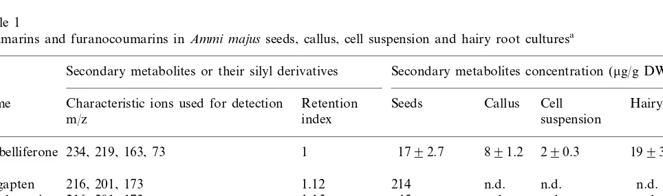
Table 1
Coumarins and furanocoumarins inAmmi majusseeds, callus, cell suspension and hairy root culturesa
Secondary metabolites or their silyl derivatives Secondary metabolites concentration (mg/g DW)
Name Characteristic ions used for detection Retention Seeds Callus Cell Hairy roots suspension
m/z index
Umbelliferone 234, 219, 163, 73 1 1792.7 891.2 290.3 1993.3
216, 201, 173 1.12 214 n.d. n.d. n.d.
Bergapten
216, 201, 173 1.15 45
Xanthotoxin n.d. n.d. n.d.
246, 231, 203 1.32 98 n.d. n.d. n.d.
Isopimpinellin
n.d. n.d. n.d. n.d.
Marmesine 259, 215, 103, 73 n.d.
aCallus culture-MS+5.0 mg/l IAA+1.0 mg/l BAP+3.0% sucrose, 0.75% agar; cell suspension-MS+0.5 mg/l GA
3+1.0 mg/l
Table 2
Growth of A. majus callus, cell suspension and hairy root cultures on MS mediuma
Growth of culture (g FW) Types of culture
10 days 20 days
0 30 days
Callus 1.7 3.1 4.1 6.9
2.7 3.8 6.4
Cell suspensions 1.3
3.9 8.0
0.1 15.0
Hairy roots
aCallus culture-MS+5.0 mg/l IAA+1.0 mg/l BAP+3.0%
sucrose, 0.75% agar; cell suspension-MS+0.5 mg/l GA3+1.0
mg/l KIN+3.0% sucrose; hairy roots-MS+3.0% sucrose.
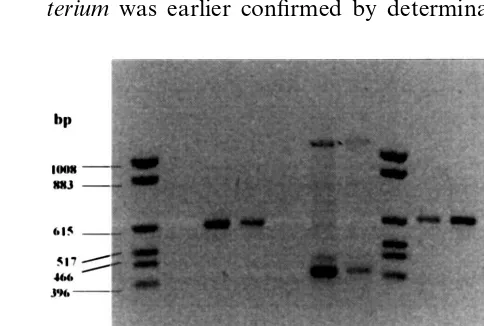
the PCR with primers constructed on the se-quences of rolB and rolC genes of A. rhizogenes. Primers based on the sequence of rolB gene am-plify the expected fragment of 383 bp and those specific for rolC gene amplify the fragment of 586 bp DNA in PCR reaction with DNA isolated from hairy root culture tissues (Fig. 1). The PCR product was absent in non-transformed tissue. Amplification of both fragments was obtained in PCR reactions performed with DNA isolated from A. rhizogenes cells (Fig. 1).
To be sure that rolB and rolC gene were not amplified from bacterial cells, the following proce-dure was performed. Hairy root cultures were maintained on a medium with antibiotics usually during the growth of four to five subcultures, then they were grown on a medium without antibiotics for several subcultures. Later on hairy root tissues were homogenised and suspensions obtained were plated on MYA and Luria Agar media. Plates were incubated for several days, but the growth of A. rhizogenes was never observed.
Transformation of plant tissue by Agrobac -terium was earlier confirmed by determination of weeks after inoculation of the explants by A4 and
LBA9402 strains. The successful infection and root formation were obtained after inoculation of the first node and only when bacteria were grown before inoculation on MYA medium supple-mented with 200mM of acetosyringone. This
phe-nomenon can be explained by the observation of Stachel [20] showing the activation of Agrobac
-terium rhizogenes 6ir genes expression by
acetosyringone.
Bacterial cells were eliminated from the co-cul-tures by incubation of hairy root culco-cul-tures on MS medium with claforan (500 mg/l) and carbenicillin (500 mg/l), [21]. After four to five passages on media with antibiotics, hairy root cultures were free of bacteria. The next few passages on MS media without antibiotics and plant growth regu-lators, enabled the obtaining of uniform hairy root cultures that displayed a typical phenotype charac-terised by plagiotropic growth and high incidence of lateral branching. These roots grew faster than normal ones. A similar phenotype was described earlier by several authors [8,22,23].
The growth rate of hairy root cultures on an MS medium without plant growth regulators was about thirty times higher than that of callus grow-ing on an MS medium supplemented with 5.0 mg/L IAA and 1.0 mg/L BAP and cell suspension cultures growing on MS medium supplemented with 0.5 mg/l GA3 and 1.0 mg/l KIN (Table 2). Fresh weight of hairy root cultures increased about 200-fold, from 0.08 to 15.0 g after 30 days (Table 2).
3.2. Detection of rol genes in transgenic tissue
Integration of the T-DNA into A. majus genome was confirmed on the molecular level by
Fig. 1. Identification of rolB and rolC gene fragments in transformed tissue of A.majus. 1 and 8, Marker pKO3/Hinf I (Gibco BRL); PCR reactions were performed using as a target DNA isolated from: 2, Non-transformed leaves of A.
majus+primer rolC; 3, hairy roots of A.majustransformed byA.rhizogenesLBA 9402+primerrolC; 4, hairy roots ofA.
majus transformed by A. rhizogenes A4+primer rolC; 5, non-transformed leaves of A. majus+primer rolB; 6, hairy roots ofA.majustransformed byA.rhizogenesLBA 9402+
primer rolB; 7, hairy roots of A. majus transformed by A.
rhizogenes A4+primer rolB; 9, A. rhizogenes LBA 9402+
primer rolC; 10, A. rhizogenes A4+primer rolC; 11, A.
rhizogenesLBA 9402+primer rolB; 12, A. rhizogenesA4+
the production of opine by plant tissue [24]. Re-cently Henzi [25] has confirmed integration of the T-DNA to the plant genome using PCR with primers for the NPTII gene cloned earlier to the Ri plasmid of the A. rhizogenes. Primers for rolBand rolC genes used in this work enable molecular confirmation of the transformation with the wid type strains of A. rhizogenes.
3.3. Determination of coumarin contents
The analysis of methanol extracts from seeds indicated the presence of umbelliferone, bergapten, xanthotoxin, imperatorin and several other cou-marins and furanocoucou-marins (Table 1). Furanocou-marins: psoralen, bergapten, xanthotoxin, imperatorin were absent in extracts of callus, cell suspension and hairy root cultures (Table 1). How-ever, in vitro cultures indicated the presence of umbelliferone. Extracts of hairy roots indicated a similar level of umbelliferone (about 19 mg/g DW)
to those of seeds, as well as twice the level of this compound than extracts of callus and nine times that of cell suspension (Table 1). The work of Hamerski [2] and Hamerski and Matern [26] indi-cated that only umbelliferone, but not furanocou-marins was present in cell suspension ofA. majus. Furanocoumarins biosynthesis in plants of the familyUmbelliferaeand Rutaceaehas been studied by several groups [27 – 30]. It was established that psoralens are derived from umbelliferone by the prenylation [26,31]. As indicated in the work of Hamerski and Matern [26] the umbelliferone prenyltransferases, marmesin and psoralen synthase are located in the membranes of the endoplasmatic reticulum of A. majus cells. Activity of these en-zymes was strongly dependent on induction by biotic elicitors [26,32]. Non-elicited cultures showed no activities of the enzymes involved in biosynthesis of psoralens. Fungal elicitors induce a specific set of endoplasmic reticulum enzyme: prenyltrans-ferase, as well as marmesin and psoralen synthase in A. majus cells [32].
In further experiments the possibility for the induction of furanocoumarins biosynthesis in hairy root cultures of A. majus by abiotic and biotic elicitors will be examined.
Acknowledgements
This work was supported by the Polish Scientific
Committee (KBN) grant 0819/PO4/98/15. The au-thors wish to thank Dr F. Bourgaud, Dr K. Kromer and Dr H. Wysokin´ska for providing strains of Agrobacterium rhizogenes.
References
[1] A. Scho3berg, A. Sina, Xanthoxin from the fruits of
Ammi majus, Nature 161 (1948) 481.
[2] D. Hamerski, R.C. Beier, R.E. Kneusel, U. Matern, K. Himmelspach, Accumulation of coumarins in elicitor-treated cell suspension cultures of Ammi majus, Phyto-chemistry 4 (1990) 1137 – 1142.
[3] S. Koul, A.K. Koul, Development of media for growth and furanocoumarin production of Ammi majus cells, Fitoterapia 5 (1993) 415 – 422.
[4] M. Purohit, D. Pande, A. Datta, P.S. Srivastava, En-hanced xanthotoxin content in regenerating cultures of
Ammi majus and micropropagation, Planta Med. 61 (1995) 481 – 482.
[5] A. Kro´licka, M. Krauze-Baranowska, E. L*ojkowska,
Production of secondary metabolites in elicitor-treated callus and cell suspension cultures ofAmmi majus, Ab-stract of 9th International Congress On Plant Tissue Cell Cultures, Jerusalem, Israel, 1998, 92.
[6] M.D. Chilton, D.A. Tepfer, A. Petit, C. David, F. Casse-Delbart, J. Tempe, Agrobacterium rhizogenes in-serts T-DNA into the genomes of the host plant root cells, Nature 295 (1982) 432 – 434.
[7] H.E. Flores, W.R. Curtis, Approaches to understanding and manipulating the biosynthetic potential of plant roots, Ann. NY Acad. Sci. 655 (1992) 188 – 209. [8] H. Wysokin´ka, A. Chmiel, Transformed root cultures
for biotechnology, Acta Biotechnol. 17 (1997) 131 – 159.
[9] T. Murashige, F. Skoog, A revised medium for rapid growth and bioassays with tobacco tissue cultures, Phys-iol. Plant 15 (1962) 473 – 497.
[10] L. Moore, G. Warren, G. Strobel, Involvement of a plasmid in the hairy root disease of plants caused by
Agrobacterium rhizogenes, Plasmid 2 (1979) 617. [11] J. Payne, J.D. Hamill, R.J. Robins, M.J.C. Rhodes,
Production of hyoscyamine by ‘hairy root’ cultures of
Datura stramonium, Planta Med. 53 (1987) 474 – 478.
[12] H. Yonemitsu, K. Shimomura, M. Satake, S. Mochida, M. Tanaka, T. Endo, A. Kaji, Lobeline production by hairy root culture ofLobelia inflanta L, Plant Cell Rep. 9 (1990) 307 – 310.
[13] C.P. Constabel, G.H.N. Towers, Thiarubine accumula-tion in hairy root cultures of Chaenactis douglasii, J. Plant Physiol. 133 (1988) 67 – 72.
[14] J.R. Porter, Host range and implications of plant infec-tion by Agrobacterium rhizogenes, Crit. Rev. Plant Sci. 10 (1991) 387 – 421.
[16] A. Petit, J. Tempe, Isolation ofAgrobacterium Ti-plas-mid regulatory mutants, Mol. Gen. Genet. 167 (1978) 147 – 155.
[17] I.J. Furner, G.A. Huffman, R.M. Amasino, D.J. Garfi-nkel, M.P. Gordon, E.W. Nester, An Agrobacterium
transformation in the evolution of the genusNicotiana, Nature 319 (1986) 422 – 427.
[18] G.F. Spencer, L.W. Tjarks, R.G. Powell, Analysis of linear and angular furanocoumarins by dual-column HPLC, J. Agric. Food. Chem. 35 (1987) 803 – 805. [19] C. Nguyen, F. Bourgaud, P. Forlot, A. Guckert,
Estab-lishment of hairy root cultures ofPsoraleaspecies, Plant Cell Rep. 11 (1992) 424 – 427.
[20] S.E. Stachel, E. Messens, M. Van Montagu, P. Zam-bryski, Identification of the signal molecules produced by wounded plant cells that activate T-DNA transfer in
Agrobacterium tumefaciens, Nature 318 (1985) 624 – 629. [21] A. Kro´licka, J. Kurlenda, E. L*ojkowska, Resistance to antibiotics of the chosen species ofAgrobacterium rhizo
-genes, Biotechnologia 45 (1999) 94 – 103.
[22] D.A. Tepfer, Transformation of several species of higher plants byAgrobacterium rhizogenes: sexual transmission of the transformed genotype and phenotype, Cell 37 (1984) 959 – 967.
[23] E.L.H. Aird, J.D. Hamill, M.J.C. Rhodes, Cytogenetic analysis of hairy root cultures from a number of plant species transformed by Agrobacterium rhizogenes, Plant Cell, Tissue Organ. Cult. 15 (1988) 47 – 57.
[24] A. Petit, C. David, G.A. Dahl, J.G. Ellis, P. Guyon, F. Casse-Delbart, J. Tempe, Further extension of the opine
concept: plasmids on Agrobacterium rhizogenes cooper-ate for opine degradation, Mol. Gen. Genet. 190 (1983) 204 – 214.
[25] M.X. Henzi, M.C. Christey, D.L. McNeil, K.M. Davies,
Agrobacterium rhizogenes-mediated transformation of broccoli (Brassica oleracea L. var. italica) with an anti-sense 1-aminocyclopropane-1-carboxylic acid oxidase gene, Plant Sci. 143 (1999) 55 – 62.
[26] D. Hamerski, U. Matern, Elicitor-induced biosynthesis of psoralens inAmmi majussuspension cultures, Eur. J. Biochem. 171 (1988) 369 – 375.
[27] H. Ekiert, Tissue culture of Ammi majus L. and its metabolites, Acta Polon. Pharm. 6 (1986) 634 – 636. [28] F. Bourgaud, M.C. Brunel, A. Guckert, P. Forlot, Effect
of nitrogen nutrition and environmental conditions on the production of pharmaceutically useful metabolites by Psoralea cinerea, Eur. J. Agron. 1 (1992) 37 – 43. [29] M.H.A. Elgamal, N.M.M. Shalaby, H. Duddeck, M.
Hiegemann, Coumarins and coumarin glucosides from the fruits of Ammi majus, Phytochemistry 34 (1993) 819 – 823.
[30] S.A. Brown, Biosynthetic studies on coumarins, Planta Med. 36 (1979) 299 – 310.
[31] S.A. Brown, M. El-Dakhakhny, W. Steck, Biosynthesis of linear furanocoumarins, Can. J. Biochem. 48 (1970) 863 – 871.
[32] D. Hamerski, U. Matern, Biosynthesis of psoralens, psoralen 5-monooxygenase activity from elicitor-treated
Ammi majus cells, FEBS Lett. 239 (1988) 263 – 265.