First evidence for improved resistance to fire blight in transgenic
pear expressing the
attacin E
gene from
Hyalophora cecropia
J.P. Reynoird
a,*, F. Mourgues
a ,1, J. Norelli
b, H.S. Aldwinckle
b, M.N. Brisset
a,c,
E. Chevreau
aaINRA Station d’Ame´lioration des Espe`ces Fruitie`res et Ornementales,BP57,49071Beaucouze´ cedex,France bDepartment of Plant Pathology,Cornell Uni6ersity,Gene6a,NY14456,USA
cINRA Station de Pathologie Ve´ge´tale et de Phytobacte´riologie,BP57,49071Beaucouze´ cedex,France
Received 15 April 1999; received in revised form 29 June 1999; accepted 13 July 1999
Abstract
Fire blight, caused byErwinia amylo6ora, is the most important bacterial disease of pear (Pyrus communisL.). Attacin E, a lytic
protein originating fromHyalophora cecropia, has been previously reported to be active againstE.amylo6orain transgenic apple. Integration of theattacin Egene under control of a derivative of the constitutive promoterCaMV35Swas accomplished using a transformation protocol developed for several pear cultivars. The integration was checked by polymerase chain reaction (PCR) for 11 lines of the cultivar Passe Crassane. Expression was determined in in vitro plantlets. The differences in transcription levels among lines revealed by comparative reverse transcription PCR correlated very well with the differences in attacin E accumulation observed by Western blot analysis. Fire blight inoculations were performed in vitro on all transgenic lines. A significant reduction of symptoms was observed for six lines, in comparison with the susceptible control Passe Crassane. © 1999 Published by Elsevier Science Ireland Ltd. All rights reserved.
Keywords:Pear; Fire blight; Resistance; Genetic transformation; Attacin
www.elsevier.com/locate/plantsci
1. Introduction
Fire blight is a major disease of the European pear (Pyrus communis L.) and other members of the Rosaceae family, caused by the necrotic bac-terium Erwinia amylo6ora. It is responsible for
serious production losses in Europe and North America. Conventional breeding requires a long time and causes recombination in the genome, which hampers the selection of high-quality com-mercial cultivars. Genetic transformation, which preserves the genetic background of the trans-formed cultivar, offers an attractive alternative method for the introduction of genes conferring resistance into specific varieties of vegetatively
propagated species. An efficient regeneration sys-tem for pear is available [1] and transgenic plants have recently been obtained from pear cultivars [2].
Many non-plant lytic proteins with antibacterial effects have been identified [3]. Among these, pep-tides secreted into the haemolymph of pupae of the Cecropia moth (Hyalophora cecropia) in re-sponse to bacterial infection have been extensively studied [4]. Besides the cecropins, a family of small basic proteins [5], and lysozymes, a group of ubiq-uitous enzymes [6], the attacins represent the largest antibacterial proteins known in haemolymph (MW$20 kDa). Six attacins were isolated [7] and displayed antibacterial activity directed against the outer membrane of Es
-cherichia coli, affecting permeability [8]. Synthesis of the outer membrane proteins has also been reported to be inhibited [9,10]. A possible synergy * Corresponding author. Present address: Department of Plant
Pathology, Cornell University, Geneva, NY 14456, USA.
1Present address: ENEA Casaccia-S.P.026.301, via Anguillarese,
00060 S. Maria di Galeria (Roma), Italy.
between attacin, cecropin and lysozyme has been suggested [8] because of the complementary mode of action of these three proteins against bacterial membranes. Minimum inhibitory concentration (MIC) of attacin E against different strains of E.
coli was 8 mM [8], whereas only 1 mM or less of cecropins was lethal [4]. While MICs of cecropins have been determined against a variety of phyto-pathogenic bacteria [11], to our knowledge, no information is available about the activity of attacin E against plant pathogens.
Introduction of antibacterial protein genes from insects to enhance plant resistance was previously suggested by Jaynes et al. [12] and Casteels et al. [13]. In particular, expression of cecropin B gene and synthetic analogues has been widely analyzed. Although enhanced resistance to bacteria was re-ported in some cases [11,14], susceptibility was not modified in others [15,16]. Degradation of ce-cropins by intercellular fluids (IF) [16 – 18] could explain the lack of resistance. In comparison with cecropins, nothing is known about activity of attacins and their stability in planta or in IF. The first transgenic plants expressing the attacin E
gene, driven by the potato proteinase inhibitor II [14] or by the CaMV35S promoter [19], were obtained from apple rootstock and displayed en-hanced resistance to fire blight [20]. In this report, we describe for the first time the successful trans-formation of pear with the attacin E gene and its expression in transgenic lines.
2. Materials and methods
2.1. Plant material
The Passe Crassane (PC) cultivar was chosen for transformation experiments because of its high susceptibility to fire blight. For the in vitro fire blight resistance test, the resistant cv. Old Home (OH) and non-transformed PC were used as con-trols. Shoots were propagated in vitro as previ-ously reported [1].
2.2. Plasmid constructions
Two binary expression vectors, pFAJ3000 and pFM3002, were used: pFAJ3000 [21] contained an
nptII-based expression cassette as selectable marker and a uidA-intron expression cassette ob-tained as a 3 kbp HindIII fragment from p35S GUS INT [22], adjacent to the right border; pFM3002 derived from pFAJ3000 by replacement of theuidA-intron cassette by an attacin E expres-sion cassette excised from pLDB11[23] as a
HindIII fragment, containing the attacin E gene [12] driven by a derivative of the cauliflower mo-saic virus promoter (Ca2MV35S) containing a tandem duplication of 250 bp of upstream se-quence [24] and the nopaline synthase transcrip-tional terminator sequence (Fig. 1). The binary vectors were transferred by electroporation to su-pervirulent Agrobacterium tumefaciensstrain EHA 101 [25].
2.3. Transformation experiments and propagation of transgenic lines
Five hundred fully expanded leaves were excised from shoots 2 weeks after subculture and transfor-mation experiments were carried out as previously reported [2], using A. tumefaciens strain EHA101 containing either pFM3002 or pFAJ3000. Trans-genic shoots were subcultured on basal medium with kanamycin (100 mg/l) as selective agent, then rooted as described in [2].
2.4. E6idence of transformation
DNA isolation from leaves of in vitro shoots and polymerase chain reaction (PCR) experiments were carried out as previously described [2]. In order to check the presence of attacin E or uidA
gene, specific primers were designed: 5%
-CT-TACGCTCAACTCCGATG-3% (forward) and 5% -AATCCGAAGTTAGGCTCCC-3% (reverse) for
amplification of a 530 bp fragment in attacin E
gene, 5%-GGTGGGAAAGCGCGTTACAAG-3%
(forward) and 5%
-GTTTACGCGTTGCTTC-CGCCA-3% (reverse) for amplification of a 1.2 kb fragment in uidA gene. Annealing was performed at 58°C.
2.5. Control of the ploidy le6el
Ploidy level in the transgenic lines and control was estimated by flow cytometry. Nuclei were isolated from leaves by manual chopping with a razor blade directly into Brown et al. buffer [26] with 2 % 4,6 diamino 2 phenyl indole dihy-drochlorode (Cheminex), then filtered through a 4
mm nylon mesh and analysed with a cytometer (Cell analyzer II; Partec, Germany). Pea leaf nu-clei were used as internal reference.
2.6. Determination of expression le6els
2.6.1. RNA extraction, Northern analysis and semi-quantitati6e re6erse transcription PCR analysis
Total RNA (50 – 100 mg) was extracted as de-scribed by Verwoerd et al. [27] from 0.5 g of young leaves excised from in vitro shoots. Purifica-tion was achieved by addiPurifica-tion of 2-butoxyethanol in order to precipitate polysaccharides [28].
For Northern blot analysis, 10mg of total RNA were denatured and separated by electrophoresis on a 1.2% agarose/formaldehyde gel in MOPS buffer, then transferred to Hybond™-N nylon membranes (Amersham, UK) as described by Sambrook et al. [29]. Filter was prehybridized for 2 h then hybridized overnight at 50°C in sodium dodecyl sulfate (SDS)-formamide buffer [30], with 50 ng of anattacin EDNA probe.Attacin EDNA was obtained by digestion of pFM3002 with
HindIII, then 32P-labelled with the ‘Ready to go’
kit (Pharmacia), purified on TE MIDI Select-D G-50 columns (5%-3% Inc., USA). After
hybridiza-tion, the filter was washed according to manufac-turer’s recommendations and finally exposed for 4 days on X-OMATTM film with one intensifying
screen at −80°C.
Reverse transcription (RT) was carried out from 1 mg total RNA according to Rosati et al. [31]. In
order to evaluate relative differences in cDNAs between transgenic lines, comparative kinetic anal-ysis was conducted by PCR as suggested by Horikoshi et al. [32]. Initial amounts of PCR substrates were adjusted for each line on the basis of an equivalent amplification of a cDNA from a member of the constitutively expressed gene family encoding the alpha subunit of translation elonga-tion factor 1 (EF1-a) [33]. In order to evaluate differences among lines proportional to differences in initial amounts, we limited the amplification to 20 cycles. EF1-a PCR reactions were carried out on 2 ml reverse transcription products. Degener-ated EF1-a primers (forward, 5% -ATTGTGGT-CATTGGYCAYGT-3%; and reverse, 5%-CCAAT
CTTGTAVACATCCTG-3%), custom designed from multiple sequence alignments [31], were used to amplify a 702 bp fragment. In a second step, comparative amplification was carried out with
attacin E primers on equivalent total cDNA amounts, adjusted in a volume of 2 ml, using the minimal number of cycles required to distinguish differences. The PCR reactions were run with products from Eurogentec (Seraing, Belgium) and 1 mM primers in a Minicycler (MJ Research, USA). The thermocycler program was: 94°C, 5 min, (20 cycles of: 94°C, 30 s; 58°C, 1 min; 72°C, 1 min); 72°C, 15 min for specific attacin E and
EF1-a amplifications. After gel electrophoresis on 0.8% (w/v) agarose-ethidium bromide gel and de-tection by ultraviolet light fluorescence, amplified products were blotted onto a Hybond™-N nylon membrane following standard procedures of the manufacturer (Amersham, UK). The filter was prehybridized for 2 h at 42°C in SDS-formamide buffer [30], then hybridized overnight under the same conditions, with attacin E and constitutively expressed EF1-a probes prepared as already de-scribed. After hybridization and washing, mem-branes were exposed for 5 – 24 h to X-OMATTM
film with one intensifying screen at −80°C.
2.6.2. Protein extraction and Western analysis
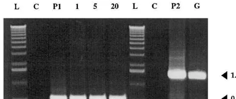
Fig. 2. PCR analysis of transgenic pear plants. Gel elec-trophoresis ofattacin EanduidAgene PCR products; lane C, non-transformed plant; lanes P1 and P2, plasmids pFM3002 and pFAJ3000; lanes 1, 5 and 20,attacin Etransgenic lines 1, 5 and 20; lane G, GUS transgenic line; lane L, molecular weight marker. Fragment sizes are indicated in kb.
severity was assessed by estimating the progression of necrosis from the inoculated leaf and apex to the base using a scale of 0 – 3: 0, no damage; 1, necrotic apex; 2, less than half of the shoot ne-crosed; 3, more than half of the shoot necrosed. For each line, susceptibility was calculated as the average of all replicates (about 75 replicates per line). Shoots of non-transformed lines of PC and OH were used as controls, and a transgenic PC line expressing theb-glucuronidase and the NPTII enzymes was also inoculated in order to estimate the effect of the expression of the transgenes that were assumed to lack antibacterial activity.
Resistance levels were compared pairwise be-tween transgenic lines and non-transformed PC using the non-parametric H test of Kruskall and Wallis. Data analysis was carried out using the
SAS/STAT 6.06 software, NPAR1WAY procedure.
3. Results
3.1. Transformation rates
Two transformation experiments with the bi-nary vector pFM3002 harbouring the attacin E
gene were performed on leaves from in vitro shoots and gave transformation rates of 1.0 and 1.3%, respectively, whereas a rate of 3.4% was obtained in transformation experiment with the binary vector pFAJ3000 harbouring the uidA
gene. These rates of transformation were similar to those obtained with other Rosaceae woody plants [39 – 41] and one order of magnitude lower than those previously reported on pear cultivar Confer-ence [2]. Transformation with the attacin E gene, as well as the uidA gene, was confirmed by PCR analysis for all the lines growing on kanamycin selective medium (Fig. 2). Eleven attacin E lines were subcultured on selective medium in the pres-ence of 100 mg/l kanamycin without cefotaxime, and absence of residual A. tumefaciens was checked by PCR before acclimatization of the plants (data not shown).
3.2. Ploidy le6els of transgenic lines
Ploidy levels were determined by flow cytome-try. Leaves from in vitro shoots or from acclima-tized plants were composed exclusively of 2C cells. All the transgenic lines including line 5, which had Protein extracts were resuspended in 50 ml 0.1 M
Tris – HCl (pH 6.8), 7 mM dithiothreitol, and quantified against a bovine serum albumin stan-dard using a colorimetric assay according to Brad-ford [35]. For Western analysis, 10 mg aliquots of protein extract from control and transgenic lines in Laemmli buffer were separated on 16.5% SDS-tricine polyacrylamide gel according to the discon-tinuous procedure of Scha¨gger and Von Jagow [36]. After electrophoresis, proteins were blotted onto Hybond C nitrocellulose membrane (Amer-sham, UK) by passive transfer. Polyclonal rabbit anti-attacin antiserum [7] was used and attacin E was detected with the enhanced chemolumines-cence Western blotting detection system (ECL, Amersham, UK) using horseradish peroxidase-la-belled secondary antibody, according to the manu-facturer’s instructions.
2.6.3. Determination of in 6itro resistance
Before inoculation, shoots were micropropa-gated on basal multiplication medium [2] without kanamycin during at least three subcultures. Shoots (2 – 3 cm high) were subcultured in baby food jars (five per jar) 2 weeks before inoculation. For each line, five replicate jars were used and experiments were repeated three times. E.
amylo6ora strain CFBP1430 was grown overnight
on King’s medium B [37] at 26°C and resuspended in water at a concentration of 5×10⁷ colony
strongly reduced growth in vitro and in the green-house, showed the 2C value of the non-trans-formed diploid PC.
3.3. Attacin E gene transcription
We analyzed the transcription of foreign DNA in the transgenic lines. Precipitation with 2-bu-toxyethanol eliminated most of the contaminants from RNA extracts, as confirmed by the high absorbance ratiosA260/A230(data not shown).
Pre-liminary Northern blot experiments were unsuc-cessful and we were not able to detect clearly the
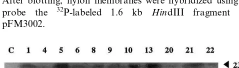
attacin E transcript (data not shown). Since levels of mRNA could be limiting, we used RT-PCR to compare the levels of transcription. After retro-transcription, the amounts of cDNA were firstly adjusted among samples based on equivalent am-plification of an EF1-a fragment, then used as templates for attacin E PCR. Amplification prod-ucts showed differences among transgenic lines after gel electrophoresis, blotting on nylon mem-brane and hybridization with a32P-labelledattacin E fragment (Fig. 3). No amplification occurred in the non-transformed control, whereas transgenic lines showed variable but always detectable ex-pression. In particular, lines 9, 13, 5, 10 and 21
displayed high transcription levels. By contrast, lines 4, 20 and 22 displayed very low transcription.
3.4. Transgenic protein detection
Transgenic lines were assayed for attacin E. Proteins extracted from leaves of in vitro shoots amounted to 4 ‰ of the fresh weight. Western analysis detected expression in all the transgenic lines (Fig. 4). The non-transformed control showed no cross-reactivity with attacin E anti-body. Another protein with a lower molecular weight (MW$17 kDa) was also detected ahead of the 23 kDa signal corresponding to attacin E. Since this protein was only present in the extracts from transgenic lines and proportional to the 23 kDa signal, it may have resulted from partial breakdown of attacin E or one of its precursors. Cross-hybridization with the putative breakdown product was not suppressed by adding or substi-tuting another protease inhibitor (E-64, trans-epoxysuccinyl-L-[4-guanidino]butane) for PMSF
(data not shown). Levels of amplified cDNA and protein amounts were in good agreement for all lines, except for line 9 which exhibited a moderate amount of attacin E. Since this line had a high level of transcription, its translation may have been reduced.
3.5. In 6itro resistance tests against E. amylo6ora
Preliminary experiments with different concen-trations of inoculum varying from 105 to 108
CFU/ml showed that 5×10⁷CFU/ml were
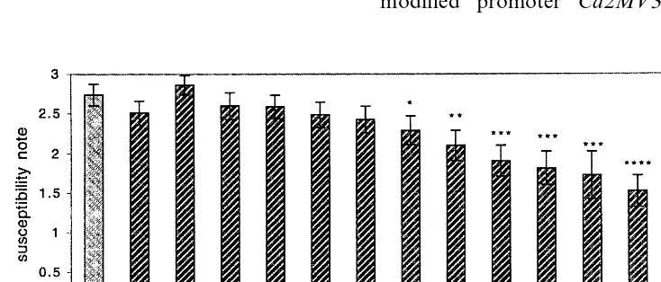
neces-sary to obtain complete infection of the susceptible control PC after 10 days (data not shown). With these conditions, shoots exhibited first symptoms on veins and petioles after only 3 days. After 10 days, less than 10% of OH shoots (resistant con-trol) were infected with low severity, whereas 90% of untransformed PC shoots showed maximum necrosis (Fig. 5). The transgenic control expressing the uidA gene showed the same symptoms as the susceptible control. Transgenic lines displayed in-termediate levels of susceptibility (Fig. 6). Six lines were significantly (PB0.05) more resistant than PC in rank comparison, whereas five lines showed little or no difference with the control. Trends were similar in the three sets of experiments, as indicated by the low standard errors. Data on in vitro disease susceptibility and attacin E levels
Fig. 3. Transcription of attacin E gene in leaves of in 6itro
shoots from control (lane C) and the transgenic lines (lanes 1 – 22) studied by comparative RT-PCR. Differences among transcription levels of transgenic clones were estimated after specific PCR (20 cycles) on equivalent total cDNA amounts. After blotting, nylon membranes were hybridized using as a probe the 32P-labeled 1.6 kb HindIII fragment from
pFM3002.
Fig. 5. Behaviour of resistant Old Home (a) and susceptible Passe Crassane (b) in vitro shoots 10 days after inoculation withE.amylo6oraCFPB 1430. Bar=1 cm.
4. Discussion
This study is the first report of successful trans-formation of pear with a gene encoding a lytic protein. The attacin E gene was introduced into the susceptible cultivar Passe Crassane. Eleven transgenic lines were obtained for which expres-sion was analyzed in in vitro plantlets. The amounts of attacin E transcripts, attacin E and levels of resistance toE.amylo6orawere compared
among the transgenic lines.
After retrotranscription of total RNA extracted from leaf samples, we used non-competitive PCR in order to compare the quantity of attacin E
cDNA among RT products. This procedure relies on the well-established observation of linear rela-tionship between the levels of cDNA before and after amplification prior to the onset of the plateau effect [42]. Amplification of EF1-a, an internal housekeeping gene uniformly expressed [43] as an internal standard, allowed adjustment of the amount of total cDNA among samples to yield comparable amplification products. We then am-plified a fragment of attacin E in the samples. After 20 cycles, detection required blotting of the electrophoresis gel and labelling with a specific probe. By this procedure, we detected differences in transcription levels among the lines.
The expression of attacin E was detected by Western blotting. Leaves of all transgenic lines expressed detectable levels of the protein. The modified promoter Ca2MV35S was previously were in good agreement for most of the lines,
especially for the resistant lines 5, 10, 13 and 21, and for the susceptible lines 4 and 20. However, the ranks according to disease susceptibility and attacin E levels were quite different in two cases. Line 22 exhibited an intermediate to high level of resistance despite a small amount of attacin, in agreement with a low transcription level. No in-crease of resistance was detected for line 9 despite a high level of transcription and a moderate level of attacin E.
Fig. 6. Disease susceptibility of non-transformed PC and OH cv. and attacin E lines, 10 days after in vitro inoculation withE.
amylo6oraCFPB 1430. A GUS transgenic line of PC (G) was also used as control. Each bar is the mean susceptibility score of three sets of experiments on 25 replicate shoots per clone. Lines on top of bars represent standard errors of the means. Susceptibility scores of transgenic plants are significantly different from those of control plants at *PB0.05, **PB0.01, or ***
used to drive high-level expression of cecropin gene [16] in tobacco. Quantification of attacin E in samples was not possible, due to the lack of purified or synthesized protein. We observed cross-hybridization with a protein of lower molec-ular weight than attacin E. This band of approxi-mately 17 kDa was only present in the transgenic samples. Since the intensities of both attacin E and the 17 kDa protein were proportional, except for line 10, we suggest that it may be a degradation product. Compared with attacin, degradation of cecropin B and its analogues has been extensively studied in transgenic tissues. Indeed, a rapid breakdown of cecropin B was observed after incu-bation with tobacco [16], peach [17] and pear [18] IFs, and attributed to the action of plant serine proteases.
Fire blight inoculations provided consistent re-sults among replicates of each line. Disease reac-tion varied with the concentrareac-tion of inoculum and 90% of non-transformed control microcut-tings were diseased 10 days after inoculation with 5×10⁷ CFU/ml, as previously reported for pear
[38] and apple [44] plantlets propagated in vitro. Since the short size of the in vitro shoots did not allow precise measurements of the length of necro-sis, disease severity was scored using three classes. This time-saving method may allow selection of promising lines before further assessment of resis-tance on rooted plants after acclimatization. Previ-ous reports indicate that in vitro fire blight screening distinguishes clearly between resistant and susceptible lines of pear [38,45] and apple [44,45], and correlates well with greenhouse [44] and field [46] evaluations. However, replicate plants of somaclones regenerated from the apple cv. Greensleeves varied in their resistance to fire blight following in vitro and greenhouse inocula-tions [47]. Since differences in growth rates be-tween replicates could partially explain these results, the authors suggested multiple screenings at different stages of development. Inoculation of adult trees is desirable to determine whether attacin also increases resistance of blossoms, since in nature, infection of flowers is common. In the present research, six lines displayed higher in vitro resistance levels than the untransformed control. Further assessment of the transgenic pear resis-tance will be performed in greenhouse. Royal Gala transgenic apple lines expressing theattacin Egene also showed increased resistance to E. amylo6ora
[48] as young plants, which was confirmed in a field trial.
The results from Western analysis were gener-ally in good agreement with those from fire blight susceptibility tests. In particular, line 5, which expressed a high level of resistance, showed the highest level of attacin E. However, this line exhib-ited dramatically reduced growth in the green-house, in contrast with other resistant lines. Growth reduction associated with increased fire blight resistance has already been observed in pear somaclonal variants with doubled chromosome numbers [49]. In our study, all the transgenic lines exhibited the same ploidy level as the control. Cryptic DNA modifications due to somaclonal variation or to the localization of the transgene could explain the abnormal phenotype of line 5. Overproduction of attacin E in this line might also cause a toxic effect of the lytic protein to the plant. Similar results have already been observed among transgenic apple lines with high levels of expression of endochitinase transgene under con-trol ofCa2MV35S[50]. ThePin2 promoter, which has been used to drive the attacin E gene in apple [14], was shown to be constitutively activated to a higher level than the Ca2MV35S promoter [51]. In addition, expression of the transgene at the RNA and protein levels showed high correlation, except in line 9, which exhibited a low to interme-diate level of attacin E. The discrepancy observed between attacin accumulation and the high level of transcription could result from reduced translation or maturation of the protein.
There are several possibilities to increase the resistance of transgenic pears expressing antibacte-rial genes. First, bacteantibacte-rial-inducible promoters could be used to obtain strong and rapid tran-scription level at the site of infection and to avoid accumulation of the transgenic protein under non-inductive conditions. Such promoters have re-cently been isolated from tobacco [52]. Second, the use of a signal peptide (SP) should direct secretion of attacin E to the intercellular spaces [53]. Fur-thermore, in tobacco, cecropin B-mRNA expres-sion was affected by the type of construct. In particular, the lowest transcription levels were ob-served without SP, suggesting positive coupling between secretion, translation and mRNA stability [16]. Finally, since synergies in inhibition ofE. coli
coordinate expression of several of these genes under the control of the same or different pro-moters might be an effective strategy.
Pear engineering with antimicrobial peptide genes also raises the questions of potential toxicity or allergenic risks when transgenic fruits are con-sumed. Specific studies will be needed to assess these risks. Additionally, the promoters specifically activated during bacterial infection can be useful to limit the amount of transgenic protein in fruits.
Acknowledgements
We would like to thank C. Rosati and D. Hultmark, who provided us with degenerated primers of the elongation factor gene EF-1a and anti-attacin antiserum, respectively, S. Mareau who analysed the ploidy levels in the laboratory of SNES (Angers, France), L. Jouanin and P. Si-moneau for critical reading of the manuscript, C.E. Durel for helpful discussion on statistics, and C. Cesbron and L. Leclout for technical support.
References
[1] C. Leblay, E. Chevreau, L.M. Raboin, Adventitious shoot regeneration from in vitro leaves of several pear cultivars (Pyrus communisL.), Plant Cell Rep. 25 (1991) 99 – 105.
[2] F. Mourgues, E. Chevreau, C. Lambert, A. de Bondt, EfficientAgrobacterium-mediated transformation and re-covery of transgenic plants from pears, Plant Cell Rep. 16 (1996) 245 – 249.
[3] F. Mourgues, M.N. Brisset, E. Chevreau, Strategies to improve plant resistance to bacterial diseases through genetic engineering, Trends Biotechnol. 16 (1998) 203 – 209.
[4] H.G. Boman, D. Hultmark, Cell-free immunity in in-sects, Ann. Rev. Microbiol. 41 (1987) 103 – 126.
[5] D. Hultmark, A. Engstro¨m, H. Bennich, R. Kapur, H.G. Boman, Insect immunity. Isolation and structure of ce-cropin D and four minor antibacterial components from Cecropia pupae, Eur. J. Biochem. 127 (1982) 207 – 217. [6] D. Hultmark, H. Steiner, T. Rasmuson, H.G. Boman,
Insect immunity. Purification and properties of three inducible bactericidal proteins from hemolymph of im-munized pupae ofHyalophora cecropia, Eur. J. Biochem. 106 (1980) 7 – 16.
[7] D. Hultmark, A. Engstro¨m, K. Andersson, H. Steiner, H. Bennich, H.G. Boman, Insect immunity. Attacins, a family of antibacterial proteins from Hyalophora ce
-cropia, EMBO J. 2 (1983) 571 – 576.
[8] P. Engstro¨m, A. Carlsson, A. Engstro¨m, Z.J. Zao, H. Bennich, The antibacterial effect of attacins from the silk
moth Hyalophora cecropia is directed against the outer membrane ofEscherichia coli, EMBO J. 3 (1984) 3347 – 3351.
[9] A. Carlsson, P. Engstro¨m, E.T. Palva, H. Bennich, Attacin, an antibacterial protein from Hyalophora ce
-cropia, inhibits synthesis of outer membrane proteins in
Escherichia coliby interferring with omp gene transcrip-tion, Infect. Immun. 59 (1991) 3040 – 3045.
[10] A. Carlsson, T. Nystrom, H. de Cock, H. Bennich, Attacin — an insect immune protein-binds LPS and trig-gers the specific inhibition of bacterial outer-membrane protein synthesis, Microbiology 144 (1998) 2179 – 2188. [11] J.M. Jaynes, P. Nagpala, L. Deste´fano-Beltran, J.H.
Huang, J.H. Kim, T. Denny, S. Ce´tiner, Expression of a cecropin B lytic peptide analog in transgenic tobacco confers enhanced resistance to bacterial wilt caused by
Pseudomonas solanacearum, Plant Sci. 89 (1993) 43 – 53. [12] J.M. Jaynes, X.M. Xanthopoulos, L. Deste´fano-Beltran,
J.H. Dodds, Increasing bacterial disease resistance in plants utilizing antibacterial genes from insects, Bioassays 6 (1987) 263 – 270.
[13] P. Casteels, C. Ampe, F. Jacobs, M. Vaeck, P. Tempst, Apidaecins: antibacterial peptides from honey-bees, EMBO J. 8 (1989) 2387 – 2391.
[14] J.L. Norelli, H.S. Aldwinckle, L. Deste´fano-Beltran, J.M. Jaynes, Transgenic ‘Malling 26’ apple expressing the attacin E gene has increased resistance to Erwinia amylo6ora, Euphytica 77 (1993) 123 – 128.
[15] R. Hightower, C. Baden, E. Penzes, P. Dunsmuir, The expression of Cecropin peptide in transgenic tobacco does not confer resistance toPseudomonas syringae pv.
Tabaci, Plant Cell Rep. 13 (1994) 295 – 299.
[16] D. Florack, S. Allefs, R. Bollen, D. Bosch, B. Visser, W. Stiekema, Expression of giant silkmothcecropin B genes in tobacco, Transgenic Res. 4 (1995) 132 – 141.
[17] D. Mills, F.A. Hammerschlag, R.O. Nordeen, L.D. Owens, Evidence for the breakdown of cecropin by proteinases in the intercellular fluid of peach leaves, Plant Sci. 104 (1994) 17 – 22.
[18] F. Mourgues, M.N. Brisset, E. Chevreau, Activity of different antibacterial peptides on Erwinia amylo6ora
growth, and evaluation of the phytotoxicity and stability of cecropins, Plant Sci. 139 (1998) 83 – 91.
[19] H.S. Aldwinckle, J.L. Norelli, J.Z. Mills, Transformation of Gala apple with lytic protein genes, Acta Hort. 411 (1996) 411.
[20] M.T. Momol, J.L. Norelli, H.S. Aldwinckle, Field evalu-ation of fire blight resistance of an Attacin E transgenic M7 apple rootstock, Phytopathology 86 (1996) 592. [21] A. De Bondt, K. Eggermont, P. Druart, M. De Vil, I.
Goderis, J. Vanderleyden, W.F. Broekaert, Agrobac
-terium-mediated transformation of apple (Malus x do
-mesticaBorkh.): an assessment of factors affecting gene transfer efficiency during early transformation steps, Plant Cell Rep. 13 (1994) 587 – 593.
[22] G. Vancanneyt, R. Schmidt, A. O’Connor-Sanchez, L. Willmitzer, M. Rocha-Sosa, Construction of an intron-containing marker gene: splicing of the intron in trans-genic plants and its use in monitoring early events in
[23] L. Destefano-Beltran, P.G. Nagpala, M.S. Cetiner, J.H. Dodds, J.M. Jaynes, Enhancing bacterial and fungal disease resistance in plants: application to potato, in: M.E. Vayda, W.D. Park (Eds.), The Molecular and Cellular Biology of Potato, CAB International Society of Plant Molecular Biology, Tucson, AZ, 1990, pp. 205 – 210. [24] R. Kay, A. Chan, M. Daly, J. McPherson, Duplication of
CaMV35Spromoter sequences creates a strong enhancer for plant genes, Science 236 (1985) 1299 – 1302.
[25] E.E. Hood, G.L. Helmer, R.T. Frayler, M.D. Chilton, The hypervirulence ofAgrobacterium tumefaciensA281 is encoded in a region of pTiBo542 outside of T-DNA, J. Bacteriol. 168 (1986) 1297 – 1301.
[26] S.C. Brown, C. Bergounioux, S. Tallet, D. Marie, Flow cytometry of nuclei for ploidy and cell cycle analysis, in: I. Negrutiu, Gharti-Chetri (Eds.), Biomethods, Vol. 4. A Laboratory Guide for Cellular and Molecular Plant Biol-ogy, Birkhaeuser Verlag, Basel, 1991, pp. 326 – 345. [27] T.C. Verwoerd, B.N.M. Dekker, A. Hoekema, A
small-scale procedure for the rapid isolation of plant RNAs, Nucleic Acids Res. 17 (1989) 2360.
[28] K. Manning, Isolation of nucleic acids from plants by differential solvent precipitation, Anal. Biochem. 195 (1991) 45 – 50.
[29] J. Sambrook, E.F. Fritsch, T. Maniatis, Molecular Cloning: A Laboratory Manual, Cold Spring Harbor Laboratory Press, New York, 1989 2nd ed.
[30] G.M. Church, W. Gilbert, Genomic sequencing, Proc. Natl. Acad. Sci. U.S.A. 81 (1984) 1991 – 1995.
[31] C. Rosati, A. Cadic, M. Duron, J.P. Renou, P. Simoneau, Molecular cloning and expression analysis of dihy-droflavonol 4-reductase gene in flower organs ofForsythia x intermedia, Plant Mol. Biol. 35 (1997) 303 – 311. [32] T. Horikoshi, K.D. Danenberg, T.H.W. Stadlbauer, M.
Volkenandt, L.C.C. Shea, K. Aigner, B. Gustavsson, L. Leichman, R. Fro¨sing, M. Ray, N.W. Gibson, C.P. Spears, P.V. Danenberg, Quantitation of thymidylate synthase, dihydrofolate reductase, and DT-diaphoroase gene expression in human tumors using the polymerase chain reaction, Cancer Res. 52 (1992) 108 – 116.
[33] A. Mahe, J. Grisvard, M. Dron, Fungal and specific gene markers to follow the bean-anthracnose infection process and normalize a bean chitinase mRNA induction, Mol. Plant Microbe Interact. 5 (1992) 242 – 248.
[34] A.M. Schuster, E. Davies, Ribonucleic acid and protein metabolism in pea hypocotyl. I: the aging process, Plant Physiol. 73 (1983) 809 – 813.
[35] M.M. Bradford, A rapid and sensitive method for the quantification of microgram quantities of protein utilizing the principle of protein-dye binding, Anal. Biochem. 72 (1976) 248 – 254.
[36] H. Scha¨gger, G. Von Jagow, Tricine-sodium dodecyl sulfate-polyacrylamide gel electrophoresis for the separa-tion of proteins in the range from 1 to 100 kDa, Anal. Biochem. 166 (1987) 368 – 379.
[37] E.O. King, M.K. Ward, D.E. Raney, Two simple media for the demonstration of pyocyanin and fluorescin, J. Lab. Clin. Med. 44 (1954) 301 – 307.
[38] M.N. Brisset, J.P. Paulin, M. Duron, Feasibility of rating fire blight susceptibility of pear cultivars (Pyrus commu
-nis) onin6itromicrocuttings, Agronomie 8 (1988) 707 –
710.
[39] D.J. James, A.J. Passey, D.J. Barbara, M. Bevan, Genetic transformation of apple (Malus pumila Mill.) using a disarmed Ti-binary vector, Plant Cell Rep. 7 (1989) 658 – 661.
[40] A. De Bondt, K. Eggermont, I. Penninckx, I. Goderis, J. Vanderleyden, W.F. Broekaert,Agrobacterium-mediated transformation of apple (Malus x domesticaBorkh.): an assessment of factors affecting regeneration of transgenic plants, Plant Cell Rep. 15 (19xx) 549 – 556.
[41] R. Scorza, M. Ravelonandro, A.M. Callahan, J.M. Cordts, M. Fuchs, J. Dunez, D. Gonsalvez, Transgenic plums (Prunus domesticaL.) express the plum pox virus coat protein gene, Plant Cell Rep. 14 (1994) 18 – 22. [42] W.C. Gause, J. Adamovicz, The use of the PCR to
quantitate gene expression, PCR Methods Appl. 3 (1994) 123 – 135.
[43] M. Axelos, C. Bardet, T. Liboz, A. Le Van Thai, C. Curie, B. Lescure, The gene family encoding the Arabidopsis thaliana translation elongation factor EF-1a: molecular cloning, characterization and expression, Mol. Gen. Genet. 219 (1989) 106 – 112.
[44] J.L. Norelli, H.S. Aldwinckle, S.V. Beer, Virulence of
Erwinia amylo6ora strains to Malus sp. Novole plants
grown in vitro and in the greenhouse, Phytopathology 78 (1988) 1292 – 1297.
[45] J. Viseur, M. Tapia y Figueroa, In6itro co-culture as a
tool for the evaluation of fire blight resistance in pears and apples, Acta Hort. 217 (1987) 273 – 281.
[46] M. Le Lezec, B. Thibault, P. Balavoine, J.P. Paulin, Sensibilite´ varie´tale du pommier et du poirier au feu bacte´rien, Phytoma (1985) 37 – 44.
[47] A.M. Donovan, R. Morgan, C. Valobra-Piagnani, M.S. Ridout, D.J. James, C.M.E. Garrett, Assessment of so-maclonal variation in apple. I. Resistance to the fire blight pathogen, Erwinia amylo6ora, J. Hort. Sci. 69 (1994)
105 – 113.
[48] J.L. Norelli, E.E. Borejsza-Wysocka, M.T. Momol, J.Z. Mills, A. Grethel, H.S. Aldwinckle, K. Ko, S.K. Brown, D.W. Bauer, S.V. Beer, A.M. Abdul-Kader, V. Hanke, Genetic transformation for fire blight resistance in apple. Acta Hort. (in press).
[49] J. Viseur, Evaluation of fire blight resistance of so-maclonal variants obtained from the pear cultivar ‘Duron-deau’, Acta Hort. 273 (1990) 275 – 284.
[50] J.P. Bolar, J.L. Norelli, H.S. Aldwinckle, G.E. Harman, S.K. Brown, Production of transgenic apple lines for scab resistance with genes coding for one or two chitinolytic enzymes, 7th International Congress of Plant Pathology, Edinburgh, ICPP-98 Abstracts (3), 1998, p. 5.3.2. [51] K. Keinon-Mettala, A. Pappinen, K. Von Weissenberg,
Comparisons of the efficiency of some promoters in silver birch (Betula pendula), Plant Cell Rep. 17 (1998) 356 – 361. [52] D. Pontier, L. Godiard, Y. Marco, D. Roby,hsr203J, a tobacco gene whose activation is rapid, highly localized and specific for incompatible plant/pathogen interactions, Plant J. 5 (1994) 507 – 521.