VOLUME 2
NOMOR 1
JUNI 2015
ISSN 2442 - 2606
JURNAL
BIOTEKNOLOGI & BIOSAINS INDONESIA
Homepage Jurnal: http://ejurnal.bppt.go.id/index.php/JBBI
PRELIMINARY CYTOTOXIC EVALUATION OF
Andrographis paniculata
IN BREAST CANCER CELL LINES
Uji Pendahuluan Sitotoksik Andrographis paniculata pada Sel Kanker Payudara
Tarwadi1,*, Churiyah2, Olivia Bunga Pongtuluran2, Fifit Juniarti2, Fery Azis Wijaya3
1
Biotech Center BPPT, Building 630 PUSPIPTEK Area, Setu, Tangerang Selatan, Banten 15314
2
Centre for Pharmaceutical and Medical Technology BPPT, Building 610, PUSPIPTEK Area, Setu, Tangerang Selatan, Banten 15314
3
PT Biogen Scientific, Rukan Tanjung Mas Raya –Jl Raya Lenteng Agung Blok B1/21, Jakarta Selatan. *E-mail: [email protected]
ABSTRAK
Sambiloto (Andrographis paniculata) banyak digunakan untuk mengobati berbagai penyakit di Indo-nesia dan negara-negara Asia lainnya. Dalam studi ini, ekstrak metanol dan etanol sambiloto yang diperoleh dari B2PTO Tawangmangu telah diuji terhadap sel lini kanker payudara T47D dan MCF-7 dan sel lini normal fibroblast HFL-1 menggunakan reaksi enzimatik 3-(4,5-dimethylthiazoyl-2-yl) 2,5-diphenyltetrazoliumbromide (MTT). Uji in vitro terhadap sel lini normal fibroblast HFL-1 menunjukkan bahwa 50 ppm ekstrak metanol sambiloto tidak menghambat pertumbuhan sel. Tetapi, ekstrak metanol dan etanolnya menghasilkan IC50 yang relatif rendah pada sel lini kanker payudara, yaitu 111
ppm dan 122 ppm pada sel lini MCF-7 dan 70 ppm dan 197 ppm pada sel lini T47D. Selain itu, campuran ekstrak sambiloto yang mengandung 25% ekstrak Thyponium divaricatum dan Anredera cordifolia memberikan daya hambat pertumbuhan pada sel kanker payudara MCF-7 yang lebih besar, dengan nilai IC50 masing-masing adalah 68 ppm dan 34 ppm. Kesimpulannya, total ekstrak metanol
atau etanol sambiloto yang diperoleh dari Tawangmangu memiliki potensi sebagai sumber senyawa anti-kanker serta perlu kajian lebih lanjut.
Kata kunci: Ekstrak Andrographis paniculata, MTT, sel lini normal, sel lini kanker, aktivitas anti kanker
ABSTRACT
Sambiloto (Andrographis paniculata) is widely used as medicine to treat various diseases in In-donesia and other Asian countries. In this study, methanolic and ethanolic extracts of sambiloto collected from B2PTO Tawangmangu have been tested againts breast cancer cell lines of T47D and MCF-7 and normal fibroblast cell line of HFL-1 using enzymatic reaction of 3-(4,5-dimethylthiazoyl-2-yl) 2,5-diphenyltetrazoliumbromide (MTT). In vitro assay performed on normal fibroblast of HFL-1 cell line showed that 50 ppm of methanolic extract of sambiloto did not inhibit cell growth. However, methanolic and ethanolic extracts of sambiloto gave relatively low of IC50
on breast cancer cell lines which were 111 ppm and 122 ppm on the MCF-7 cell lines and 70 ppm and 197 ppm on the T47D cell lines, respectively. In addition, the mixture of sambiloto ex-tract containing 25% of Thyponium divaricatum and Anredera cordifolia exex-tracts confered greater growth inhibition on breast cancer cell line of MCF-7, where IC50 values were 68 ppm and 34
ppm, respectively. In conclusion, the total methanolic or ethanolic extract of sambiloto collected from Tawangmangu has potency as a source of anti-cancer compounds and needs further study.
INTRODUCTION
The genus Andrographis is a widely distributed in South Asia and medicinally important member of Acanthaceae, consisting of approximately 40 species. Andrographis paniculata Nees (known as “Sambiloto” in Indonesia) is an herb well known in the South East Asian as traditional medicine and its beneficial actions are many and varied. Over the century, mostly the leaves and the roots have been used traditionally not only in Asia but also in Europe as folklore remedy for wide spectrum of ailments or supplements for health support as listed in Table 1 (Jarukamjorn and Nemoto 2008).
Phytochemical studies resulted in isolation of flavonoid and andrographolides from A. paniculata (Rao et al. 2004). Furthermore, three new compounds of ent -labdane diterpenoids, namely 19-norandrographolides A–C (compounds 1–3), were also isolated from the ethanolic extract of A. paniculata. (Zhang et al. 2006). Recent studies demonstrated that of A. paniculata
has a potency as an antimicrobial activity (Sule et al. 2010). Previous research also reported that A. paniculata has anticancer
and immunostimulatory effect (Kumar et al.
2004), anti-hyperglycemic and renal protective activities (Rao 2006).
A. paniculata are the most common
traditional herbal that is used to achieve lower blood glucose in diabetic pasients by exhibits insulin-releasing actions (Wibudi et al. 2008). Hydroalcoholic extract of A. paniculata was reported as antioxidant, antilipid peroxidative and antiischemic activity and was used in ischemic heart diseases (Ojha 2009). In addition, leaf extract of A. paniculata has ability to suppress arsenic -provoked toxicity in human peripheral lymphocyte culture (Ghopalkrisnan and Rao 2008).
In this present study, we evaluated the methanolic and ethanolic extracts of A. paniculata, collected from Medicinal Plant Research Centre, Tawangmangu, Central Java, againts breast cancer cell lines of T47D and MCF-7 and normal fibroblast cell line of HFL-1 using enzymatic reaction of 3-(4,5-dimethylthiazoyl-2-yl) 2,5 diphenyl-tetrazolium bromide (MTT). In addition, we also tested the mixture extract of A. paniculata Ness with
Thyponium divaricatum (known as Keladi
tikus) as well as with Anredera cordifolia
(known as Binahong).
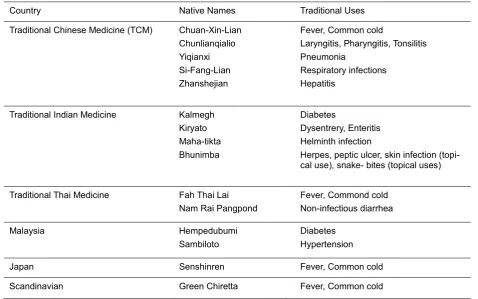
Table 1. Traditional Uses of A. paniculata
Country Native Names Traditional Uses
Traditional Chinese Medicine (TCM) Chuan-Xin-Lian Chunlianqialio
Traditional Indian Medicine Kalmegh Kiryato
Herpes, peptic ulcer, skin infection (topi-cal use), snake- bites (topi(topi-cal uses)
Traditional Thai Medicine Fah Thai Lai Nam Rai Pangpond
Japan Senshinren Fever, Common cold
MATERIALS AND METHODS
Reagents
Unless stated otherwise, all reagents used in this experiment were purchased from Sigma Aldrich (USA). RPMI-1640 medium, heat-inactivated fetal bofine serum (FBS), EDTA-trypsin was purchased from Gibco BRL (NY, USA). MTT powder (3-(4,5-dimethylthiazol-2-yl)-2,5-diphenyltetrazolium bromide) was purchased from Promega (USA).
Extract preparation
The aerial parts (stems and leaves) of A. paniculata Nees, T. divaricatum and leaves of
A. cordifolia used in this study were collected from Medicinal Plant Research Centre, Tawangmangu, Central Java, Indonesia. The plant samples were dried at room temperature and finely powdered. Suitable amounts of the powdered materials were soaked in 95% ethanol or methanol (1 L per 100 g), and the solvent was evaporated at 40°C to dry up under reduced pressure using a rotary evaporator to produce crude extracts. The extract was then collected and stored at -20°C for further testing. The High Performance Liquid Chromatography (HPLC) analysis HPLC profile of A. paniculata Ness
was carried out to detect the presence of active compound of Andrographolide (C18 volumn, λ = 223 nm, flowrate = 0.5-1 mL/min, 20 minutes).
Cell culture
The human breast adenocarcinoma cells (MCF-7 and T-47D) and the human normal lung fibroblast cell lines (HFL-1) were used to determine the cytotoxicity of the ex-tract. Cells were cultured in T-flask contain-ing RPMI 1640 medium supplemented with 10% heat inactivated foetal bovine serum (FBS), 50 IU/mL penicillin and 50 μg/mL streptomycin. The cells were maintained in the CO2 incubator at 37ºC in a 5% CO2 with
95% humidity. After reaching the confluency of approximately 70-80%, exponentially growing of the cells were washed twice with magnesium and calcium free phosphate buffer saline (PBS). The buffer solution was decanted and cells were detached with 0.025% trypsin-EDTA solution by incubating the cells at 37ºC in a 5% CO2 with 95%
hu-midity for couple of minutes, then cell culture growth medium were added to a volume of 5 mL. The cell suspension was then trans-ferred to the falcon tube and the cell pellet was obtained by centrifugation (1000 g, 5 min). The cells were then resuspended in 10 mL of medium to make single cell suspen-sion. The viable cells were counted by trypan blue exclusion assay in haemocytometer.
Cytotoxicity assay
Cells were seeded in 96-well plate in 100 µl of cell culture growth medium to a fi-nal concentration of 5x10³ cells/well and in-cubated to allow for cell attachment. After 24 h incubation, a partial monolayer was formed (confluency of 70-80%) then 100 μL of the medium containing the plant extract (initially dissolved in DMSO, those were: 250, 100, and the MTT (3-(4,5-dimethylthiazol-2-yl)-2,5-diphenyltetrazolium bromide) (Promega, USA) solution ( 5 µg) in culture media of 100 µl was added. The incubation time for cells-MTT solution was 4 hours in which blue crys-tal were formed, then 100 μL of the stop so-lution (SDS 10%) were added and cells were then incubated further for the next 24 h. Re-duced MTT was assayed at 550 nm using a microplate reader. Culture medium contain-ing 0.1% DMSO was used as a solvent con-trol and untreated cells were used as a negative control.
RESULTS AND DISCUSSION
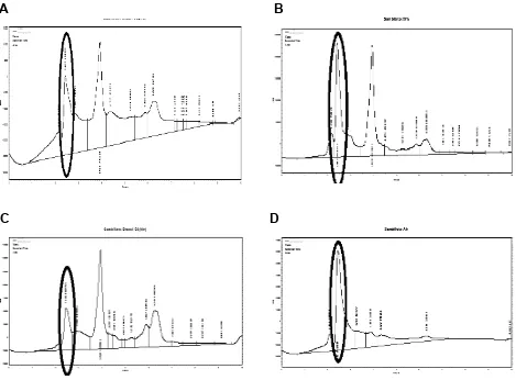
Table 2. Andrographolide content of samples extracted using various solvent compositions
Solvent Composition Area under curve [Andrographolide, ppm] % Andrographolide
96% ethanol 490398 15.97 1.6
Ethanol : H2O = 70 : 30 3851355 96.25 9.6
Ethanol : H2O = 50 : 50 1469925 69.85 7
H2O (100%) 2032409 6.75 0.7
Note: Sample extract injected: 1000 ppm at λ= 223 nm
A
B
C
D
Figure 1. HPLC profile of Andrographis paniculata Ness extracts containing andrographolide; A. ethanolic extract of 96%, B. etahnolic extract of 70%, C. ethanolic extract of 50% and D. Decoction (H2O extract)
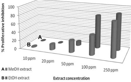
In vitro cytotoxicity assay performed on normal fibroblast of HFL-1 cell line showed that 50 ppm of methanolic and ethanolic extract of A. paniculata did not inhibit cell growth, where the cell viability was about 101% on the methanolic extract and 94% on the methanolic extract. In this regards, we could simply assume that the extract has no cytotoxic effect on normal cell line of HFL-1. However, methanolic and ethanolic extracts of the sample gave IC50
relatively low on breast cancer cell lines of
MCF7 which were 122 ppm and 111 ppm, respectively. The growth inhibition of the extract in MCF7 was dose dependent manner. As shown in Figure 2, the proliferative inhibition activity indicated that increasing extract concentration resulted in an increasing the percent proliferative inhibition. Furthermore, cytotoxicity of the methanolic and ethanolic extracts on other breast cancer cell line of T-47D showed an IC50 value of and 70 ppm and 197 ppm
activity pattern of the extracts on T-47D cell line similar with the proliverative inhibition activity on MCF-7 cell line, done dependent manner, where increasing of the extract concentration treated followed with an increased the proliferative inhibition activity (Figure 3).
Our present study on A. paniculata Ness extract was in agrement with the re-sults from previous researcher using another cell line, which reported that 10 ug/mL methanolic extract of A. paniculata inhibited proliferation of HT-29 (colon cancer cell) by cluding flavonoids and labdane diterpenoids, where the compound 6 was rich source for the active compound of andrographolide. The bioactivity assays showed that metabo-lites 1-4 and 6-8 exhibited moderate cytotox-ic activity against Jurkat, PC-3 and Colon 205 cell lines, where compound 6 had IC50
values of 0.05, 0.07 and 0.05 mm, respec-tively. Further, among these effective com-pounds, 3 and 6 selectively blocked the cell cycle progression at G0/G1, while 1, 2, 4, 7 and 8 metabolites blocked the same at G2/M phase of the Jurkat cell line (Geethangili et al.
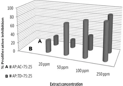
2008). Subsequently, the mixture of 75% A. paniculata extract with 25% of T. divaricatum
confered greater proliferative inhibition on MCF-7 cell with gave IC50 values of 68 ppm.
Moreover, the combination of 75% A. paniculata extract with 25% A. cordifolia ex-tracts resulted in lowest IC50 value of 34 ppm.
The pattern of proliverative inhibition of the mixture of A. paniculata with T. divaricatum
and A. cordifolia was shown on Figure 4. This result revealed possibility any synergis-tic action between the constituents resulted from the A. paniculata and from the T. cell line significantly, with IC50<15μg/mL, and
several fractions from this extract were also found to inhibit the growth of non-tumorigenic BALB/c 3T3 mouse fibroblast cell line. This particular fraction was not only less cytotoxic to the non-tumorigenic cells, where the IC50 was 48.6 μg/mL compared to
IC50 7.5 μg/mL for NCI-H23, but it was also
found to induce apoptosis in the cancer cell line. GC–MS analysis revealed that D/F21 contains hexadecanoic acid, 1-hexadecene, phytol and a derivative of phytol (Lai et al.
2008). Subsequent studies reported that pu-rification of the chemical constituents was guided by the antiproliferative activity using MTT. reagent on NCI-H23 (lung cancer) and HS578T (breast cancer) cell lines. Four pheophorbide related compounds, namely pheophorbide-a, pheophorbide-a′, pyropheo -phorbide-a and methyl pyropheo-phorbide-a were identified in the most active fraction, D/F19. These constituents exhibited
Figure 2. Effect of A. paniculata methanolic and ethanolic extract on proliferation of MCF-7 cancer cell
Figure 3. Effect of A. paniculata methanolic and ethanolic extract on proliferation of T-47D
Figure 4. Effect of the mixture of A. paniculata with T. divaricatum and A. cordifolia ethanolic extract on proliferation of MCF-7 cancer cell line. AP :
A. paniculata, AC: Anredera cordifolia, TD: T. divaricatum
antiproliferative activity against cancer cells and the activity increased following photoactivation. The inhibitory effect of the fractions was apoptotic in the absence of light. Other chemical constituents that have been identified in this study include hexadecanoic acid, oleic acid, linoleic acid, linolenic acid, campesterol, stigmasterol
and β-sitosterol (Lai et al. 2010).
CONCLUSIONS
The total methanolic or ethanolic ex-tract of medicinal plant A. paniculata col-lected from Tawangmangu, Central Java have potency as a source of anti cancer compounds and need to be further studied to understand the mechanism of action of the extracts against cancer cell.
ACKNOWLEDGEMENT
The authors would like to kindly ac-knowledge to the Centre for Pharmaceuti-cal and MediPharmaceuti-cal Technology-Deputy for Agroindustry and Biotechnology-BPPT for the funding of this research.
REFERENCES
Gopalkrishnan A, Rao MV (2008) Sup-pression of arsenic-provoked toxici-ty by Andrographis paniculata leaf extract. Asian J Tradit Med 3:104-109
Jarukamjorn K, Nemoto N (2008) Pharma-cological Aspects of Andrographis paniculata on Health and Its Major
Diterpenoid Constituent
Andrographolide. J Health Sci 54:370-381
Kumar RA, Sridevi K, Kumar NV, Nanduri S, Rajagopal S (2004) Anticancer and immunostimulatory compounds from
Andrographis paniculata. J
Ethnopharmacol 92:291–295
Ojha SK, Nandave M, Kumari S, Arya DS (2009) Antioxidant Activity of
Andrographis paniculata in Ischemic
Myocardium of Rats. Global J Pharmacol 3:154-157
Rao NK (2006) Anti-hyperglycemic and renal protective activities of Andrographis paniculata roots chloroform extract. Jordan J Pharmacol Terapeutic 5:47-50
Sule A, Ahmed QU, Samah OA, Omar MN (2010) Screening for Antibacterial Ac-tivity of Andrographis paniculata Used in Malaysian Folkloric Medicine: A Possible Alternative for the Treatment of Skin Infections. Ethnobot Leaflets 2010:8
Wibudi A, Kiranadi B, Manalu W, Winarto A, Suyono S (2008) The Traditional Plant,
Andrographis paniculata (Sambiloto)
Exhibits Insulin-Releasing Actions in Vitro. Acta Med Indones-Indones J In-tern Med 40:63-68
Zhang XQ, Wang GC, Ye WC, Li Q, Zhou GX, Yao XS (2006) New diterpenoids from Andrographis paniculata (Burm. f.) Nees. J Integr Plant Biol
48:1122−1125
A
B
A