M. Mansyur Romi, Department of Anatomy, Embryology and Anthropology, Faculty of Medicine Gadjah Mada University, Yogyakarta
Preliminary study on congenital anomaly
in DR. Sardjito General Hospital Yogyakarta
M. Mansyur Romi
Department of Anatomy, Embryology & Anthropology Faculty of Medicine Gadjah Mada University
DR. Sardjito General Hospital, Yogyakarta
ABSTRACT
M. Mansyur Romi - A preliminary study on congenital anomalies at Dr. Sardjito General Hospital, Yogyakarta.
Background: Congenital anomalies and genetic diseases tend to increase and dominate hospital admittance, especially in the pediatric wards in developed countries. The spectrum of diseases in regions with low infant mortality rate is likely to go with such tendency It is necessary to study congenital anomalies and genetic diseases at Dr. Sardjito General Hospital, locating in a region with much lower infant mortality rate than that of the national average.
Objective: To know the occurrence of congenital anomalies and its pattern among DR. Sardjito General Hospital in-patients.
Method: An desricptive study was done on secondary data taken from data-base of the Hospital in 1998 – 2002 period. All of admittances in that period diagnosed as primary or secondary diagnosis, with Q00 – Q99 congenital malformations, deformations, and chromosomal abnormalities according to International Classisifaction of Diseases (ICD), were included. The pattern of anomaly at the Hospital was compared to that of different population and period.
Results: There were 1968 1070 males and 898 females) admittances with congenital anomalies at Dr. Sardjito General Hospital in 1998 – 2002. The proportion compared to the whole admittance was 2.06%. The most prevalent was Q35 – Q37 cleft lip and cleft palate (21.14%), followed by Q38 – Q45 other congenital malformations of the digestive system (18.45%), Q20 – Q28 malformations of the circulatory system (15.65%), Q50 – Q56 malformations of genital organs (12,55%), Q00 – Q07 malformations of the nervous system (11.89%), Q65-Q79: malformations nd deformations of musculoskeletal system (6.40%), Q80-Q89: other congenital malformations (4.06%), Q10-Q18: malformations of eye, ear, face and neck (3,91%), Q60-Q64: malformations of the urinary system (2.54%), Q90-Q99: Chromosomal anomalies (2,44%), and Q30-Q34: malformations of the respiratory system (0.97%).
Conclusion: Congenital anomalies at Dr. Sardjito General Hospital were dominated by cleft lip and palate, and other congenital malformations of the digestive system. The pattern was similar to that of other regions of Indonesia, and differed from Indian and European populations
Key words: congenital anomalies – genetic diseases – malformations – Dr. Sardjito General Hospital
ABSTRAK
M. Mansyur Romi – Kajian awal terhadap anomaly congenital di RSUP Dr. Sardjito
Latar belakang: Anomali kongenital dan penyakit genetik cenderung makin banyak dilaporkan dan tampak mendominasi pasien rawat inap, khususnya bangsal anak di negara maju. Di kawasan dengan angka kematian bayi rendah mungkin terjadi pergeseran ke arah pola penyakit tersebut. Perlu kajian yang membahas anomali kongenital dan penyakit genetik di RSUP Dr. Sardjito, Yogyakarta yang berada di wilayah dengan angka kematian bayi jauh di bawah rerata nasional.
Tujuan: Kajian ini bertujuan untuk mengetahui angka kejadian anomali kongenital dan gambaran polanya pada pasien yang dirawat di RSUP. Dr. Sardjito.
data pasien rawat inap dengan diagnosis utama maupun sekunder yang termasuk kelompok congenital malformations, deformations, and chromosomal abnormalities (Q00 – Q99) menurut International Classisifaction of Diseases (ICD) diikutsertakan dalam kajian ini. Pola anomali yang ada dibandingkan dengan pola dari populasi dan kurun waktu yang berbeda.
Hasil: Anomali kongenital yang masuk ke RS. Dr. Sardjito antara 1998 sampai 2002 tercatat 1968 kasus, terdiri atas 1070 lelaki dan 898 perempuan, dengan proporsi terhadap seluruh pasien yang dirawat terhitung 2,06%. Bibir sumbing (Q35-Q37) merupakan kasus terbanyak mencapai (21,14%), diikuti Q38-Q45: malformasi lain pada sistem digesti (18,45%), Q20-Q28: system sirkulasi (15,65%), Q50-Q56: organ genital (12,55%), Q00-Q07: sistem saraf (11,89%), Q65-Q79: muskuloskeletal (6,40%), Q80-Q89: malformasi lain-lain (4,06%), Q10-Q18: mata, telinga, wajah dan leher (3,91%), Q60-Q64: sistem urinaria (2,54%), Q90-Q99: abnormalitas kromosom, tidak masuk kelompok lain (2,44%) dan Q30-Q34: sistem respirasi (0,97%). .
Simpulan: Anomali kongenital di RS. DR. Sardjito didominasi oleh bibir sumbing dan malformasi lain pada sistem digesti. Pola anomali mempunyai kesamaan dengan wilayah lain di Indonesia dan berbeda dengan populasi di India maupun Eropa.
INTRODUCTION
Congenital anomalies and genetic diseases are more commonly reported along with the advance of medical knowledge. There is a paradox that the better health service thar decreases the infant mortality rate the longer life expectancy of children with congenital anomalies and the greater genetic burdens, with all consequences. Because of the fact, in areas where the infant mortality rate is low, a change in health problems needs to be anticipated, one of them is the shift of disease pattern. It can be observed, for example, in the occupants of pediatric wards in North America are dominated by patients with genetic problems or congenital anomalies. The frequency of genetic disorders admitted into the pediatric ward in Seattle, United States, were: chromosomal anomalies (0.6%), single gene (3.9%), polygenic (48.9%); while in Montreal, Canada, the proportion were 0.4%, 6.9%, and 29%, respectively.1 Other reports showed that in general,
congenital anomalies were responsible for 50% admission to the hospital, 10% perinatal death, and 40% death in the first year of life.2
Such data in Indonesia are limited. Studies showed that the proportion of congenital abnorma-lity cases in patients admitted to RS Ibu dan Anak Harapan Kita Jakarta in 1990-1994 were 5.21%, 6.46%, 6.79%, and 8.84%, respectively, or as a whole was 3.16%.3 Eventhough the rate was lower
than that in North America, apparently there had been an increased rate.
RSUP Dr Sardjito as a referral hospital and main teaching hospital in Yogyakarta which has
infant mortality rate lower than national average is assumed to have many congenital anomalies and genetic problem patients. This study is a preliminary one to reveal the pattern of congenital anomaly for 5 years, comparing the data with another period and populations, discussing various potential etiology and risk factors. With further and better studies, it 15 expected that the anticipation of holistic health problem dynamics are considered, including the increase in the knowledge and competence of experts, from molecular to clinical, software of policy, instruments and logistics needed for community health service, as one of the efforts to develop translational medicine.
METHOD
RESULTS AND DISCUSSION
Pattern of congenital anomaly
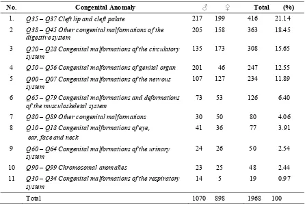
For 5 years, from 1998 until 2002, there were 1968 admissions to RS Dr Sardjito with diagnosis category of Q00-Q99 congenital malformations, deformations, and chromosomal abnormalities, according to the International Classisifaction of Diseases (ICD-10). Based on the whole patients admitted in the year, the proportion of congenital anomaly was 2.06%, comprising 1070 male and 898 female patients.
The most common diseases were Q35-Q37 cleft lip and cleft palate (416 cases, 21.14%), followed by Q38-Q45 other congenital malformations of the digestive system (363 cases, 18.45%), Q20-Q28 congenital malformations of the circulatory system (308 cases, 15.65%), Q50-Q56 congenital malformations of genital organ (247 cases, 12.55%), and Q00-Q07 congenital malformations of the nervous system (234 cases, 11.89%). Other group was less than 10%.
TABLE 1. The list of congenital anomaly according to ICD-10 in DR Sardjito Hospital 1998-2002
In 1990-1994, in Ibu & Anak Harapan Kita Hospital, there were 2442 cases of congenital abnormalities, or 6.78% of all patients. Based on similar diagnosis classification, Q35-Q37 cleft lip and cleft palate was in the second position (19.5%) after Q38-Q45 other congenital malformations of the digestive system (24.15%). The next positions
were Q65-Q79 congenital malformations and deformations of the musculoskeletal system (14.67%), Q20-Q28 congenital malformations of the circulatory system (14.12%), and Q50-Q56 congenital malformations of genital organ (10.54%). Based on the birthrate in this hospital, the proportion of congenital anomaly was 3.16%.3
No. Congenital Anomaly ♂ ♀ Total (%)
1. Q35 – Q37 Cleft lip and cleft palate 217 199 416 21.14
2 Q38 – Q45 Other congenital malformations of the digestive system
205 158 363 18.45
3 Q20 – Q28 Congenital malformations of the circulatory system
135 173 308 15.65
4 Q50 – Q56 Congenital malformations of genital organ 201 46 247 12.55
5 Q00 – Q07 Congenital malformations of the nervous system
107 127 234 11.89
6 Q65 – Q79 Congenital malformations and deformations of the musculoskeletal system
73 53 126 6.40
7 Q80 – Q89 Other congenital malformations 30 50 80 4.06
8 Q10 – Q18 Congenital malformations of eye, ear, face and neck
41 36 77 3.91
9 Q60 – Q64 Congenital malformations of the urinary system
24 26 50 2.54
10 Q90 – Q99 Chromosomal anomalies 23 25 48 2.44
11 Q30 – Q34 Congenital malformations of the respiratory system
14 5 19 0.97
TABLE 2 The frequency of congenital anomaly according to ICD-10 in Harapan Kita Hospital
No. Congenital Anomaly
1. Q38 – Q45 Other congenital malformations of the digestive syste 2 Q35 – Q37 Cleft lip and cleft palate
3 Q65 – Q79 Congenital malformations and deformations of the musculoskeletal system
4 Q20 – Q28 Congenital malformations of the circulatory system 5 Q50 – Q56 Congenital malformations of genital organ 6 Q00 – Q07 Congenital malformations of the nervous system 7 Q90 – Q99 Chromosomal anomalies
8 Q60 – Q64 Congenital malformations of the urinary system 9 Q80 – Q89 Other congenital malformations
10 Q10 – Q18 Congenital malformations of eye, ear, face and neck 11 Q30 – Q34 Congenital malformations of the respiratory system
Total The pattern of congenital anomalies in DR
Sardjito Hospital, with cleft lip and palate as the most common, was similar with the observation in Manado, although the next position in the list was different, that is, talipes, multiple malformation, anal atresia, omphalocele, and congenital heart diseases. This data were of babies born in Gunung Wenang Hospital in 5 year period, which showed a total incidence of congenital malformations of 0.9% and 0.5%, some of them were in major category.4 In RS Pirngadi Medan
in 1981-1984, there were 77 cases of congenital anomalies from 15.185 births (0.51%), and the most common were pes equinovarus (7 cases, 9.1%), labiognathopalatoschisis, hydrochepalus, and anencephalus, each was 6 cases (7.7%).5
A study in South India in September 1989 until December 1992 included 12.797 births, it was found a total incidence of 3.7%, consisted of 3.2% in liveborns and 15.7% in stillbirths. The anomalies were musculosceletal cases (9.69 per 1000), cutaneus (6.33 per 1000), genitourinary (5.47 per 1000), gastrointestinal (5.47 per 1000), central nervous system (3.99 per 1000), and cardiac anomalies (2.03 per 1000). Musculosceletal, cutaneus, and genito-urinary malformation cases were commonly found in live births, while gastrointestinal and central nervous system malformations were commonly found in stillbirths. In the study, antenatal infection and drug consumption were not found as significant causal factors of defects.6 In Indira Gandhi Medical College,
there were 180 babies with congenital malformations, with overall incidence of 1.78%. In 311 stillbirths, there were 47 cases (15.1%), while in livebirths 1.3%. The most common malformations were central nervous system (40%), followed by musculosceletal system (23.8%), while the less common was genitourinary system (3.8%). In this study malformations were more often found in babies with birthweight < 2500 gm (2.6%).7
In European population congenital anomaly occurred in 2-4% births, depended on the inclusion criteria and diagnostics. This report showed cardiac defects were the most common (25%), followed by limb anomalies (17%), chromosomal syndromes and urinary system malformations (each was 15%), central nervous system and neural tube malformations (10%), and oral cleft (6%).8
TABLE 3. Description of the relative occurrence of congenital anomaly in Europe
1. Cardiac defect 25% 2. Limb anomalies 17% 3. Chromosomal syndromes 15% 4. Urinary system 15% 5. Central nervous system and
neural tube 10%
6. Oral cleft 6%
Eskimo, talipes was found in the skeletons from ancient Egypt more than 4500 years ago.10,11,12 The
understanding of its etiology has revolutionized from mystic irrational to the latest scientific explanations developing to the molecular level.
In pregnancy, a unique event is occurred, affecting all biochemical, physiological, and anatomical processes. Exposure of xenobiotic factors and natural agents may change the growth and development process with consequences probably obvious since birth and/or all through life; the consequences can be cured partly or wholly, but may also caused a fixed defect. Organogenesis occurs in a short period, that is : 10 days (day 6-15) in rat 22 day pregnancy, 13 days (day 6-19) in rabbit 29 day pregnancy, and 7 weeks (7 x 7 = 49 days, that is, day 9-58) in human 9 month pregnancy. Generally, only this short growth and development stage is susceptible to the induction of malformations.13
Congenital anomaly is a broad concept, including all types of structural and functional defects. Structural abnormality can be classified into malformations, malformation syndromes, deformations, and disruptions. Functional anomaly may be caused by the hormonal imbalance while the babies in growth and developmental stage since pregnancy which caused health consequences several years later; nervous system disorder while in intrauterine stage also caused congenital anomaly.14
severity affecting the phenotype, age effect, expansion and contraction.9 Based on the above
discussion, the proportion of congenital anomaly in RS Dr Sardjito in 1998-2002 based on inpatients was 2.06%, while in RSIA Harapan Kita in 1990-1994 was 6.78%. Possible factors affecting this result was the more general scope of patients in RS Dr Sardjito compared to RS Harapan Kita who admitted mothers and children only. A more urban life pattern in Jakarta compared to in Yogyakarta needs to be studied, although there was an increase in proportion each year in Jakarta, while in Yogyakarta, the increase was not observed. In Indonesian population, admitted into RS Harapan Kita and RS Dr Sardjito, the anomaly of gastrointestinal system and cleft lip and palate were the most common (around 40%), while circulation system anomaly was around 15%. In Indian population, the anomalies of musculosceletal and nervous system were more common. European population was dominated by heart defects (25%), while cleft lip and palate was only 6%. This difference is probably caused by various factors, including the etiology and existence of risk factors, and its relationship with critical window in current growth and development process.
Concerning the etiology and risk factors, congenital anomaly as an abnormality known since the ancient times can be traced in paleopathology studies. Palatoschisis was noted in Nubian residence more than 3000 years ago and in ancient
TABLE 4. Definitions and examples of classic cases of each anomalies.15 (Seller, 2004)
Category Definition Exam
Malformasi Local growth and development anomaly in the morphogenesis stage of tissue or organ
Some various malformations due to the same etiology occur in an individu
Trisomi 21
Deformation The abnormalities of form or structure of the body part after or at the time of its normal morphogenesis
Club foot, oligohydramn Disruption The abnormalities or apparent damage of
part of the body that has been normally grown or potentially able to grow normally
Congenital anomaly is caused by genetic and or environmental factors in growth and development process since pregnancy. Generally, it was known that most congenital anomalies have genetic background. The role of environment as the causal factor has been known more commonly found (2-37%). Drugs, radiation, infection, and alcohol were believed as the etiology in 6-8% of cases.15 The
variations may be explained by the difference in the definition of “environment”. Some expert
Various study results on potentially teratogenic agents were summarized from electronic and printed sources. The discussions include: i). cigarettes have a role in the occurense of heart defects, neural tubes defect, oral-facial cleft and club foot,16 ii). lithium medication was considered
decreasing the risk, iii). glucocorticoids are related to facial cleft, iv) trimetophrim, v). methimazole syndrome.17,18 In infertile couples, either in
treatment or not, there was a higher congenital malformation prevalence; but the association with hormonal therapy is needed to be studied further.19
There was also reports suggested that certain diets, toxin exposure, endocrine disrupting chemicals (EDC), and insecticides had roles in the incidence of congenital anomaly.13,20,21
Another factor often discussed was hyperthermia as teratogenic source. The threshold of temperature for teratogenic effect of
over the core values for a long exposure, and probably for ~5 minutes or more in 4°C temperature increase. Various pathogenic mechanisms leading to defects in development suggested include cell death and cell proliferation deceleration, disruption of normal gene activity, vascular disruption, destruction of cell membrane and intracellular structures, and enzyme inhibition. Usually, the effect of heat is received directly by embryo and foetus in the uterus, the defect is not caused by maternal reaction to toxic exposure, although the maternal reaction has an effect on foetal response.22,23,24
Other data showed that pregnant rats who exposed to hyperthermia would have pathological change in their placenta, and this might be responsible for the growth and development disorders.25
The previous discussion generally analyzed various evidence obtained from experimental studies. Several reports from clinical studies are considered enviroment in broad meaning, that is, every non-genetic factor increasing the risk of congenital anomaly in exposed individual, consisted of biological factors (such as rubella infection), physical factors (X ray, ionizing radiation), and chemical agents (such as drugs used in pregnancy). Some other experts limited the environment definition on chemical and physical exposures in the air, water, soil, and food.
TABLE 5. Chemical agents and exposure conditions associated with congenital anomaly in humans14
1. Chemical agents
z Pharmaceuticals, for example: DES, thalidomide, warfarin
z Hair colouring agents
z Pesticides
z Non-pesticide endocrine disrupter (ED, for example: bisphenol A, phthalate, vinyl chloride, TCDD
z Heavy metal (Pb, Hg, Cd, As, Cr and Ni)
z Organic solvents, for example: styrene
2. Exposure conditions associated to congenital anomaly
z Drink water (heavy metals, nitrates, chlorinated agents)
z Residence near dangerous waste disposal
z Pesticides in farming or gardening area
z Air pollution
z Food contamination (dioxin, PCB)
TABLE 6. Malformation types reported from clinical studies 14,15
Infectious agents
1. rubella: before 10 weeks pregnancy: cataract and heart defect; 10-16 weeks pregnancy, hearing loss and retinopathy
2. varicella : extremity hypoplasia, microcephaly, chorioretinitis
3. CMV : hydrocephalus, periventricular calcification, neurological problems 4. toxoplasmosis : hidrocephalus, microcephaly, cerebral calcification
Maternal diseases
1. insulin-dependent diabetes : macrosomy, caudal regression syndrome, neural tube defect, heart defect, especially VSD and transposition of great arteries
2. phenylketonuria (with no diet control): microcephaly, micrognathia, heart defect, mental retardation 3. folate deficiency : neural tube defect, cleft lip and palate
4. epilepsy : cannot be differentiated from teratogenic effect of treatment
Physical agents
1. radiation : high dose on foetus in midterm until the end of pregnancy would cause microcephaly
2. hyperthermia: neural tube defect, particularly anencephaly, microcephaly, micropthalmia, cleft lip and palate; difficult to differentiate from another agents of hyperpyresis.
Drugs
1. diethylstilbestrol: female – vaginal adenosis; male – micropenis, hypospadia, cryptorchidismus 2. warfarin: nose hypoplastic and bone dysplasia, choanal atresia, microcephaly, hydrocephalus 3. phenytoin: brachicephaly, cleft lip and palate, kuku and finger hypoplasia, short neck, hirsutism
4. retinoic acid (vitamin A congeners): hydrocephalus, microcephaly, heart defect, especially conotruncal malformation, aortal arc hypoplasia, microtia/anotia, micrognathia, urogenital anomaly
5. alcohol: microcephaly, ‘long face’, ‘flat lip’, short distal finger, heart defect, mental retardation
CONCLUSION
In this study, the proportion of congenital anomaly in RS Dr Sardjito inpatients was 2.06%. The most common anomalies were cleft lip and palate (21.14%), followed by other malformations in gastrointestinal system (18.45%). This pattern is similar to another conditions in other areas in Indonesia, but different with the pattern in India and Europe. This difference needs to be studied further to find out the possible etiology and risk factors, so that an appropriate management may be developed.
ACKNOWLEDGEMENT
A deep gratitude and high praise are given to the late Prof Radiopoetro, the late Prof Soemiati Ahmad, and the late Prof T Jacob who had become models of integrity as teachers and scientists in the fields of Anatomy, Embryology, and Anthropology. The auther
also wants to thank the head and all staffs of RS Dr Sardjito who gave permission to use their data for this study.
REFERENCES
1. Gelehrter TD, Collins FS, Ginsdurg D. Principles of medical genetics. 2nd ed. Williams & Wilkins 1998.
2. Seashore MR, Weppner RS. Genetics in primary care & clinical medicine. 1st ed. London: Prentice Hall 1996.
3. Indrasanto E. Congenital abnormalities: HARAPAN KITA Children and Maternitiy Hospital’s experiences. In: Muljono DH, Sudoyo H, Harahap A.. (eds). Recent Advances in medical genetics. Jakarta 1995:41-44. 4. Masioman N, Mustadjab I, Munir M. Congenital
malformation at Gunung Wenang Hospital Manado: a five-year spectrum. Paediatr Indones 1991Nov-Dec; 31(11-12):294-302.
5. Lubis N, Tjipta GD, Panjaitan AJ, Raid N, Siregar H. Congenital malformation among newborns at Dr. Pirngadi Hospital Medan during 1981-1984. Paediatr Indones 1989 Jan-Feb; 29(1-2):1-7.
7. Grover N. Congenital malformations in Shimia. Indian J Pediatr 2000Apr;67 (4):249-51.
8. Dolk H, Vrijheid M. The impact of environmental pollution on congenital anomalies. Br Med Bull 2003; 68:25-45. 9. Baraister M. Uses of databases in dysmorphology. In:
Ferretti P, Copp A, Tickle C, Moore G Editors. Embryos, genes and birth defects. 2nd ed. John Wiley & Sons
2006:19-32.
10. Jacob T. Kesehatan dikalangan manusia purba. B I Ked 1969; 1(2):143-57.
11. Jacob T. Manusia makhluk gelisah melalui lensa bioantropologi. UMS Press 2006.
12. Zimmerman MR. Paleopathology and the study of ancient remains. In: Ember CR & Ember M (eds). Encyclopedia of medical anthropology. Kluwer 2004:49-57.
13. Mantovani A, Maranghi F. Endpoints for prenatal exposures in toxicological studies. In: Nicolopoulou-Stamati P, Hens L, Howard CV. (Eds). Congenital Diseases and the Environment. Springer 2007:21-36.
14. Hens L. Environmental impacts on congenital anomalies – information for the non-expert professional. In: Nicolopoulou-Stamati P, Hens L, Howard CV. (Eds). Congenital Diseases and the Environment. Springer 2007:409-50.
15. Seller M, Genetic causes of congenital anomalies and their interaction with environmental factors, in Eurocat Special Report: A review of environmental risk factors for congenital anomalies 2004 (on line) www. Eurocat.ulster.ac.uk/pubdata 16. Wasserman CR, Shaw GM, O’Malley CD, Tolarova MM, Lammer EJ. Parental cigarette smoking and risk for congenital anomalies of the heart, neural tube, or limb. Teratology 1996;53 (4):261-67.
17. Shepard TH, Brent RL, Friedman JM, Jones KL, Miller RK, Moore CA, et al.. Update on new developments in
the study of human teratogens. Teratology 2002; 65(4):153-61.
18. Shepard TH. Annual commentary on human teratogens. Teratology 2002:66:275-77.
19. Zu JL, Basso O, Obel C, Bille C, Olsen J. Infertility, infertility treatment, and congenital malformations: Danish national birth cohort. Br Med J 2006;333 ::679-84. 20. Nicolopoulou-Stamati P. Concepts in the relationship of
congenital diseases with the environment. In: Nicolopoulou-Stamati P, Hens L, Howard CV. (Eds). Congenital Diseases and the Environment. Springer 2007:1-20.
21. Wattiez C. Links between in utero exposure to pesticides and effect on the human progeny. In: Nicolopoulou-Stamati P, Hens L, Howard CV. (Eds). Congenital Diseases and the Environment. Springer 2007:183-206.
22. Finnell RH, Waes JGV, Endy JD, Rosenquist TH. Molecular basis of environmentally induced birth defects. An Rev Pharmacol Toxicol 2002; 42:181-208.
23. Mirkes PE, Little SA. Teratogen-induced cell death in postimplantation mouse embryos: differential tissue sensitivity and hallmarks of apoptosis. Cell Death and Diff 1998; 5:592-600.
24. Miller MW, Nyborg WL, Dewey WC, Edwards MJ, Abramowicz JS, Brayman AA. Hyperthermic teratogenicity, thermal dose and diagnostic ultrasound during pregnancy: implication of new standards on tissue heating. Int J Hypertherm 2002; 18(5):361-84.