A New Benzoyl Compound Isolated from the Endophytic Fungi of Kandis Gajah
(
Garcinia griffithii
) and Asam Kandis (
Garcinia cowa
)
Elfita
1*, Munawar
2, Muharni
1, Gusti Pratiwi
1, and Rahmadania
11. Department of Chemistry, Faculty of Mathematics and Natural Sciences, Universitas Sriwijaya, Indralaya, Kabupaten Ogan Ilir 30662, Indonesia
2. Department of Biology, Faculty of Mathematics and Natural Sciences, Universitas Sriwijaya, Indralaya, Kabupaten Ogan Ilir 30662, Indonesia
*
E-mail: elfita_kimia@unsri.ac.id
Received March 11, 2015 | Accepted February 22, 2016
Abstract
Garcinia griffithiiandGarcinia cowabelong to the genusGarcinia. The genusGarcinia has been known to be a rich source of secondary metabolites, such as xanthones, benzophenones, flavonoids, steroids, terpenoids, and other phenolic derivatives. Previous investigations of endophytic fungi fromG. griffithiirevealed the presence of three compounds not found in the host. In order to the continue the phytochemical work on endophytic fungi ofG. griffithii, the constituent of the endophytic fungi ofG. griffithiiwas re-examined. In this study, a benzoyl compound similar to that found in the endophytic fungus of G. cowa was observed. The same benzoyl compound was also isolated from the endophytic fungusAcremonium spofG. griffithiiandAspergillus spofG. cowawith cultivation of eight weeks in static conditions at room temperature. The culture medium was partitioned using ethyl acetate and evaporated to obtain the concentrated extract. Isolation of compounds was performed using the chromatography method. The chemical structure was proposed on the basis of spectroscopic data, including ultraviolet (UV), infrared (IR), mass spectrometry (MS), proton nuclear magnetic resonance (1H-NMR), carbon nuclear magnetic resonance (13C-NMR), heteronuclear single-quantum correlation spectroscopy (HSQC), heteronuclear multiple-bond correlation spectroscopy (HMBC), and correlation spectroscopy (COSY).
Abstrak
Suatu Senyawa Benzoil Baru Hasil Isolasi dari Jamur Endofit Tumbuhan Kandis Gajah (Garcinia griffithii) dan Asam Kandis (Garcinia cowa).TumbuhanGarcinia griffithii danGarcinia cowatermasuk ke dalam genusGarcinia. Genus Garcinia dikenal kaya dengan kandungan metabolit sekunder, seperti santon, benzofenon, flavonoid, steroid, terpenoid, dan turunan fenolik lainnya. Penelitian sebelumnya terhadap jamur endofit dariG. griffithiitelah menemukan tiga senyawa yang tidak ditemukan dalam tumbuhan inangnya. Sebagai lanjutan dari penelitian fitokimia mengenai jamur endofitik tumbuhanG. griffithii, kandungan kimia dari jamur endofitik ini kembali diteliti. Dalam studi ini, juga diperoleh senyawa benzoil yang sama dengan yang ditemukan dalam jamur endofit tumbuhan G. Cowa. Senyawa benzoil yang sama juga dapat diisolasi dari jamurAcremonium sptumbuhanG. griffithiidan dari jamurAspergillus sp tumbuhan G. cowa dengan masa kultivasi delapan minggu dalam kondisi statis pada suhu kamar. Medium kultur dipartisi menggunakan etil asetat dan dievaporasi untuk mendapatkan ekstrak pekatnya. Isolasi senyawa dilakukan dengan metode kromatografi. Struktur kimia diusulkan berdasarkan data spektroskopi yang meliputi ultraviolet (UV), infrared (IR), mass spectrometry (MS), proton nuclear magnetic resonance (1H-NMR), carbon nuclear magnetic resonance(13C-NMR),heteronuclear single-quantum correlation spectroscopy(HSQC),heteronuclear multiple-bond correlation spectroscopy(HMBC), dancorrelation spectroscopy(COSY).
Keywords: benzoyl, endophytic fungi, Garcinia griffithii, Garcinia cowa
Introduction
Endophytic fungi are a new source of bioactive compounds being explored recently. They live in plant tissue in a certain period of time, can form colonies in plant tissues
Plants of the genusGarciniaare known as a rich source of diversity for secondary metabolites including xanthones, benzophenone, flavonoids, steroids, terpenoids, and other phenolic derivatives [6-8]. The extract of natural products have been used as drugs and important sources of traditional medicines, such as cytotoxic, antibacterial, antiviral, antifungal, antioxidant, anti-inflammatory, antidepressant, and anti-HIV [9-11].
Phongpaichitet al.(2007) and Rumaet al.(2013) reported endophytic fungal isolated from the Garcinia genus, includingGarcinia atroviridis, Garcinia dulcis, Garcinia mangostana, Garcinia nigrolineata, Garcinia Scortechinii, Garcinia gummigutta,Garcinia indica,Garcinia morella, andGarcinia xanthochymus. The endophytic fungi are shown to have antimycobacterial, antimalarial, antiviral, antioxidant, anti-inflammatory, and cytotoxicity activities [12-13]. Several strategies used to isolate endophytic fungi have been reported recently to search for potential bioactive compounds, and one of them is based on ethnobotanical history specifically used by indigenous peoples [14].Garcinia griffithiiandGarcinia cowaare traditionally used by the local communities of Sarasah Bonta, Lembah Arau, and West Sumatra to treat various diseases, including gout, diarrhea, and malaria.
In previous work, we reported three compounds from endophytic fungi ofG. griffithii, namely, 4,6-dihydroxy, 3,8a-dimethyl-1-oxo-5-(3’-oxobutan-2’-yl)-1,4, 4a, 5, 6, 8a-hexahydronaphthalen-2-yl-1”, 2”-di-methyl- 5”- (2”’-methyl -prop -1”’-enyl) -cyclopentane - carboxylate from Aspergillus fumigatus [15]; 3,5-dihydroxy-2,5-dimethyl-trideca-2,9,11-triene-4,8-dione from Acremonium sp [16]; and 10, 12-trihydroxy-9-methoxy-7a-methyl-7, 7a, 12a, 13- tetrahydrobenzo-cyclohepta-oxocin-6-one from Aspergillus niger[17].
In the current study, we reported another secondary metabolite called benzoyl compound, which is extracted from Acremonium sp of G. griffithii and found in the endophytic fungusAspergillussp ofG. cowa.
Materials and Methods
Source of endophytic fungi. Acremonium sp of G. griffithii obtained from stock fungus was stored in the Microbiology Laboratory, Department of Biology, Faculty of Mathematics and Natural Sciences of Sriwijaya University. The fruit ofG. cowa was collected in May 2012 from Lembah Arau, West Sumatra.
Isolation and identification of endophytic fungi.The isolation of Acremonium sp from twigs of G. griffithii was reported previously by Elfita et al. 2012 [16] and Debbabet al. [18]. The isolation method ofAspergillus sp from the fruit ofG. cowa grown in Sarasah Bonta, Lembah Arau, Kabupaten Lima Puluh Kota, and West Sumatra was conducted according to the procedure by
Elfitaet al. 2011 [15]. The fungal strain was identified on the basis of the morphological method by the School of Hayati Science and Engineering, Bandung Institute of Technology, Indonesia. The voucher of specimen was stored in the Microbiology Laboratory, Department of Biology, Faculty of Mathematics and Natural Sciences of Sriwijaya University.
Cultivation of endophytic fungi.Potato dextrose broth (PDB) medium of 300 mL was used for the cultivation of endophytic fungi and was placed into 30 flasks (1 L each). Fungal suspension containing 106spores/mL was inoculated under sterile conditions to each 300 mL PDB medium (ratio 1:10). The cultures were incubated for eight weeks in static conditions at room temperature [19-20]. Extraction, exploration, and structure elucidation. Mycelia were removed from the endophytic fungus culture after eight weeks of incubation and the medium was filtered. The medium was extracted three times using ethyl acetate (1:1) followed by evaporation under vacuum to obtain the concentrated extract. The concentrated extract was separated by column chromatography over silica gel60 (70–230 mesh) at thestationary phase (1:30) and eluent, which was previously determined by thin-layer chromatographysilica gel 60 F254. The chosen eluent with
increased polarity wasn-hexane:EtOAc at a ratio of 10:0 to 0:10 (v/v). An eluate was collected and then combined using thin-layer chromatography into column fractions. Each fraction was evaporated and purified using the chromatography technique to obtain the purified compound. The ethyl acetate extract from Acremonium sp of G. griffithii(3.16 g) was subjected to column chromatography over silica gel (60 x 1.5 cm) and was eluted with n -hexane-ethyl acetate gradient (10:0–0:10) and collected in 60 vials each containing 10 mL. The thin layer chromatography (TLC) analysis showed the presence of five column fractions (F1–F5): F1 (724 mg), F2 (433 mg), F3 (406 mg), F4 (821 mg), and F5 (891 mg). Fraction F3 showed a major compound. It was further separated by column chromatography over silica gel (30 × 0.7 cm) and eluted withn-hexane-ethyl acetate (5:5– 0:10) to give 37 vials. The fractions, which gave the same Rf on TLC, were combined and yielded four column fractions (F3.1–F3.4): F3.1 (81 mg), F3.2 (52 mg), F3.3 (143 mg), and F3.4 (105 mg). Fraction F3.3 was purified by rechromatography to yield compound1 as a white crystal (87 mg). Its melting point is 109C– 111C. Further separation and purification of fraction F4 by column chromatography yielded compound2 as previously reported by Elfitaet al. 2012 [16].
presence of three column fractions (F1–F3): F1 (778 mg), F2 (653 mg), and F3 (906 mg). Fraction F2 showed a major compound. Thus, it was further separated by column chromatography over silica gel (30 × 0,7 cm) and eluted withn-hexane- ethyl acetate (5:5 – 0:10) to yield compound 1* as a white crystal (228 mg). Its melting point is 109C–111C.
The chemical structure of the compound was determined using the following spectroscopy methods: UV, IR, 1D-NMR (¹H-1D-NMR and ¹³C-1D-NMR), 2D-1D-NMR (HMQC, HMBC and COSY), NMR spectra (recorded at 500 MHz (1H) and 125 MHz (13C) on JEOL JNM ECA-500 spectrometer), and high resolution electrospray ionization mass spectrometry (HRESI-MS).
3. Result and Discussion
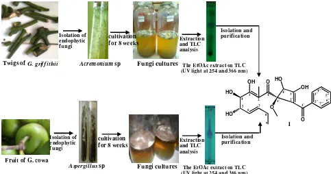
In this study,Acremoniumsp was cultivated on 9 L of PDB medium for eight weeks at room temperature. The culture broth was extracted by solvent partition with EtOAc (1:1), followed by evaporation. The extract showed two major spots on TLC.
The fungal strain from the fruit ofG. cowawas identified as Aspergillus sp by the School of Hayati Science and Engineering, Bandung Institute of Technology, Indonesia. Aspergillus sp was cultivated on 9 L of PDB medium for eight weeks at room temperature. The culture broth was extracted by solvent partition with EtOAc (1:1),
followed by evaporation. The extract showed one major spot on TLC. Figure 1 briefly illustrates the procedure for isolating the pure compound from the endophytic fungiAcremoniumsp from the twigs ofG. griffithiiand Aspergillussp from the fruit ofG. cowa.
Elucidation of compound 1 fromAcremonium sp of G. griffithii. Compound1 exhibited UV absorbance at
λmax(MeOH) 258 nm (logε= 4.54), which indicates the
presence of an aromatic chromophore. These bands did not show a bathochromic shift with NaOH, thus indicating the absence of a hydroxyl group in the aromatic ring. The IR spectrum of 1 showed intense carbonyl absorption bands at 1701.1 and 1732.0 cm-1 because of the two ketone groups. The broad spectrum at 3436 cm-1indicated the presence of a hydroxyl group. The characteristic vibrations at 3030.1, 1627.0, and 1598.9 caused by the aromatic group and stretching vibration at 2873.7–2964.4 cm-1indicated the presence of C-H aliphatic. The HRESI-MS exhibited the [M+H]+ ion peak at m/z 419.280 (calc. 419,1628), which is consistent with the molecular formula C22H26O8 and
implies 10 DoU (degree of unsaturation).
The 1H-NMR spectrum of compound 1 recorded in CDCl3 (1H-500 MHz) revealed the presence of three
proton sp3signals and two methyl proton signals at δH
0.97 ppm (3H, t, J=7.2) and 1.66 ppm (3H, s), respectively, and one methylene signal at δH2.11 ppm
(2H, m).
Figure 1. Brief Procedure for Isolating the Pure Compound from the Endophytic Fungi Acremoniumsp from Twigs G. griffithiiandAspergillussp from the Fruit ofG. cowa
Isolation of endophytic f ungi
Twigs ofG. grif f ithii Acremoniumsp cultivation for 8 weeks
Fungi cultures
Extraction and TLC analysis
Isolation and purification
Fungi cultures Fruit of G. cowa
Aspergillussp
Isolation of endophytic f ungi
cultivation
for 8 weeks Extractionand TLC analysis
1
Isolation and purification
The EtOAc extract on TLC (UV light at 254 and 366 nm)
The EtOAc extract on TLC (UV light at 254 and 366 nm)
OH
O HO O OH
HO
HO
O 1 2 3 4 5
6 7
8 9
1' 2' 3' 4'
1'' 2'' 6" 3''
7" 4''
The presence of five aromatic protons showed the signal atδH7.48 ppm (2H, d, J=7.8), 8.32 ppm (2H, d, J=7.8),
and 7.64 ppm (1H, t, J=7.5). A signal was also found for methoxy proton atδH3.45 ppm (3H, s) and two vinylic
protons atδH5.56 ppm (1H, m) and 5.23 ppm (1H, m).
The 1H-NMR revealed the presence of signals to three methyne protons sp3 as oxygenated carbon at δH 4.69
ppm (1H, s), 4.74 ppm (1H, dd, J=4.5; 3.9), and 4.59 ppm (1H, d, J=3.9).
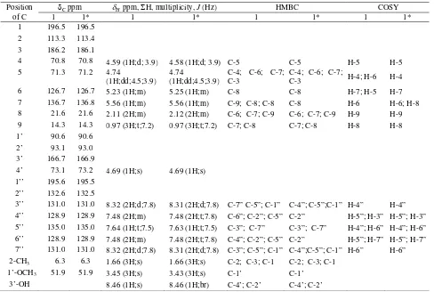
The13C-NMR spectrum showed 22 signals, including 8 carbon sp3and 14 carbon sp2. The13C-NMR and DEPT 135 spectrum, which were supported by HSQC spectrum, showed eight carbon sp3signals consisting of two methyl, one methylene, three methine, one methoxy, and one quaternary carbons.
Three signals of methine atC70.8, 71.3, and 73.1 ppm
were assigned to carbon atoms bearing hydroxyl groups and one quaternary carbon bearing a methoxyl group. Furthermore, 14 carbon sp2signals included 6 aromatic carbons at C 128.9 and 135.0, and 2 carbons with the
same chemical shift at 131.0 ppm and 132.6 ppm. Two of these signals were assigned to the ketone carbons at
C195.6 ppm and 196.5 ppm. Six other sp2carbon signals
were present atC 93.1, 113.3 126.7, 136.7, 166.7, and
186.2 ppm.
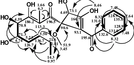
In the HMBC spectrum (Table 1.), correlations from H-3” (δH8.32) to C-1” (C195.6) , C-4” (C128.9), and
C-5” (C 135.0) suggested that C-1” was attached to the
aromatic ring as a benzoyl group. Correlations from the hydroxyl proton at C-3’ (δH8,46) to C-2’ (C93.1) and
C-4’ (C 73.1) and from1’-OCH3(δH3.45) to C-1’ (C
90.6) demonstrated the presence of a cyclobuthene ring bearing methoxyl and hydroxyl groups. The methoxyl resonance connecting to C-1’ in HMBC indicated C-1’ with a high chemical shift (C90.6). Moreover, oxygenated
carbon was attached to C-1 (carbonyl carbon). Correlations from the hydroxyl proton at C-3’ (δH8,46) to C-2’ with
a low chemical shift as sp2 C=C (C 93.1) showed an
anisotropy effect of carbonyl groups (C-1”). HMBC correlations from2-CH3(δH1.66) to C-1 (C196.5), C-2
(C113.3), and C-3 (C186.2) indicated that 1 and
C-3 were located at the carbonyl group and hydroxyl group, respectively. Correlations from H-4 (δH4.59) to
C-5 (C 71.3) and H-5 (δH 4.74) to C-4 (C 70.8)
revealed that C-4 and C-5 were attached to the hydroxyl groups. Figure 2 illustrates the HMBC correlation and
δ-assignment of compound1. Table 1. NMR Data of Compound 1 recorded at1H-500MHz,13C-125 MHz in CDCl3
Position of C
Cppm Hppm,H, multiplicity,J(Hz) HMBC COSY
1 1* 1 1* 1 1* 1 1*
1 196.5 196.5 2 113.3 113.4 3 186.2 186.1
4 70.8 70.8 4.59 (1H;d; 3.9) 4.58 (1H;d; 3.9) C-5 C-5 H-5 H-5 5 71.3 71.2 4.74
(1H;dd;4.5;3.9) 4.74
(1H;dd;4.5;3.9)
C-4; C-6; C-7; C-3
C-4; C-6; C-7;
C-3 H-4; H-6 H-4 6 126.7 126.7 5.23 (1H;m) 5.25 (1H;m) C-8 C-8 H-7; H-5 H-7 7 136.7 136.8 5.56 (1H;m) 5.56 (1H;m) C-9; C-8; C-8 C-8 H-6 H-6; H-8 8 21.6 21.6 2.11 (2H;m) 2.12 (2H;m) C-6; C-7; C-9 C-6; C-7; C-9 H-9 H-9 9 14.3 14.3 0.97 (3H;t;7.2) 0.97 (3H;t;7.2) C-7; C-8 C-7; C-8 H-8 H-8 1’ 90.6 90.6
2’ 93.1 93.0 3’ 166.7 166.9
4’ 73.1 73.2 4.69 (1H;s) 4.69 (1H;s) 1’’ 195.6 195.5
2’’ 132.6 132.5
3’’ 131.0 131.0 8.32 (2H;d;7.8) 8.31 (2H;d;7.8) C-7” C-5”; C-1” C-4”; C-5”;C-1” H-4” H-4” 4’’ 128.9 128.9 7.48 (2H;m) 7.48 (2H;t;7.8) C-6”; C-2”; C-5” C-2” H-5”; H-3” H-5”; H-3” 5’’ 135.0 135.0 7.64 (1H;t;7.5) 7.63 (1H;t;7.5) C-3”; C-7” C-3”; C-7” H-4”; H-6” H-4”; H-6” 6’’ 128.9 128.9 7.48 (2H;m) 7.48 (2H;t;7.8) C-4”; C-2”; C-5” C-2” H-5”; H-7” H-5”; H-7” 7’’ 131.0 131.0 8.32 (2H;d;7.8) 8.31 (2H;d;7.8) C-3”; C-5”; C-1” C-4”;C-5”; C-1” H-6” H-6” 2-CH3 6.3 6.3 1.66 (3H;s) 1.66 (3H;s) C-2; C-3; C-1 C-2; C-3; C-1
1’-OCH3 51.9 51.9 3.45 (3H;s) 3.43 (3H;s) C-1’ C-1’
3’-OH 8.46 (1H;s) 8.46 (1H;br) C-4’; C-2’ C-4’; C-2’
14.3 21.6 136.7 126.7 71.3
70.8186.2113.3196.5 90.6
In the1H-1H COSY spectrum, the correlations from H-4 to H-5 indicated the presence of vicinal hydroxyl groups. The next correlations from H-6 to H-5, H-7 and from H-8 to H-9 revealed the presence of long-chain aliphatic subunits. The correlation of 1H-1H aromatic indicated a mono-substituted aromatic ring. The COSY correlation of compound1is illustrated in Figure 2. Carbon C-1' of compound1 had a similar position and chemical environment to C-1' of talaroflavone [21]. Carbon C-1'of the two compounds is C sp3 (quarterner), which binds two C sp2, one oxygenated C sp3, and one oxygen atom. Each carbon appeared atδC90.6 and 91.8
ppm. Furthermore, carbon C-2' of compound 1 had a position and chemical environment similar to those of C-3 of methyltriacetic lactone [21], each of which appeared onδC 93.1 and 95.0 ppm. The chemical shift
of C-1' (δC 90.6) was shown to approach the calculated data using Chem Draw Ultra 10 (δC 94.8)
Combining the results obtained by the spectrometric methods (UV, IR, MS,1H-NMR,13C-NMR, HSQC, HMBC, and COSY) and comparing the results of the literature data [21] with the calculated data of Chem Draw Ultra 10, we determined C22H26O8 as the molecular formula
for compound 1 with a molecular weight of 418. The structure of compound 1 is 1-(2’-benzoyl-3,4-dihydroxy-1’-methoxycyclobut-2’-enyl)-3, 4, 5-trihydroxy- 2-methyl-nona-2,6-dien-1-one. The confirmation structure of 1 will be followed by an X-ray single crystal, and this work is still in progress.
Elucidation of compound 1* from Aspergillus sp of G. cowa.The1H-NMR spectra recorded in CDCl3(1
H-500 MHz) indicated that the spectroscopy data of1and 1*are identical (see Table 1).
Based on the Dictionary Natural Products database (December 18, 2015), the benzoyl derivative could be a new compound. However, the identical benzoyl compounds (1 and1*) were produced by two different endophyte fungi could further raise the question of whether it is related to the host of the same genus or assumed as a secondary metabolite typically produced by the fungi. Based on these phytochemical studies, further studies on the metabolite profiling of these endophytic fungi and plants should be performed for confirmation.
Figure 2. The HMBC Correlation
Conclusions
The endophytic fungiAcremonium spfrom the twigs of G. griffithii produced the newly proposed benzoyl compound, which is identical to that found from the endophytic fungus Aspergillus sp from the fruit of G. cowa.
Acknowledgements
The authors are grateful to the Directorate General of Higher Education for funding this research through the Strategis Nasional Grant in 2011 entitled “Produksi Sediaan Obat Tradisional Terstandar untuk Asam Urat dari Mikroba Endofitik Tumbuhan Kandis Gajah (Garcinia griffithii T. Anders)” with contract No. 397/SP2H/PL/Dit.Litabmas/IV/2011.
References
[1] Xue, H., Lu, C., Liang, L., Shen, Y. 2012. Secondary metabolites of Aspergillus sp. CM9a, an endophytic fungus ofCephalotaxus mannii.Rec. Nat. Prod. 6(1): 28-34, http://acgpubs.org/RNP/ 2012/Volume%206/2012_issue_ 6_1_1_list.htm. [2] Kalyanasundaram, I., Nagamuthu, J.,
Muthukumaras-wamy, S. 2015. Antimicrobial activity of endophytic fungi isolated and identified from salt marsh plant in vellar estuary. J. Microbiol. Antimicrob. 7(2): 13-20, doi: 10.5897/JMA2014.0334.
[3] Elfita, E., Munawar, M., Muharni, M., Ivantri, I. 2015. Chemical constituens from an endophytic fungusAspergillusSp (Sbd5) isolated from sambiloto (Andrographis paniculata Nees). Microbiol. Indonesia. 9(2): 82-88, DOI: 10.5454/mi.9.2.5. [4] Zhao, J., Zhou, L., Wang, J., Shan, T., Zhong, L.,
Liu, X., Gao, X. 2010. Endophytic fungi for producing bioactive compounds originally from their host plants. Curr. Res., Technol. Educ. Top. Appl. Microbiol. Microb. Biotechnol. A. Mendez-Vilaz (Ed.): 567-576, http://formatex.info/micro biology2/567-576.pdf.
[5] Strobel, G. 2006. Harnessing endophytes for industrial microbiology.Current Opinion in Micro-biology. 9:240–244, DOI 10.1016/j.mib.2006.04.001. [6] Ritthiwigrom, T., Laphookhieo, S., Pyne, S.G. 2013. Chemical constituents and biological activities of Garcinia cowa Roxb. Maejo Int. J. Sci. Techol. 7(02), 212-231, http://www.mijst.mju.ac.th/vol7/ 212-231.pdf.
[7] Irene See, I., Lian Ee, G.C., Teh, S.S., Kadir, A.A., Daud, S. 2014. Two new chemical constituents from the stem bark ofGarcinia mangostana.Molecules. 19: 7308-7316, doi:10.3390/molecules19067308. [8] Wahyuni, F.S., Shaari, K., Stanslas, J., Lajis, N.,
[9] Elfita, E., Muharni, M., Madyawati, L., Darwati, D., Ari, W., Supriyatna, S., Bahti, H. H., Dachriyanus, D., Cos, P., Maes, L., Foubert, K., Apers, S., Pieters, L. 2009. Antiplasmodial and other constituents from four Indonesian Garcinia spp. Phytochemistry. 70(7): 907-912, doi: 10.1016/ j.phytochem.2009.04.024.
[10] Elfita, E., Supriyatna, S., Bahti, H.H., Dachriyanus, D. 2008. Diprenylated xanthone from the stem bark of kandis gajah (Garcinia griffithii). Indo. J. Chem. 8(1): 97-100, http://pdm-mipa.ugm.ac.id/ojs/index. php/ijc/article/ viewFile/347/364.
[11] Ritthiwigrom, T., Laphookhieo, S., Pyne, S.G. 2013. Chemical constituents and biological activities ofGarcinia cowaRoxb. Maejo Int. J. Sci. Technol. 7(2): 212-231, http://www.mijst.mju.ac.th/vol7/212-231.pdf.
[12] Phongpaichit, S., Nikom, J., Rungjindamai, N., Sakayaroj, J., Towatana, N.H., Rukachaisirikul, V., Kirtikara, K. 2007. Biological activities of extracts fromendophytic fungi isolated from Garcinia plants. FEMS Immunol. Med. Microbiol. 51: 517-525, doi:10.1111/j.1574-695X.2007.00331.x. [13] Ruma, K., Sunil, K., Prakash, H. S. 2013.
Antioxidant, antiinflammatory, antimicrobial and cytotoxic properties of fungal endophytes from garcinia species. Int. J. Pharm. Pharm. Sci. 5(3): 889-897, http://ijppsjournal.com/Vol5Suppl3/7618. pdf.
[14] Strobel, G., Daisy, B., Castillo, U. 2005. The biological promise of microbial endophytes and their natural products. Plant Pathol. J. 4(2):161-176, http://dx.doi.org/10.3923/ppj.2005.161.176. [15] Elfita, E., Muharni, M., Indah, T. 2011. Secondary
metabolite ofAspergillus fumigatus, an endophytic fungi of the medicinal plant Garcinia griffithii.
Makara Sci. Ser. 15(2): 124-128, http://journal.ui. ac.id/science/article/viewFile/ 1061/974.
[16] Elfita, E., Muharni, M., Munawar, M., Rizki, R. 2012. Isolation of antioxidant compound from endophytic fungi Acremonium sp. from the twigs of kandis gajah (Garcinia griffithii). Makara Sci. Ser. 16(1): 46-50, http://journal.ui.ac.id/science/ article/viewFile/1280/1177.
[17] Elfita, E., Muharni, M., Munawar, M., Aryani, S. 2012. Secondary metabolite from endophytic fungi Aspergillus nigerof the stem bark of kandis gajah (Garcinia griffithii T. Anders). Indo. J. Chem. 12(2): 195-200, http://pdm-mipa.ugm.ac.id/ojs/index. php/ijc/article/view/685/734.
[18] Debbab, A., Aly, A.H., Ebel, R.A.E., Muller, W.E.G., Mosaddak, M., Hakiki, A., Ebel, R., Proksch, P. 2009. Bioactive secondary metabolites from the endophytic fungus Chaetomium sp. isolated from Salvia officinalisgrowing in Morocco. Biotechnol. Agron. Soc. Environ. 13(2): 229-234, http://www. pressesagro.be/base/text/ v13n2/229.pdf.
[19] Elfita, E., Munawar, M., Muharni, M., Suprayetno, S. 2013. New pyran of an endophytic fungus Fusariumsp. isolated from the leaves of brotowali (Tinaspora crispa). Indo. J. Chem. 13(3): 209-2015, http://pdm-mipa.ugm.ac.id/ojs/index.php/ijc/ article/view/757.
[20] Elfita, E., Munawar, M., Muharni, M., Sudrajat, M.A. 2014. Identification of new lactone derivatives isolated from Trichoderma sp., an endophytic fungus of brotowali (Tinaspora crispa). Hayati J. Biosci. 21(1): 15-20, doi: 10.4308/hjb.21.1.15. [21] Hassan, A.E.H.A. 2007. Novel natural products from