Indo. J. Chem., 2009, 9 (1), 142 - 145
Hendig Winarno and Ermin Katrin W
142
O
R3 R6
1' 5
4' 3
4
R2
R4
7
R1
R5
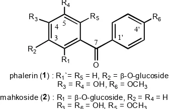
phalerin (1) : R1`= R5= H, R2=-O-glucoside, R3= R4= OH, R6= OCH3
m ahkoside (2) : R1=-O-glucoside, R2= R4= H
R3= R6= OH, R5= OCH3
* Corresponding author. Tel/Fax : +62-21-7690709/7691607 Email address : [email protected]
BENZOPHENONE GLUCOSIDE ISOLATED FROM THE ETHYL ACETATE EXTRACT OF
THE BARK OF
MAHKOTA DEWA
[
Phaleria macrocarpa
(Scheff.) Boerl.] AND ITS
INHIBITORY ACTIVITY ON LEUKEMIA L1210 CELL LINE
Hendig Winarno* and Ermin Katrin W.
Center for the Application of Isotopes and Radiation Technology, BATAN, Jakarta Jl. Cinere Pasar Jumat, Lebak Bulus, Jakarta 12440
Received August 1, 2008; Accepted October 15, 2008
ABSTRACT
Isolation and elucidation of benzophenone glucoside from ethyl acetate extract of Phaleria macrocarpa bark and its inhibitory activity test against leukemia L1210 cell line have been done. The Phaleria macrocarpa bark were macerated using n-hexane, ethyl acetate, and ethanol, respectively. The ethyl acetate extract was then chromatographed on silica gel column and gradiently eluted by n-hexane - ethyl acetate – ethanol with the composition from 20:1:0 until 0:0:1, gave eight fractions. Separation of fraction 6 using semipreparative HPLC on reverse phase column (Capcell Pak C-18 SG120, 15 mm I.D. x 250 mm) using methanol – water (40:60, 5 mL/min) gave a brown powder, with the melting point of 182.3 ºC. Spectroscopic analysis and comparison of its physico-chemical data, this compound was clarified as 2,4’-dihydroxy-4-methoxy-benzophenone-6-O--D-glucopyranoside (3). Inhibitory activity of its compound against leukemia L1210 cell line showed that this compound exhibited inhibitory activity with IC50 was 5.1g/mL.
Keywords: Phaleria macrocarpa, 2,4’-dihydroxy-4-methoxybenzophenone-6-O--D-glucopyranoside, cytotoxic activity, leukemia L1210
INTRODUCTION
Mahkota dewa [Phaleria macrocarpa (Scheff.) Boerl] is the one of the medicinal plant which grows in Indonesia. Empirically, Phaleria macrocarpaare used for medical treatment. The stems are used for treatment of bone cancer, the leaves are used for impotency, blood diseases, allergies, diabetes mellitus, and tumor treatments. Egg shell of seeds are used for breast cancer, cervix cancer, lung diseases, liver and heart diseases [1]. The fruits consisting of alkaloid, saponin, flavanoid, and polyphenol [2].
Wahyuningsih, et al. [3] found a new benzophenone glucoside from the leaves of Phaleria macrocarpa, so called phalerin (4,5-dihydroxy,4’-methoxybenzophenone-3-O--D-glucoside (1) (Fig. 1), while another new benzophenone glucoside derivative, mahkoside A (4,4’-dihydroxy-6-methoxybenzophenone-2-O--D-glucopyranoside) (2) (Fig. 2) was isolated by Zhang, et al.[4] besides six known compounds including mangiferin, kaempferol-3-O--D-glucoside, dodecanoic acid, palmitic acid, ethyl stearate and sucrose. Biological activity test on phalerin showed that this compound was non toxic and has a potent immunostimulant [3].
Preliminary study indicated that ethyl acetate extract of the bark of Phaleria macrocarpa exhibited IC50
of 10.15 g/mL against mouse leukemia L1210 cell as anticancer screening model [5]. Therefore, this study was aimed to isolate the mayor compound present in the
Fig. 1. The structure of phalerin (1) and mahkoside A (2)
ethyl acetate extract and examined its inhibitory activity against leukemia L1210 cell line.
EXPERIMENTAL SECTION
Material
The plant of Phaleria macrocarpa is collected from Cibeuteung village, Parung, West Java, Indonesia in May 2006, and determined by Herbarium Bogoriensis, Research Center for Biology, Indonesian Institute of Sciences, Bogor, Indonesia [5]. Another materials were silica gel 60 (70–230 mesh ASTM, Merck), Ce(SO4)2 (Merck), sulfuric acid (Merck), KBr, n
Indo. J. Chem., 2009, 9 (1), 142 - 145
Hendig Winarno and Ermin Katrin W
143
minimum essential medium, dimethyl sulfoxide-d6, tetramethyl silane.
Instruments
LC-MS/DIP-MS (Mariner Biospectrometry), FT-IR spectrophotometer (Shimadzu 8400) UV-Visible spectrophotometer (Shimadzu 1601), NMR spectrometer (JEOL JNM-ECA 500), melting point apparatus (Buchi B-540), HPLC (Shimadzu LC-6A equipped with UV-detector), semipreparative HPLC column: Capcell Pak C18 SG120 (15 mm i.d. x 250 mm length), analytic HPLC column: Capcell Pak C18 (4.6 mm i.d. x 250 mm length), flush column chromatography (8.0 cm i.d x 90 cm length), thin-layer chromatography chamber, UV light cabinet (254 nm wavelength), laminar air flow, microscope.
Procedure
Isolation
Amount of 746 g of the dried bark of Phaleria macrocarpa (3.5% of water content) was macerated with
n-hexane, ethyl acetate, and ethanol, respectively. The maceration was done three times for each solvent. After removing of the solvent by rotary evaporator, the dried each injection), detector: UV at 254 nm, column: Capcell Pak C18 SG120 (15 mm i.d. x 250 mm length), mobile phase: methanol – water (4:6), flow rate = 5 ml/min. The separated peaks were collected and gave mayor isolates, so called isolate A (retention time = 12,3 min) in 43.9 mg (22% from Fr-6; 0.23% from the dried bark of
Phaleria macrocarpa), isolate B (39.9 min) in 20.2 mg, and isolate C (46.2 min) in 4.3 mg. The purity of those isolates were determined by analytic HPLC system [detector: UV at 254 nm, column: Capcell Pak C18 4.6
C- and 2D-NMR spectra (solvent: dimethyl
sulfoxide-d6, internal standard: tetramethyl silane), and melting point.
Physico-chemical data of Isolate A
Isolate A appeared as reddish-brown flakes, melting point 182.3 °C. HRESI-MS: m/z 422.85 [M+H]+,
Inhibitory activity test against mouse leukemia L1210 cell line
Exponentially growing leukemia L1210 cells at a density of 2 x 10⁵ cells/mL in Eagle’s minimum essential medium was incubated in a 24 well microtiter plate for 48 h (37 °C, 5% CO2) with various
concentrations of isolate A were 2.5, 5, 10, 15, and20
g/mL. Control consisted of exposed to fresh medium only. Duplicate wells were prepared for each concentration of isolate A and for control.
The sum of growth cells were counted using haemocytometer under the microscope. The inhibitory activity of isolate A is the ability of the isolate A to inhibit the growth of leukemia L1210 cells which calculates by comparison of live cell in the sample with live cell in control. The IC50 value (inhibitory concentration fifty) is the concentration of sample that inhibits the growth of leukemia cells in 50% can be obtained from linear regression of log sample concentration vs probit of inhibitory activity.
RESULT AND DISCUSSION
Isolation of isolate A from the Fr-6 using HPLC
In the previous work, among the Fr-1 ~ Fr-8 obtained from ethyl acetate extract, Fr-6 showed the most potent inhibitory activity against mouse leukemia L1210 with an IC50 11.6 µg/mL and collected in highest
yield (52.1%). Based on the fact, Fr-6 was separated showed that only isolate A was collected in high purity (43.9 mg, 87.8% from Fr-6, 0.23% from dried bark).
Mass spectrum analysis could be clarified that this isolate has molecular weight of 422 and calculated as C20H22O10. Based on
13
Indo. J. Chem., 2009, 9 (1), 142 - 145
Hendig Winarno and Ermin Katrin W
144
HO
O
HO
OH O OH
X
X X
6
2 4
H
H X
X
-position
1' 4'
X
X
H
H
H
H
ort ho
coupling
or tho
coupling
metacoupling
(a)
(b) (c)
which consisting of one CH3, one CH2, eleven CH, six C,
and one C=O. The existing of C=O at δC 192,6 ppm
supported by IR data at 1597 cm-1 and UV spectrum at
max 291 nm indicating the C=O was ketone-substituted
aromatic ring [6]. From the chemical shifts of proton and carbon showed that the CH3 was –OCH3. δC at 100.7,
77.3, 76.7, 73.2, 69.8, and 60.8 ppm were specific for
glucoside moiety and δC 100.7 ppm and δH 4,8 ppm with
J coupling = 8 Hz showed that glucoside moiety was O
-glycosylated at anomeric position in β-linkage (Fig. 2a).
1
H-NMR spectra showed the specific meta
-coupling of aromatic ring at δH 6.13 (1H, d, J = 1.8 Hz)
and 6.29 ppm (1H, d, J = 1.8 Hz) (Fig. 2b), and δH 7,57
(2H, dd, J = 8,5 Hz) and δH 6,78 (2H, dd, J = 8,5 Hz) was
specific for orto-coupling (Fig. 2c). Twelve carbon atoms consisted of six =CH and six =C- was two aromatic rings, so consider with its proton and carbon chemical shifts, suggested that the backbone structure was 2,4,4’,6 substituted-benzophenone. From the molecular formula, it could be decided that the substituents were 2 x OH, -OCH3, and glucoside moiety.
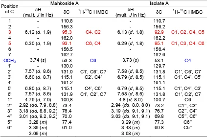
HMBC experiment and comparison of NMR data with mahkoside A, (4,4’-dihydroxy-6-methoxybenzo phenone-2-O-β-D-glucopyranoside) [4] (Table 1), it was clarified that isolate A was 6,4’-dihydroxy-4-methoxy-benzophenone-2-O--D-glucopyranoside (3) (Fig. 3). This compound has been isolated from Gnidia involucrata by Ferrari, el al.[7]. The structure was similar with mahkoside A except the positon of 6-hydroxy and
4-methoxy in mahkoside A to be a 4-hydroxy and 6-methoxy. This position was confirmed by HMBC experiment as shown in Fig. 3. This 1H- and 13C-NMR data were similar with benzophenone glycoside isolated from the leave of Phaleria macrocarpa by Kusmardiyani et al. [8], but OCH3, -OH, and gucoside
positions were not elucidated, since the 2D-NMR experiment did not examinized. Similar compound, but
Fig. 2. (a). The -glucopyranoside moiety (b). Aromatic ring with meta-coupling (c). Aromatic ring with ortho-coupling
Table 1.1H, 13C, and HMBC data of Mahkoside A [5] and isolate A, both in DMSO-d6
Mahkoside A Isolate A
Position
of C H
(mult, J in Hz) C
1
H-13C HMBC H
(mult, J in Hz) C
1
H-13C HMBC
1 - 110.8 - 110.7
2 - 156.3 - 156.2
3 6.12 (d, 1.9) 95.3 C4, C2 6.13 (d, 1.8) 92.9 C1, C2, C4, C5
4 - 162.0 - 162.2
5 6.30 (d, 1.9) 93.1 C6, C4 6.29 (d, 1.8) 95.1 C1, C3, C4, C6
6 - 156.5 - 156.4
7 192.7 192.6
OCH3 3.74 (s) 53.3 C6 3.73 (s) 53.1 C4
1’ - 130.0 - 129.7
2’ 7.57 (d, 8.6) 131.9 C1’, C6’, C7 7.58 (d, 8.5) 131.8 C1’, C6’, C7 3’ 6.60 (d, 8.7) 115.1 C2’, C4’ 6.79 (d, 8.5) 115.1 C1’, C4’, C5’
4’ - 161.2 - 161.1
5’ 6.80 (d, 8.7) 115.1 C4’, C6’ 6.79 (d, 8.5) 115.1 C1’, C4’, C3’ 6’ 7.57 (d, 8.6) 131.9 C1’, C2’, C7 7.58 (d, 8.5) 131.8 C1’, C2’, C7 1’’ 4.79 (d, 7.9) 100,8 4.8 (d, 8.0) 100.7 C6 2’’ 2.92 (dd, 7.9, 8.8) 73.4 2.94 (dd, 8.0, 8.0) 73.2 C1’’, C3’’ 3’’ 3,18 (dd, 8.8, 9.2) 76.4 3.19 (dd, 9.1, 9.1) 76.7 C2’’, C4’’ 4’’ 3.01 (dd, 9.2, 9.2) 70.0 3.03 (dd, 9.1, 9.1) 69.8 C5’’, C6’’
5’’ 3.28 (m) 77.4 3.29 (m) 77.3 C6’’
6’’ 3.39 (m) 3.69 (m)
61.0 3.43 (m)
3.68 (m)
Indo. J. Chem., 2009, 9 (1), 142 - 145
Hendig Winarno and Ermin Katrin W
145
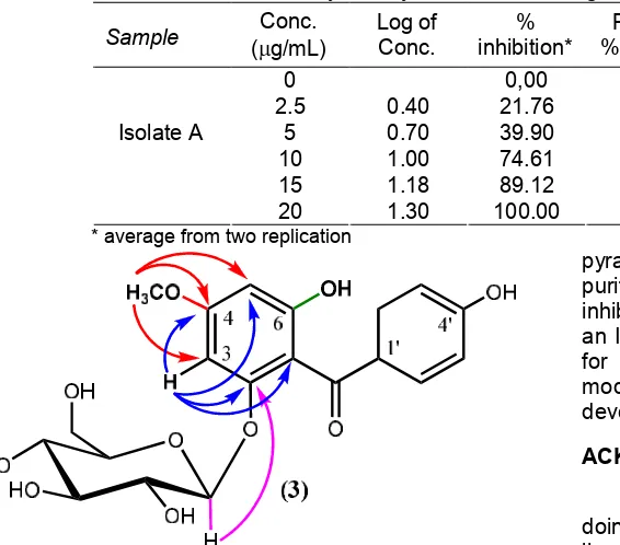
Table 2. Inhibitory activity test of isolate A against mouse leukemia L1210 cell line
Sample Conc.
(g/mL)
Log of Conc.
% inhibition*
Probit of
% inhibiton Regr. Linier eq.
IC50
(µg/mL)
0 0,00
-2.5 0.40 21.76 4,23
5 0.70 39.90 4,75
10 1.00 74.61 5,67
15 1.18 89.12 6.23
Isolate A
20 1.30 100.00 8.09
y = 3.769x + 2.35 5.1
* average from two replication
Fig. 3. 6,4’-dihydroxy-4-methoxybenzophenone-2-O-β -D-glucopyranoside (3) (arrow: HMBC correlation)
the glucoside moiety was an -anomeric, namely 6,4’-dihydroxy-4-methoxy-benzophenone-2-O--D-glucopyra noside have been found from the fruit of Phaleria macrocarpa by Tambunan, et al. [9].
Inhibitory activity test
Inhibitory activity test of isolate A against mouse leukemia L1210 cell line showed that this isolate A exhibited inhibitory activity with an IC50 5.1 µg/mL (Table
2). This IC50 value of isolate A was lower than that its
fraction, but it was still > 4.0 µg/mL. According to Swanson et al. [10], the isolate or compound has a potent anticancer agent if it has an IC50 ≤ 4.0 µg/mL in
inhibitory activity test against cancer cell line in vitro. So, for further study, the structure and function group modifications of the isolate are needed for developing the better activity. Similar benzophenone glucoside, namely phalerin isolated by Wahyuningsih, et al. [3] from the leaves of Phaleria macrocarpa exhibited the less cytotoxic effect against myeloma NS-1 with an IC50 83
µg/mL.
Isolate B (20.2 mg) and isolate C (4.3 mg) were not collected in pure, and it is not enough for structure elucidation, so the collection and purification are necessary.
CONCLUSION
From the bark of Phaleria macrocarpa have been isolated a benzophenone glucoside compound, namely 6,4’-dihydroxy-4-methoxybenzophenone-2-O--D-gluco
pyranoside (3) and isolate B and C which were not purified yet. This benzophenone glucoside showed an inhibitory activity against mouse leukemia L1210 with an IC50 5.1 µg/mL, it is higher rather than activity limit
for isolate. So, the structure and function group modifications of the isolate are necessary for developing the better activity.
ACKNOWLEDGEMENT
The authors would like to thank Mr. Suhanda for doing bioassay experiment using leukemia L1210 cell line.
REFERENCES
1. Aditama, T.Y., Kanker, 2001, Medisinal Jurnal Kedokteran, 2, 1-5.
2. Anonymous, Mahkota Dewa Nusantara (Phaleria macrocarpa (Scheff.) Boerl.). http://www.trubus-online.com, to be accessed on March 11, 2008. 3. Wahyuningsih, M.S.H., Mubarika, S., Gandjar, I.G.,
Hamann, M.T., Rao, K.V., and Wahyuono, S., 2005, Indonesian J. Pharm.,16 (1), 51-57.
4. Zhang, Y-B., Xu, X-J., and Liu, H-M., 2006, J. Asian Nat. Prod. Res., 8 (1-2), 119-123.
5. Winarno, E.K. and Winarno, H., 2008, J. Traditional Medicine. Submitted.
6. Silverstein, R.M., Bassler, G.C, and Morrill, T.C., 1991, Spectrometric Identification of Organic Compounds, 5th ed., John Wiley & Sons, Inc., New York.
7. Ferrari, J., Terreaux, C., Sahpaz, S., Msonthi, J.D., Wolfender, J.L., and Hostettmann, K., 2000,
Phytochemistry, 54 (8), 883-889.
8. Kusmardiyani, S., Nawawi, A., Rahmi, K., 2004,
Acta Pharmaceutica Indonesia, 29 (4), 150-152. 9. Tambunan, R.M. and Simanjuntak, P., 2006,
Indonesian J. Pharm.,17 (4), 184-189.
![IN VITRO BIOACTIVITY TEST OF IRRADIATED MAHKOTA DEWA BARK [ Phaleria macrocarpa ( Scheff. ) Boerl.] AGAINST HUMAN CANCER CELL LINES | Winarno | Indonesian Journal of Chemistry 21370 40456 1 PB](data:image/gif;base64,R0lGODlhAQABAIAAAP///wAAACH5BAEAAAAALAAAAAABAAEAAAICRAEAOw==)